Loading video…


 17 Concepts
 1.3M views

Video Summary: What Is Labeling DNA Probes
How do researchers detect a single gene among the billions of base pairs in human DNA? Labeling DNA probes makes this molecular detective work possible by attaching detectable markers to complementary DNA sequences. Scientists at the CDC use these labeled probes to rapidly identify pathogenic bacteria in clinical samples, transforming diagnosis from weeks to hours. What is labeling DNA probes involves chemically modifying single-stranded DNA with radioisotopes, fluorophores, or biotin for precise target detection. Watch the full video on JoVE Coach to master this concept with expert-led visuals and step-by-step explanations.
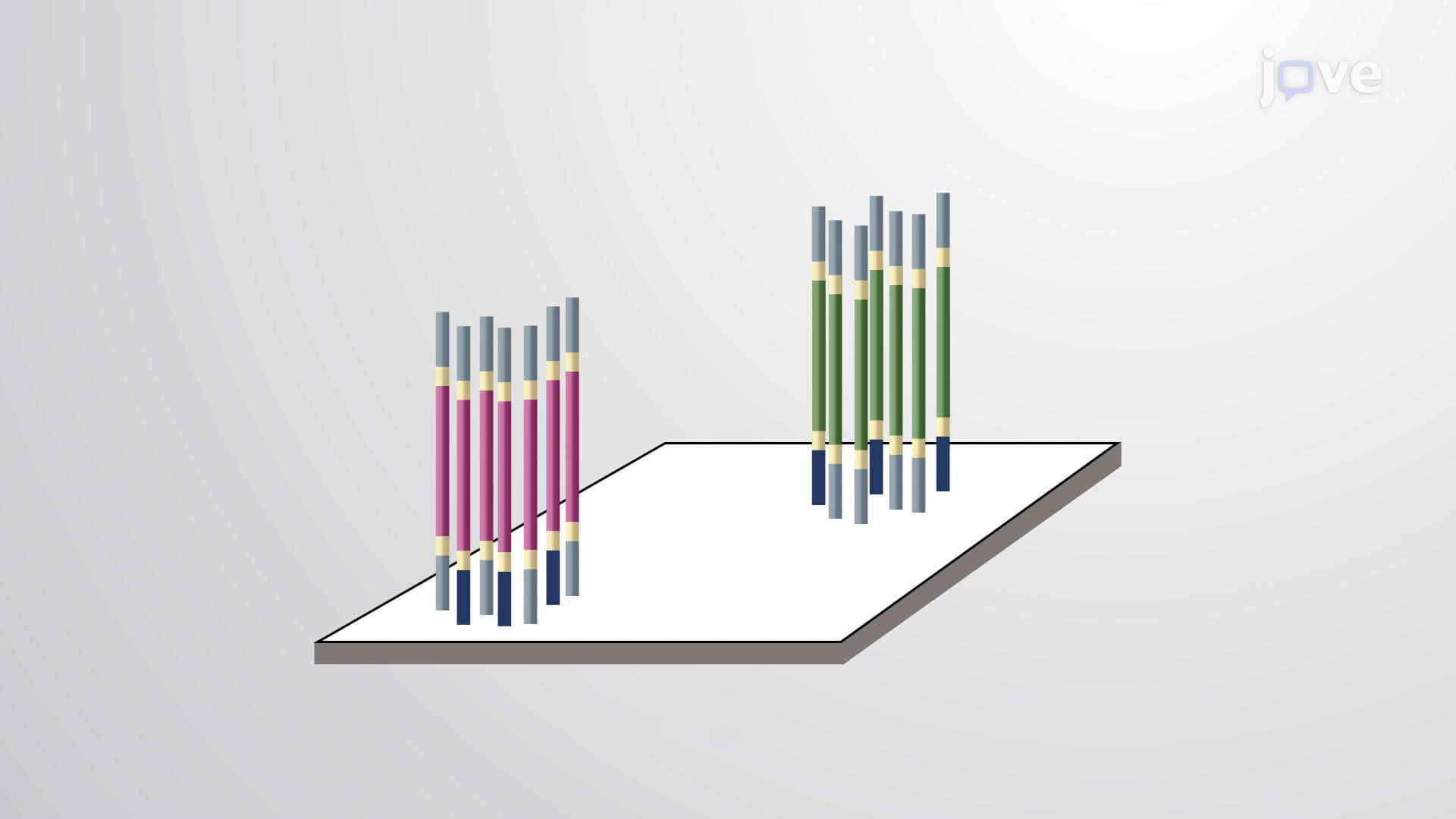
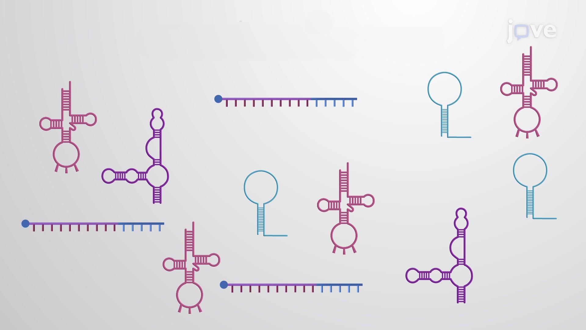
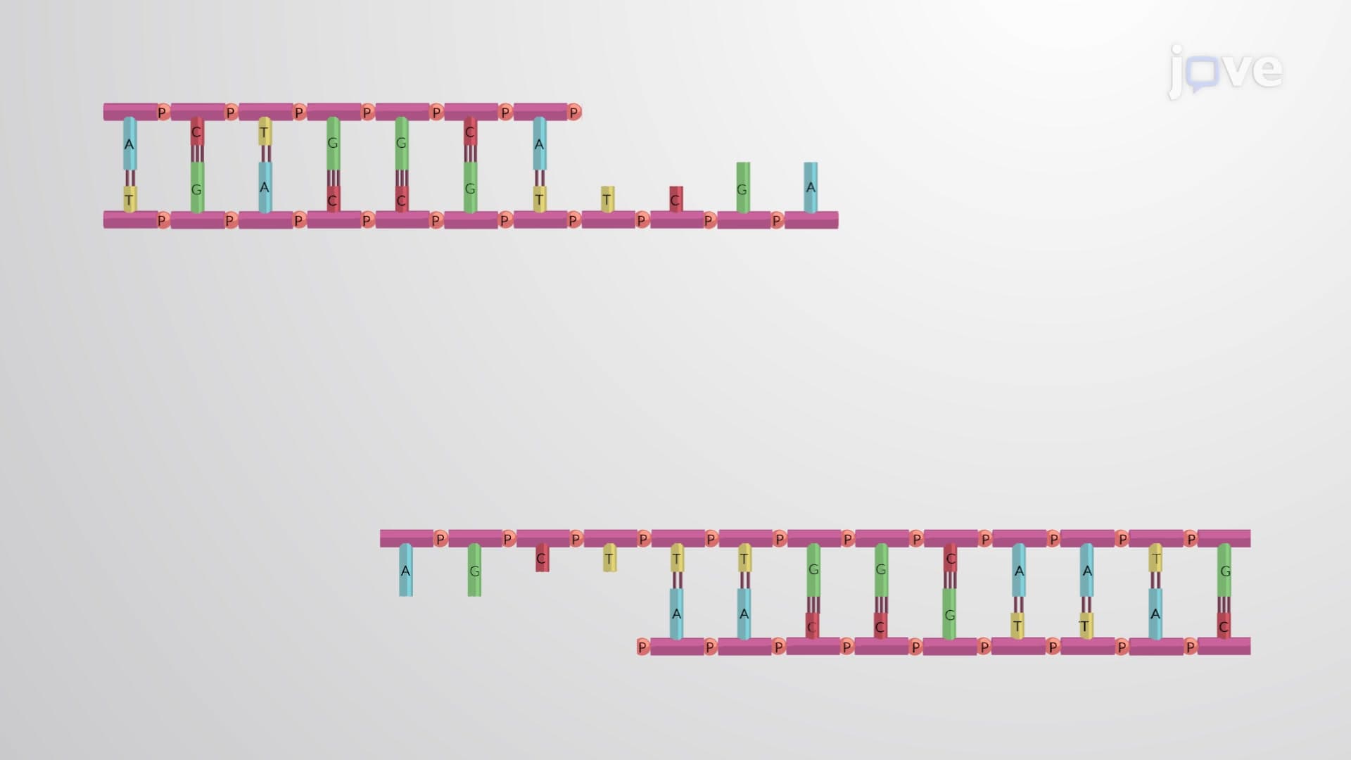
Labeling DNA probes represents a cornerstone technique in molecular biology that transforms simple DNA sequences into powerful detection tools. This process involves chemically modifying single-stranded DNA molecules with detectable markers, enabling researchers to locate specific target sequences within complex genomic samples. The concept builds on Watson-Crick base pairing rules, where complementary sequences hybridize with high specificity, making probe-based detection both reliable and precise.
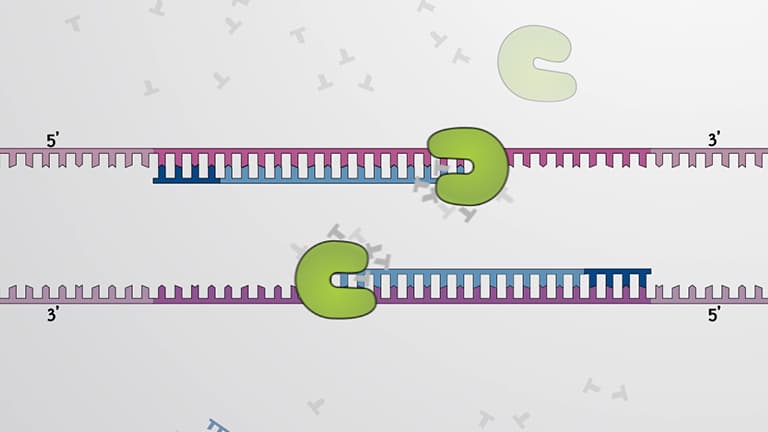
Two primary approaches dominate DNA probe labeling: end-labeling and internal labeling. End-labeling attaches detection molecules to the 5' or 3' termini of probe sequences, typically through phosphate or hydroxyl group modifications. This method preserves the probe's internal structure while providing clear detection signals. Internal labeling, exemplified by nick translation, uses DNase I to create single-strand breaks followed by DNA polymerase I incorporation of labeled nucleotides throughout the sequence.
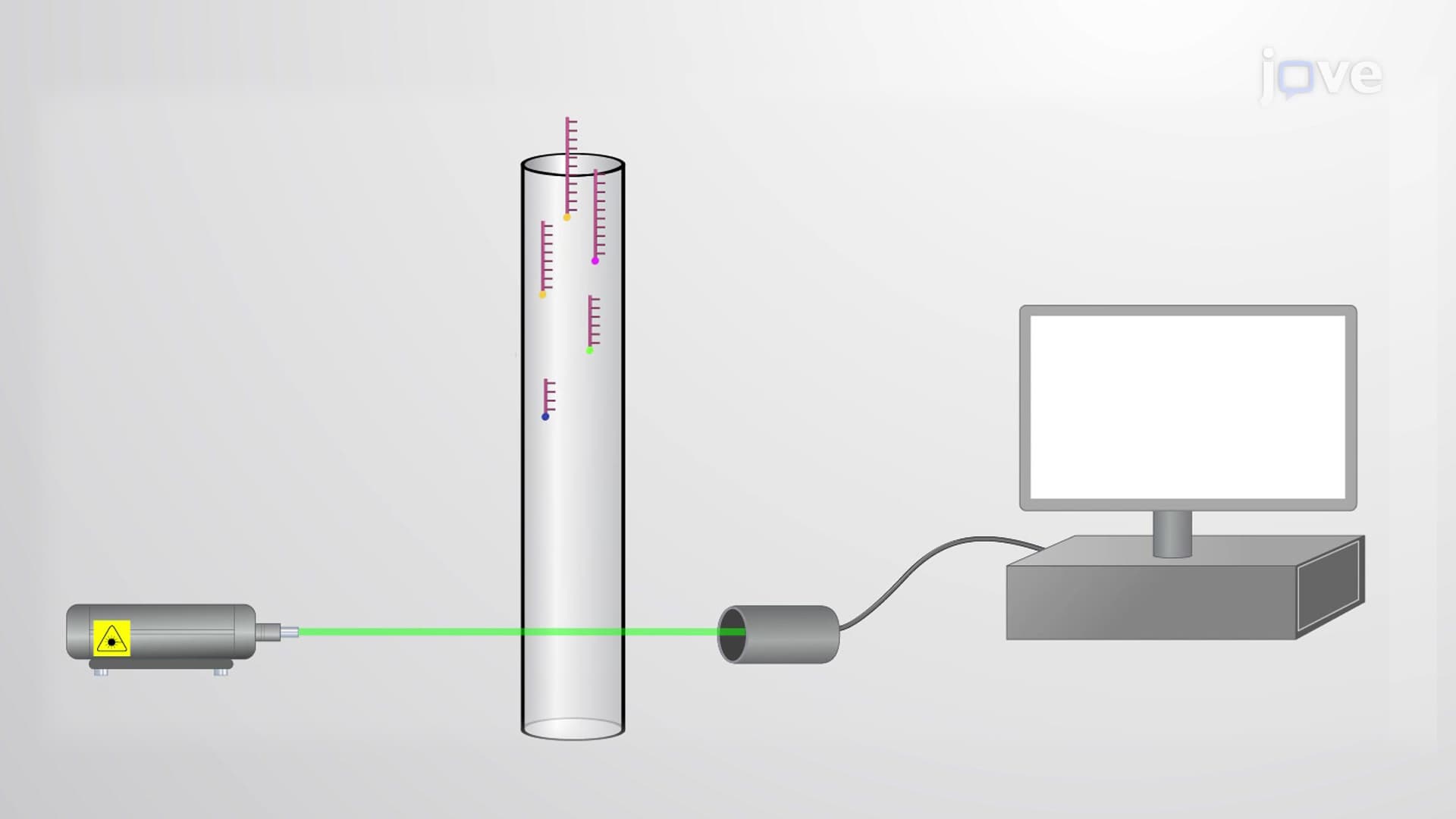
The choice of label significantly impacts experimental success. Radioisotopes like Phosphorus-32 offer exceptional sensitivity through autoradiography but require specialized facilities and have limited shelf life. Fluorophores such as fluorescein provide stable, safe alternatives detectable through fluorescence microscopy at specific wavelengths. The biotin-streptavidin system represents the most versatile approach, combining biotin's small molecular size with streptavidin's incredibly strong binding affinity (Kd ≈ 10^-15 M).
DNA probe labeling finds extensive application in US clinical laboratories and research institutions. The FDA-approved GenProbe assays used in hospitals nationwide rely on chemiluminescent-labeled probes for rapid pathogen identification. Stanford Medical Center's microbiology lab, for example, uses labeled probes to detect Mycobacterium tuberculosis in patient samples within hours rather than the weeks required for traditional culture methods.
Students preparing for the MCAT encounter DNA probe labeling within molecular biology and biochemistry sections, particularly regarding hybridization techniques and detection methodologies. AP Biology students study this concept when learning about gene expression analysis and genetic engineering applications. College biochemistry courses typically cover probe labeling in the context of Southern blotting, Northern blotting, and in situ hybridization protocols.
Understanding probe denaturation requirements proves crucial for exam success. Double-stranded probe DNA must be heat-denatured or chemically treated to create single-stranded molecules capable of target binding. The temperature and timing parameters directly influence hybridization efficiency and experimental reproducibility.
Related Micro-courses