Loading video…


 17 Concepts
 1.3M views

Video Summary: What Is Real Time Rt Pcr
Did you know that COVID-19 tests rely on the same technology used to detect genetic diseases and monitor cancer treatment? Real time RT PCR revolutionized molecular diagnostics by allowing scientists to both amplify and quantify RNA in a single process. The FDA-approved Abbott RealTime SARS-CoV-2 assay exemplifies how what is real time rt PCR transforms patient care through rapid, precise genetic analysis. This powerful technique combines reverse transcription with polymerase chain reaction, enabling real-time monitoring of amplification through fluorescent detection methods. Watch the full video on JoVE Coach to master this concept with expert-led visuals and step-by-step explanations.
Real time RT PCR represents a sophisticated molecular technique that combines reverse transcription with polymerase chain reaction, enabling simultaneous amplification and quantification of RNA targets. Unlike traditional PCR that only amplifies DNA, this method first converts RNA into complementary DNA (cDNA) through reverse transcriptase enzyme activity. The "real-time" component distinguishes this technique from endpoint PCR by monitoring amplification as it occurs, rather than analyzing products after completion.
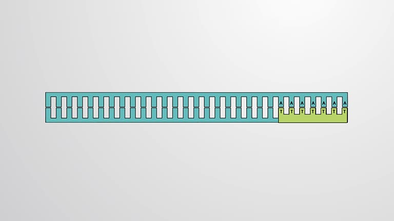
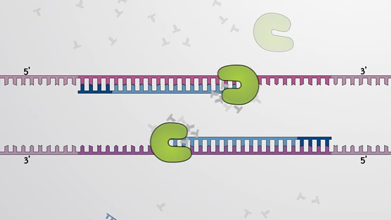
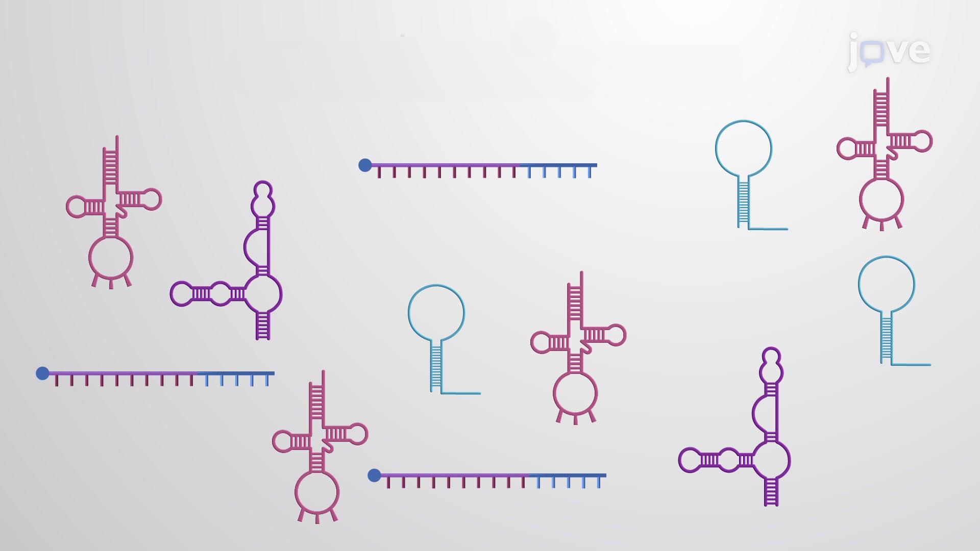
The process begins when reverse transcriptase synthesizes cDNA from the RNA template, followed by RNase H degradation of the original RNA strand. DNA polymerase then creates the complementary strand, establishing the double-stranded template required for PCR amplification. This conversion step is crucial because RNA's inherent instability and single-stranded nature make it unsuitable for direct PCR amplification.
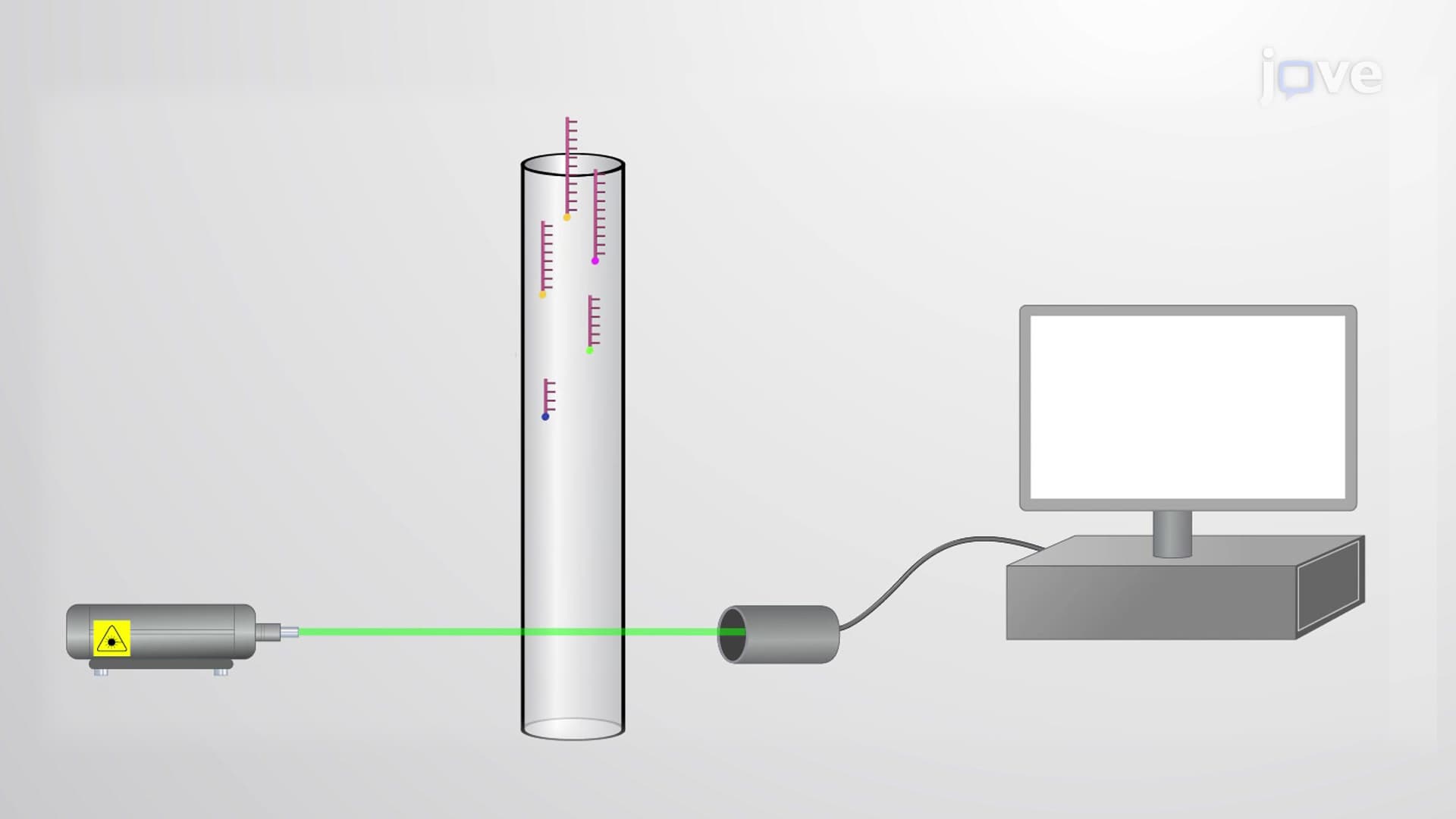
Two primary detection methods enable real-time monitoring of amplification. DNA-binding dyes, such as SYBR Green, intercalate between double-stranded DNA bases and fluoresce only when bound. While cost-effective and simple, this non-specific method detects all double-stranded DNA products, including primer dimers and non-target amplicons.
Probe-based detection offers superior specificity through sequence-specific oligonucleotides labeled with fluorophores and quencher molecules. The TaqMan probe system, widely used in clinical diagnostics, employs fluorescence resonance energy transfer (FRET). When the probe remains intact, the quencher molecule suppresses fluorescence. During amplification, DNA polymerase's 5' exonuclease activity cleaves the probe, separating fluorophore from quencher and generating detectable fluorescence proportional to amplicon formation.
The threshold cycle (Ct) serves as the fundamental quantitative parameter, representing the PCR cycle where fluorescence exceeds background levels by a predetermined threshold. This inverse relationship between Ct values and initial RNA concentrations enables precise quantification: lower Ct values indicate higher target concentrations, while higher Ct values suggest lower initial amounts.
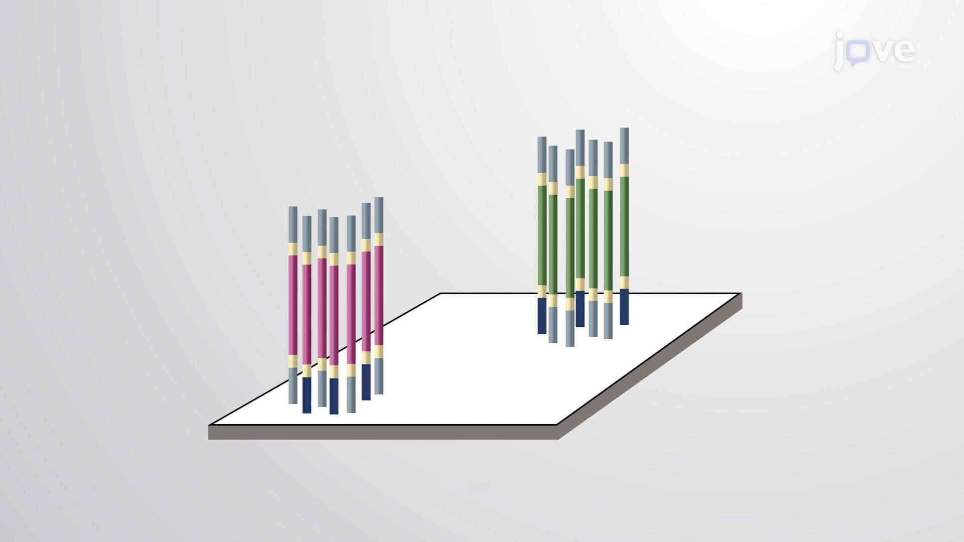
Absolute quantification requires standard curves generated from known DNA concentrations, enabling determination of exact target copy numbers. Relative quantification compares sample fluorescence to reference controls, making it ideal for gene expression studies where fold-changes matter more than absolute values. This approach proves invaluable in cancer research, where oncogene expression levels indicate treatment responses.
Understanding real time RT PCR proves essential for students pursuing healthcare careers, particularly those preparing for the MCAT's biochemistry sections or AP Biology exams covering molecular techniques. College-level courses in molecular biology, clinical chemistry, and diagnostic laboratory science extensively cover these principles. Students should focus on distinguishing between detection methods, interpreting Ct values, and recognizing applications across infectious disease diagnostics, genetic testing, and pharmaceutical development.
Related Micro-courses