Loading subject page…


1,346,738 views


Video Summary: What Is Fish Fluorescent
Ever wondered how researchers at Johns Hopkins can pinpoint a single gene causing cystic fibrosis among thousands of chromosomes? Fish fluorescent, or Fluorescence In Situ Hybridization (FISH), makes this molecular detective work possible by using fluorescent DNA probes that light up specific genetic targets like a GPS for genes. This powerful technique helps diagnose genetic disorders in patients at leading US medical centers by revealing chromosome abnormalities invisible to the naked eye. Watch the full video on JoVE Coach to master this concept with expert-led visuals and step-by-step explanations.
Fish fluorescent, scientifically known as Fluorescence In Situ Hybridization (FISH), represents a revolutionary molecular biology technique that transforms invisible genetic information into vivid, observable fluorescent signals. This method serves as a bridge between abstract DNA sequences and tangible visual evidence, making it invaluable for both research and clinical diagnostics across major US medical institutions.
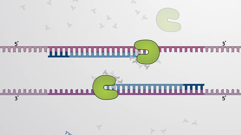
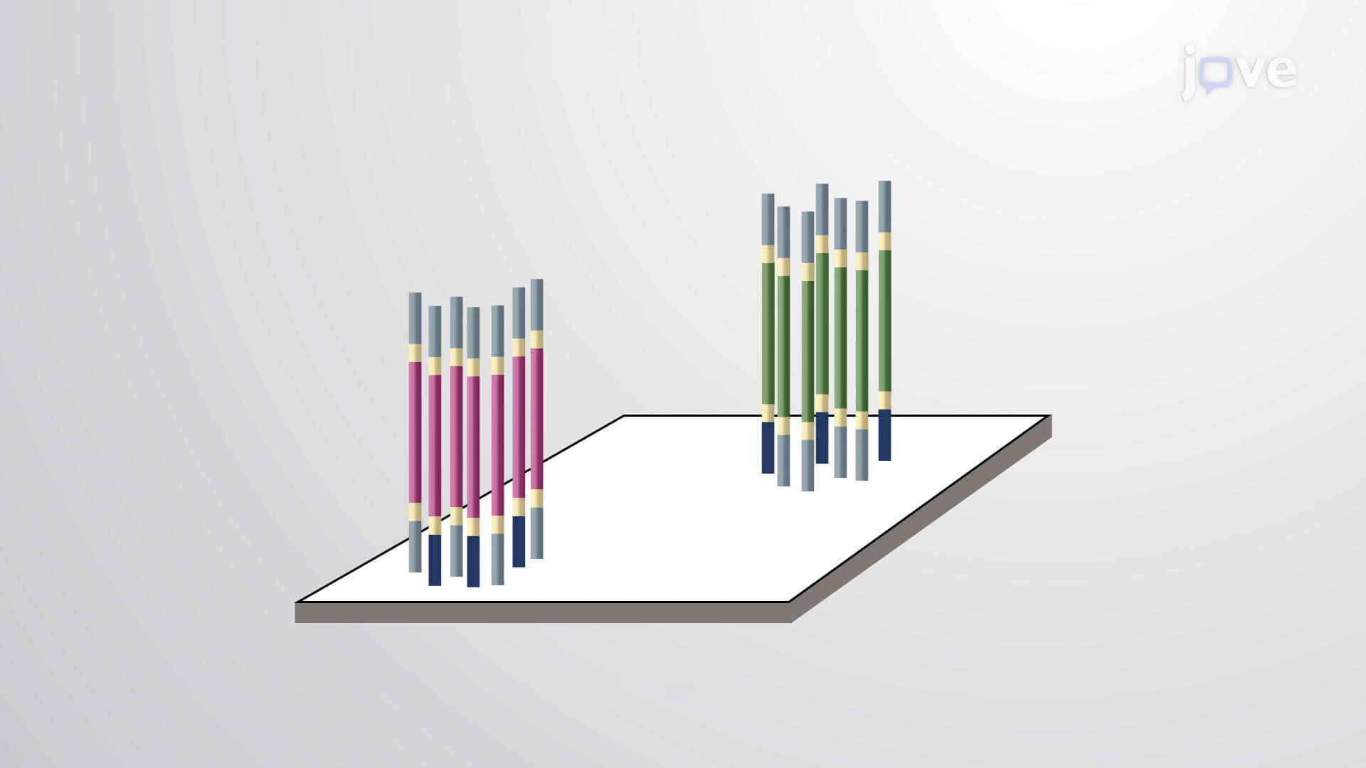
The fish fluorescent process operates on the fundamental principle of DNA base pairing, where complementary sequences naturally attract and bind to each other. Scientists design single-stranded DNA probes that match specific target sequences within chromosomes. These probes carry fluorescent markers-either directly attached fluorophores or indirect hapten molecules that later bind fluorescent antibodies.
The technique begins with cell preparation, where researchers arrest cells during interphase or metaphase to capture chromosomes in their most accessible state. Heat or chemical denaturation then separates double-stranded DNA into single strands, creating opportunities for the fluorescent probes to find and bind their complementary targets. This hybridization process occurs during overnight incubation, allowing sufficient time for stable probe-target binding.
In US healthcare systems, fish fluorescent serves as a cornerstone diagnostic tool for identifying genetic abnormalities. Major medical centers like Mayo Clinic and Cleveland Clinic routinely use FISH to diagnose chromosomal disorders including Down syndrome (trisomy 21), DiGeorge syndrome, and various cancer-associated genetic changes. The technique excels at detecting numerical chromosomal abnormalities (aneuploidy), structural rearrangements, and gene amplifications that indicate disease progression.
Prenatal genetic counselors frequently employ FISH testing on amniotic fluid samples to screen for chromosomal abnormalities in developing fetuses. Cancer geneticists use specialized FISH panels to identify oncogene amplifications and tumor suppressor gene deletions that guide targeted therapy decisions.
For students preparing for advanced placement biology exams, the MCAT, or college genetics courses, understanding fish fluorescent connects theoretical genetics knowledge to practical laboratory applications. AP Biology frequently includes questions about molecular techniques, while pre-med students encounter FISH principles in genetics and cell biology coursework. The technique demonstrates key concepts including DNA structure, complementary base pairing, and the relationship between genotype and phenotype that appear across multiple standardized assessments.
Related Micro-courses