Loading subject page…

- Molecular Biology
- Studying DNA and RNA
- Maxam-Gilbert Sequencing
Micro-course: Studying DNA and RNA
1,346,737 views
Learn key concepts and earn your certificate.







Maxam-Gilbert Sequencing

00:00
01:05
Video Summary: What Is Maxam Gilbert Sequencing
Ever wondered how scientists decoded the first complete DNA genome at Harvard in the 1970s? Maxam Gilbert sequencing revolutionized molecular biology by using chemical cleavage to read DNA sequences base by base. This groundbreaking technique, developed by Allan Maxam and Walter Gilbert at Harvard University, earned Gilbert a Nobel Prize and paved the way for modern genomics research used in US medical centers today. Watch the full video on JoVE Coach to master this concept with expert-led visuals and step-by-step explanations.
- Understand what is Maxam Gilbert sequencing and its fundamental principle of chemical DNA cleavage
- Identify the four specific chemical reagents used and their selective nucleotide targets in the sequencing process
- Analyze how radiolabeling with Phosphorus-32 enables fragment detection through autoradiography
- Compare the chemical specificity of formic acid (purines) versus hydrazine (pyrimidines) in base modification
- Evaluate the step-by-step workflow from DNA denaturation to gel electrophoresis and sequence interpretation
- Apply knowledge of fragment length patterns to decode DNA sequences from autoradiographic results
- Recognize the historical significance of this method in early genome sequencing projects
- Explore the relationship between chemical cleavage sites and resulting fragment migration patterns
Understanding Maxam Gilbert Sequencing Fundamentals
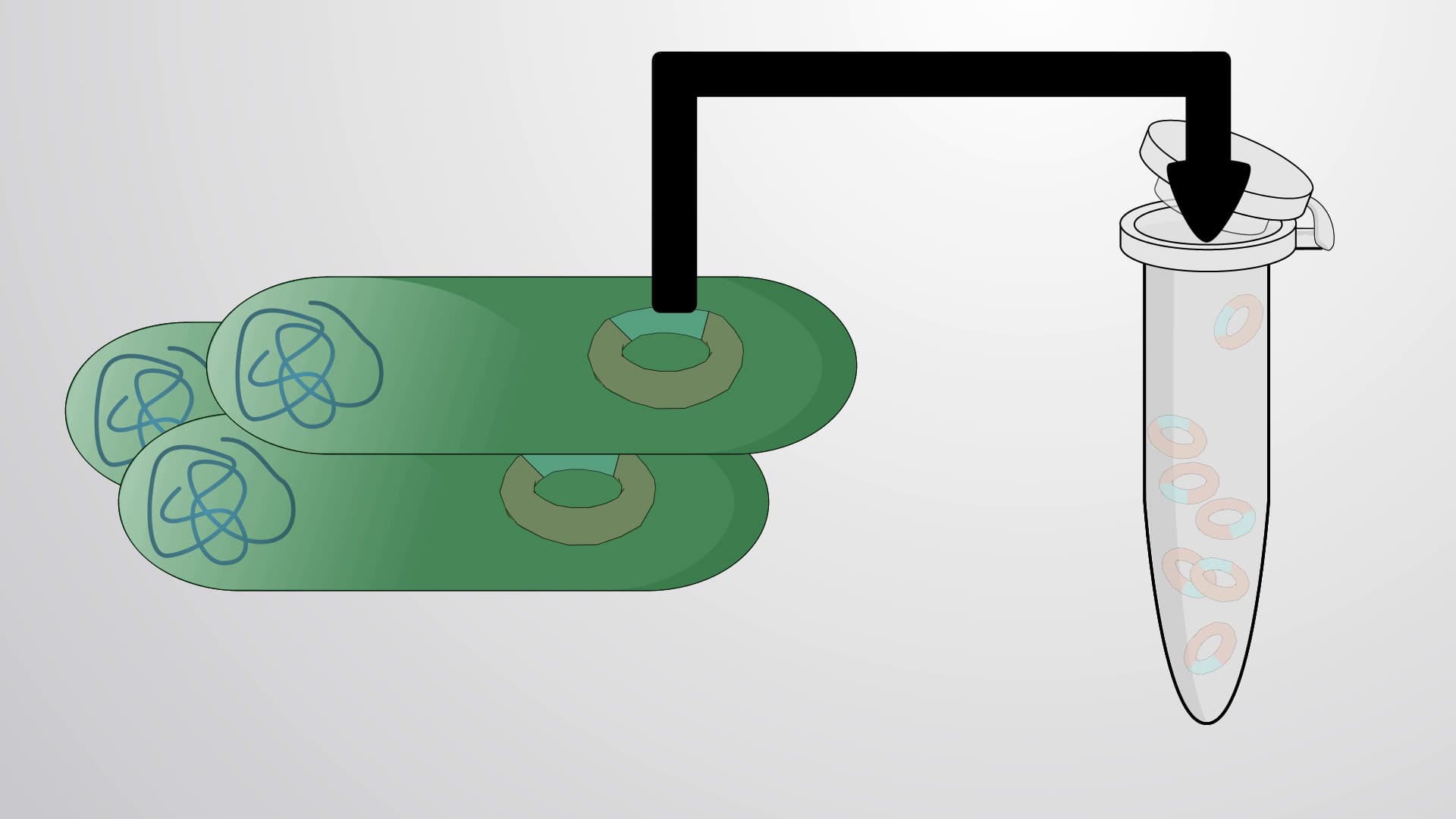
What is Maxam Gilbert sequencing represents one of the first successful methods for determining DNA sequences, developed in the mid-1970s at Harvard University. Unlike modern automated sequencing, this chemical cleavage approach relies on the selective modification and cleavage of specific DNA bases using targeted chemical reagents. The method's elegance lies in its systematic approach: each of four parallel reactions targets different nucleotides, creating a unique "fingerprint" of DNA fragments that reveals the original sequence.
The Chemical Arsenal: Reagent Specificity and Base Targeting
The power of Maxam Gilbert sequencing stems from its precise chemical toolkit. Formic acid specifically attacks purine bases (adenine and guanine), while hydrazine targets pyrimidines (cytosine and thymine). More refined reagents like dimethyl sulfate can even distinguish between individual nucleotides, providing single-base resolution. This chemical specificity creates predictable cleavage patterns that, when combined across all four reactions, generate a complete sequence readout. Students preparing for AP Biology or college biochemistry courses should understand how each reagent's selectivity contributes to the overall sequencing strategy.
From Radiolabeling to Autoradiography: The Detection System
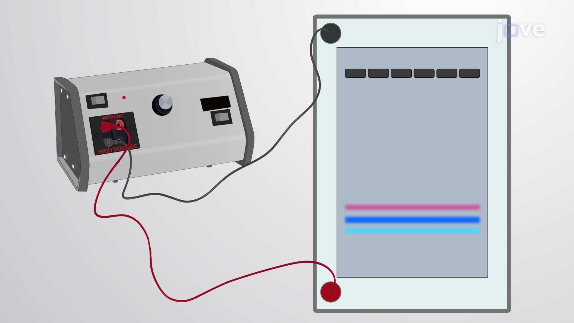
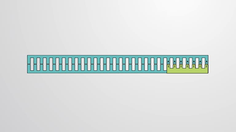
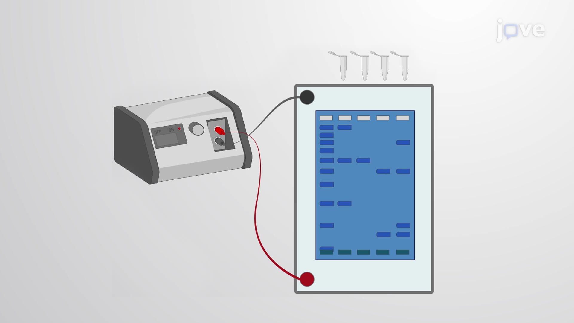
The method begins with 5'-end radiolabeling using Phosphorus-32, a radioactive isotope that serves as the detection mechanism. After chemical modification at specific bases, piperidine cleaves the DNA backbone at these modified sites, generating fragments of varying lengths. The radioactive label allows these fragments to be visualized on X-ray film through autoradiography after gel electrophoresis separation. This detection system was revolutionary for its time, enabling researchers to "see" DNA sequences directly.
Reading the Sequence: Interpretation and Applications
Sequence determination requires careful analysis of the autoradiographic pattern, reading from bottom (shortest fragments) to top (longest fragments). Each band represents a cleavage site, and comparing patterns across the four chemical reactions reveals the nucleotide identity at each position. This technique was instrumental in early genome projects, including sequencing viral genomes and smaller bacterial sequences. Students encountering this concept in MCAT preparation should focus on understanding the logical flow from chemical specificity to fragment pattern interpretation.
Frequently Asked Questions
Maxam Gilbert sequencing is a chemical method for determining DNA sequences by selectively cleaving DNA at specific nucleotides using targeted chemical reagents. The process involves radiolabeling DNA, treating it with base-specific chemicals, cleaving at modified sites, and analyzing fragment patterns through gel electrophoresis and autoradiography.
MCAT questions often focus on the chemical specificity of reagents and sequence interpretation from gel patterns. Students should understand which chemicals target which bases (formic acid for purines, hydrazine for pyrimidines) and how to read autoradiographic results from bottom to top.
AP Biology may contrast Maxam Gilbert's chemical cleavage approach with modern chain-termination methods like Sanger sequencing. Key differences include the use of radioactive labeling versus fluorescent tags, chemical modification versus enzymatic incorporation, and manual gel reading versus automated detection.
While largely replaced by modern methods, the principles are still taught in biochemistry programs at institutions like MIT and Stanford, and the chemical modification techniques influence current DNA damage research and forensic analysis methods used by the FBI and medical examiners.
The basic concept is accessible to students with general chemistry knowledge of acids, bases, and chemical specificity. Focus on understanding the logic of selective chemical cleavage rather than memorizing complex chemical structures, making it manageable for honors biology or AP Chemistry students.
Create a flowchart showing the step-by-step process from DNA preparation to sequence reading. Practice interpreting autoradiographic patterns and focus on understanding the chemical basis for reagent specificity. Many professors test pattern interpretation more than detailed chemical mechanisms.
Explore Sanger sequencing to understand the evolution of DNA sequencing technology, then investigate modern next-generation sequencing platforms. This progression shows how scientific methods advance and provides context for current genomics applications in personalized medicine.
Understanding Maxam Gilbert sequencing builds critical thinking skills about chemical specificity and experimental design that apply to modern molecular biology techniques. It also provides historical context for appreciating current high-throughput sequencing capabilities used in research hospitals and genomics companies.
Related Micro-courses
19 Concepts

11 Concepts

18 Concepts