Loading video…


 17 Concepts
 108.3K views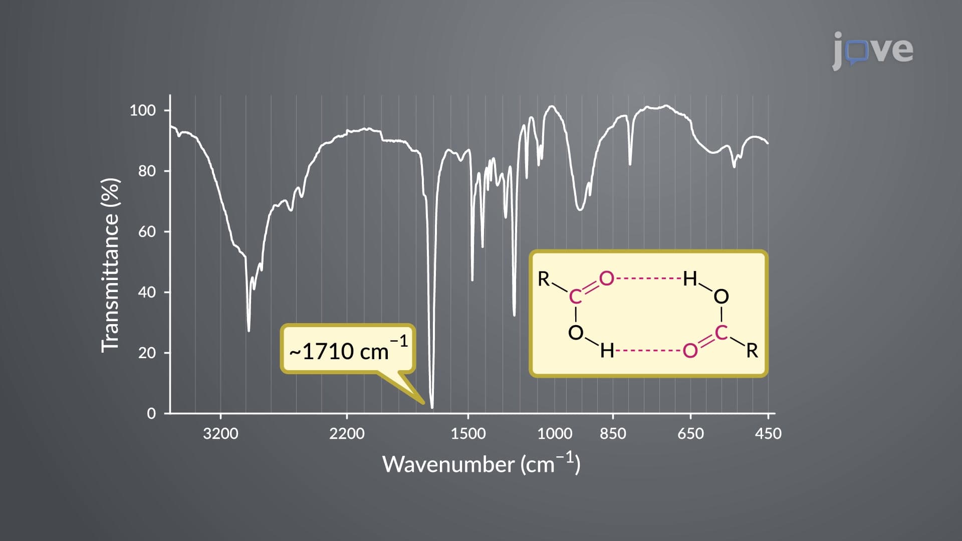

Video Summary: Ir and Uv Vis Spectroscopy Explained
Did you know that forensic labs can identify unknown white powders using just light wavelengths? IR UV VIS spectroscopy combines infrared and ultraviolet-visible light analysis to reveal molecular fingerprints, much like how the FBI's crime labs identify drugs and explosives. IR and UV VIS spectroscopy explained shows how carboxylic acids create distinctive absorption patterns-from the sharp C=O peaks around 1710 cm⁻¹ to broad O-H stretches masking other signals. Watch the full video on JoVE Coach to master this concept with expert-led visuals and step-by-step explanations.
IR and UV VIS spectroscopy explained represents two complementary analytical techniques that pharmaceutical companies, environmental agencies, and research institutions across the United States use daily. While infrared spectroscopy reveals molecular vibrations and functional groups, UV-visible spectroscopy uncovers electronic transitions and conjugation patterns. Together, they provide a comprehensive molecular fingerprint that's essential for drug development at companies like Pfizer and Merck.
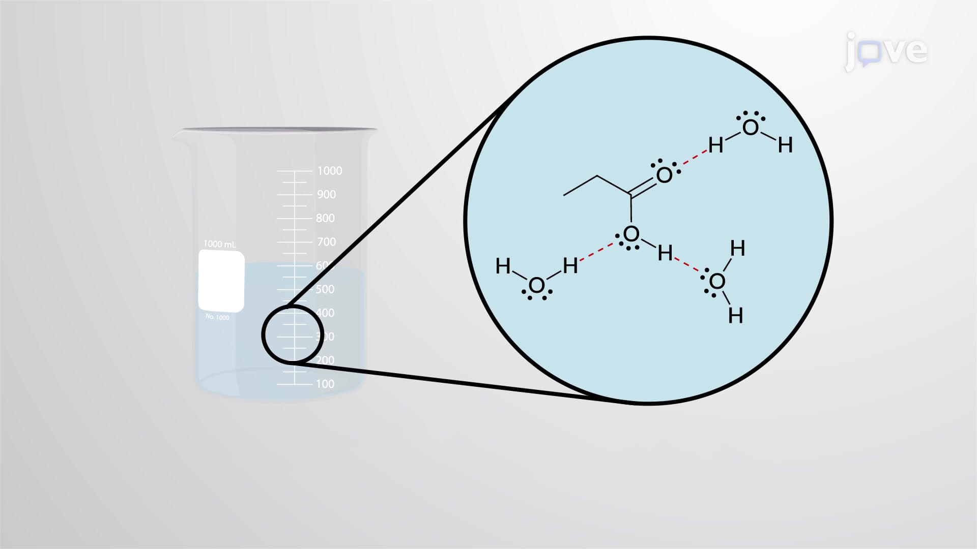
Carboxylic acids demonstrate classic IR UV VIS spectroscopy behavior through two unmistakable absorption patterns. The carbonyl (C=O) stretch appears as a sharp, intense peak, but its exact position tells a structural story. Monomeric carboxylic acids, existing as single molecules, show C=O absorption at 1760 cm⁻¹. However, most carboxylic acids form hydrogen-bonded dimers in concentrated solutions, causing the C=O frequency to drop to 1710 cm⁻¹. This shift occurs because hydrogen bonding weakens the C=O bond strength, requiring less energy to vibrate.
The broad O-H stretch spanning 2500-3500 cm⁻¹ creates another diagnostic feature, often overwhelming nearby C-H peaks. This breadth results from extensive hydrogen bonding networks that create multiple slightly different O-H environments. Students preparing for AP Chemistry or college organic chemistry exams should remember this broad peak as the "carboxylic acid signature" that immediately identifies this functional group.
In the ultraviolet region, carboxylic acids exhibit weak absorption around 200-215 nm corresponding to n→π* electronic transitions. This involves promoting a non-bonding electron from oxygen to an antibonding π orbital of the carbonyl group. The low molar absorptivity (ε ≈ 100) reflects the "forbidden" nature of this transition in quantum mechanical terms.
Conjugated carboxylic acids, like those found in pharmaceutical compounds, show dramatically different behavior. Resonance delocalization not only lowers IR C=O frequencies to 1690 cm⁻¹ but also shifts UV absorption to longer wavelengths with molar absorptivities reaching 25,000. The FDA routinely uses this principle in drug analysis-aspirin's conjugated structure produces characteristic spectroscopic signatures that confirm pharmaceutical purity and identity.
Related Micro-courses