Loading video…


 17 Concepts
 108.3K views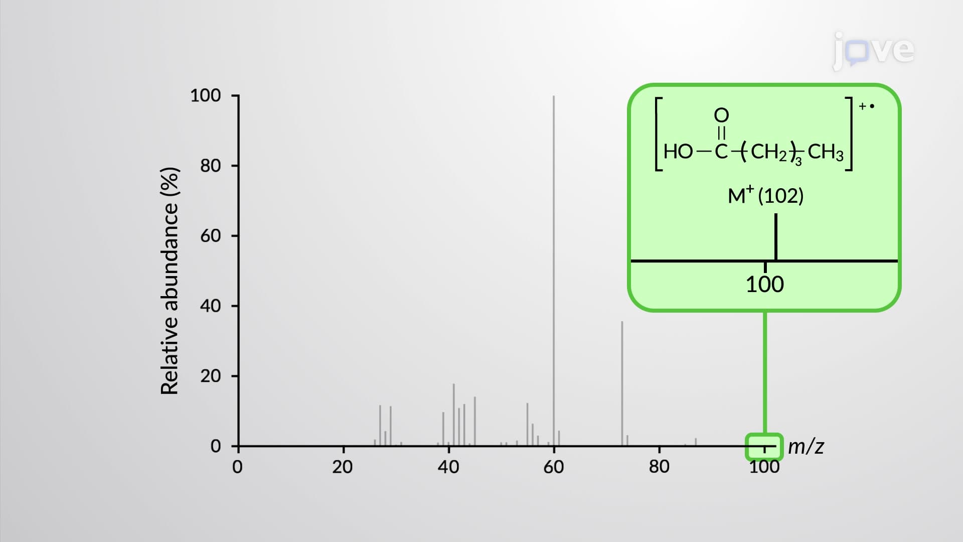

Video Summary: Nmr and Mass Spectroscopy of Explained
Ever wondered how forensic scientists identify unknown substances found at crime scenes? NMR mass spectroscopy provides the molecular fingerprints that solve these mysteries. This powerful analytical technique combines nuclear magnetic resonance with mass spectrometry to reveal both structural details and molecular weights of compounds. The FDA uses these methods daily to verify pharmaceutical purity and identify contaminants in food products. Watch the full video on JoVE Coach to master this concept with expert-led visuals and step-by-step explanations.
Nuclear magnetic resonance (NMR) and mass spectroscopy represent two cornerstone analytical techniques that, when combined, provide unparalleled insight into molecular structure and composition. While NMR reveals the electronic environment and connectivity of atoms within molecules, mass spectroscopy determines molecular weights and fragmentation patterns. Together, these methods form the backbone of modern analytical chemistry used across pharmaceutical, forensic, and research laboratories throughout the United States.
The 1H NMR spectra of carboxylic acids exhibit distinctive features that make identification straightforward. The carboxyl proton (COOH) appears dramatically downfield at 9-12 ppm due to the combined deshielding effects of the electronegative oxygen atoms and the resonance within the carboxyl group. This extreme downfield position distinguishes carboxylic acids from other functional groups like aldehydes (9-10 ppm) or aromatic protons (7-8 ppm). Alpha protons adjacent to the carboxyl group typically resonate at 2-2.5 ppm, while more distant alkyl protons appear further upfield. A particularly useful diagnostic tool involves deuterium exchange: when carboxylic acids are dissolved in deuterated solvents like D2O, the acidic proton exchanges with deuterium, causing its NMR signal to disappear completely.
13C NMR spectroscopy provides complementary structural information by revealing the carbon framework. The carbonyl carbon in carboxylic acids resonates at 160-180 ppm, reflecting significant deshielding from the electron-withdrawing oxygen atoms. This region is characteristic and well-separated from other carbon signals, making it an excellent diagnostic tool. Alkyl carbons appear upfield at 20-40 ppm, following predictable patterns based on their substitution patterns.
Mass spectroscopy reveals distinct fragmentation behaviors between aliphatic and aromatic carboxylic acids. Aliphatic carboxylic acids typically show weak molecular ion peaks due to facile fragmentation processes. The McLafferty rearrangement represents the most common fragmentation pathway, producing stable neutral molecules and characteristic fragment ions that often appear as base peaks. Additionally, loss of alkyl radicals generates resonance-stabilized acylium ions (RCO+), which typically produce the second most intense peaks in the spectrum.
These spectroscopic techniques are fundamental to organic chemistry courses at universities like UCLA, MIT, and Stanford, where students learn structure determination through hands-on laboratory experiences. The MCAT frequently tests understanding of NMR chemical shifts and mass spectral interpretation, making these concepts crucial for pre-medical students. In industrial settings, pharmaceutical companies like Pfizer and Merck rely on these methods for drug development, quality control, and metabolite identification in clinical trials.
Related Micro-courses