Loading subject page…


323,108 views

Video Summary: Bone as Supporting Connective Tissue Explained
Did you know that your bones are actually living tissues that constantly rebuild themselves throughout your life? Bone supporting connective tissue serves as your body's internal framework, much like the steel beams in skyscrapers across cities like New York or Chicago. Understanding bone as supporting connective tissue explained reveals how compact and spongy bone work together to protect vital organs while maintaining structural integrity. Watch the full video on JoVE Coach to master this concept with expert-led visuals and step-by-step explanations.
Bone classified as connective tissue represents one of nature's most sophisticated engineering marvels. Unlike other connective tissues such as cartilage or tendons, bone tissue combines organic flexibility with inorganic strength through its unique matrix composition. The osseous tissue matrix consists of approximately 35% organic components (primarily collagen fibrils) and 65% inorganic mineral salts, creating a composite material stronger than reinforced concrete.
This structural design makes bone tissue ideally suited for its dual role as both protective barrier and load-bearing framework. Consider how the ribcage protects vital organs like the heart and lungs while simultaneously supporting the weight of the upper body during activities ranging from typing at a desk to playing basketball.
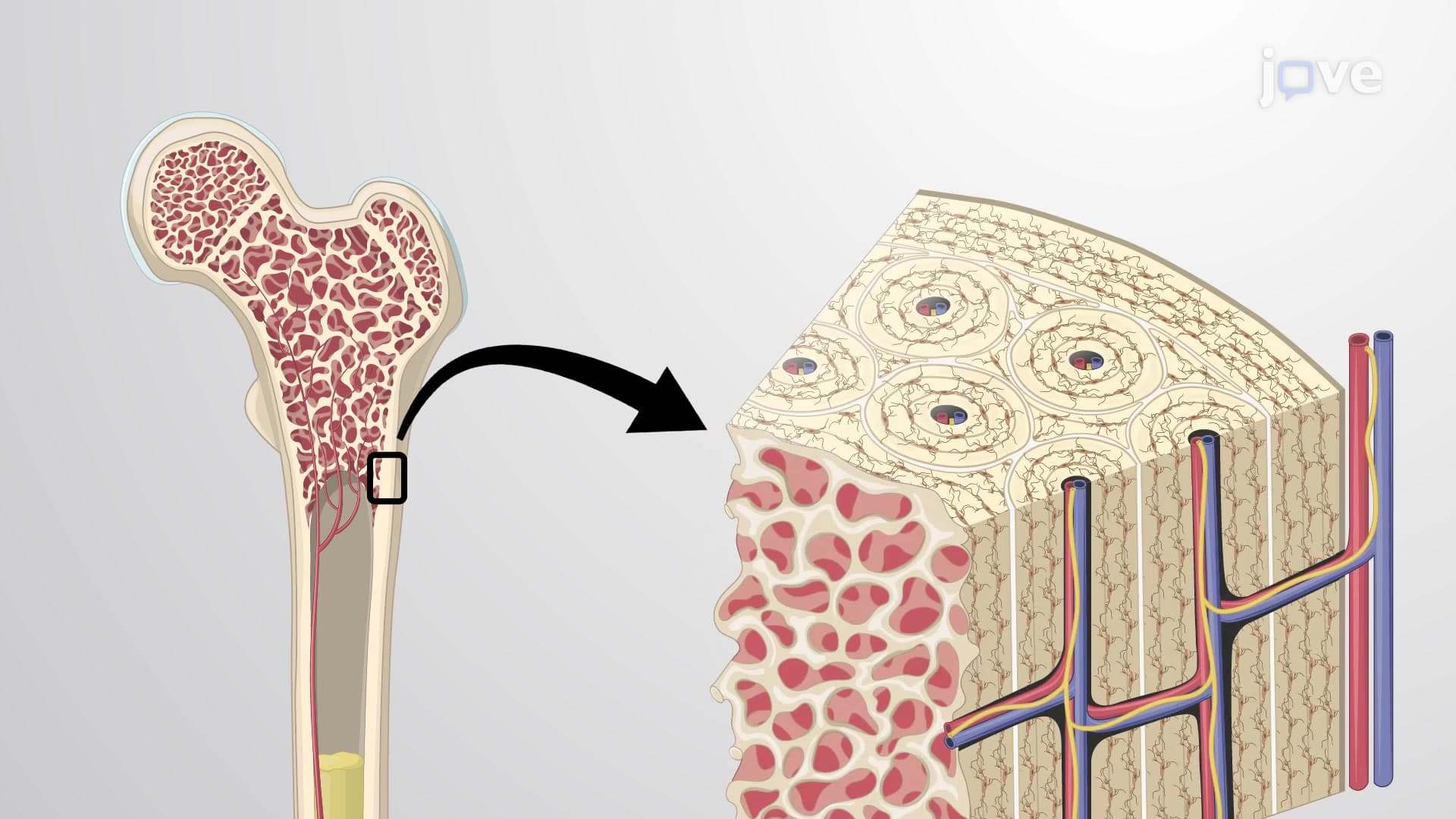
The classification system for bone as connective tissue type divides osseous tissue into two distinct forms based on density and organization. Compact bone, forming the outer shell of all bones, exhibits a highly organized structure with concentric lamellae arranged around central canals. This dense architecture provides maximum strength with minimal weight-a crucial factor for bones like the femur, which must support body weight during activities like running or jumping.
Spongy bone presents a contrasting architecture optimized for different functions. Its trabecular network creates a honeycomb-like structure that houses bone marrow while maintaining strength through strategic placement along stress lines. This design principle appears in structures ranging from the vertebrae in your spine to the ends of long bones, where shock absorption becomes critical.
How bone functions as supporting connective tissue depends heavily on three specialized cell types working in coordination. Osteoblasts function as the construction crew, secreting the osteoid bone organic matrix that forms the foundation for new bone tissue. These cells eventually mature into osteocytes, which act as the bone's maintenance supervisors, monitoring mechanical stress and chemical changes.
Osteoclasts serve as the demolition specialists, breaking down old or damaged bone tissue through a process called resorption. This cellular teamwork enables bones to adapt to changing demands-explaining why astronauts experience bone loss in zero gravity or why weight-bearing exercise strengthens skeletal structure.
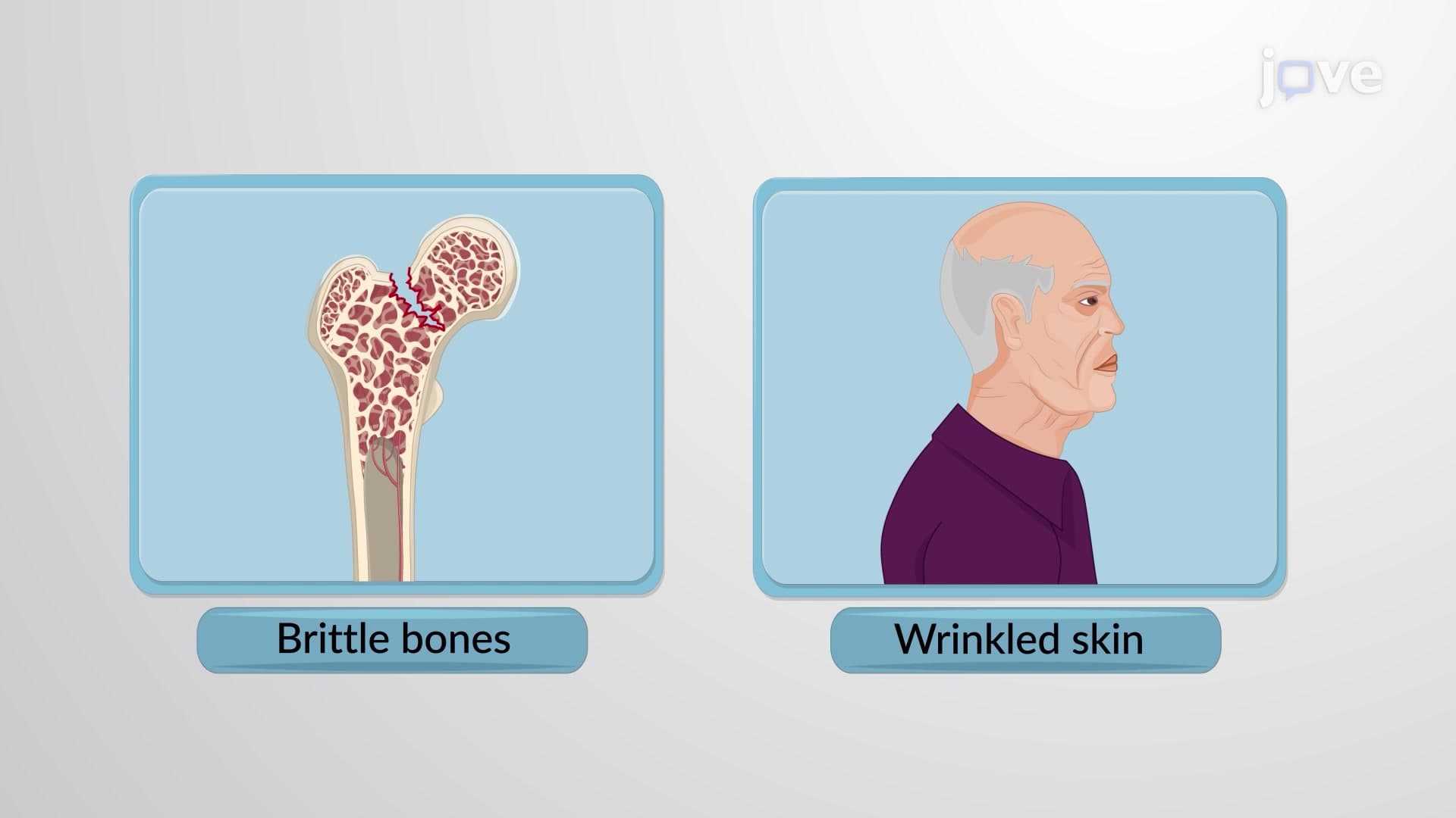
Understanding bone mineral connective tissue composition proves essential for students preparing for exams like the MCAT or AP Biology. Questions frequently focus on how diseases like osteoporosis disrupt the balance between bone formation and resorption, or how fractures heal through accelerated remodeling processes. In clinical settings, this knowledge helps healthcare professionals understand conditions ranging from stress fractures in athletes to age-related bone density changes.
Related Micro-courses