Loading video…


 26 Concepts
 323.1K views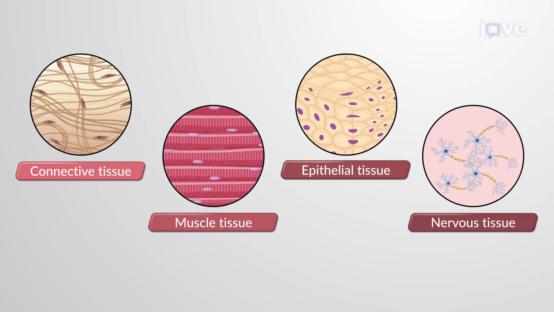

Video Summary: What are Tissues
Ever wonder how your heart beats over 100,000 times daily without you thinking about it? Understanding body tissues biology reveals how specialized cell groups create this incredible coordination. From the epithelial lining protecting your skin to the muscle tissue powering your movements, the four main tissue types work together like departments in a well-organized hospital system. Consider how surgeons at Johns Hopkins rely on precise tissue identification during operations. Watch the full video on JoVE Coach to master this concept with expert-led visuals and step-by-step explanations.
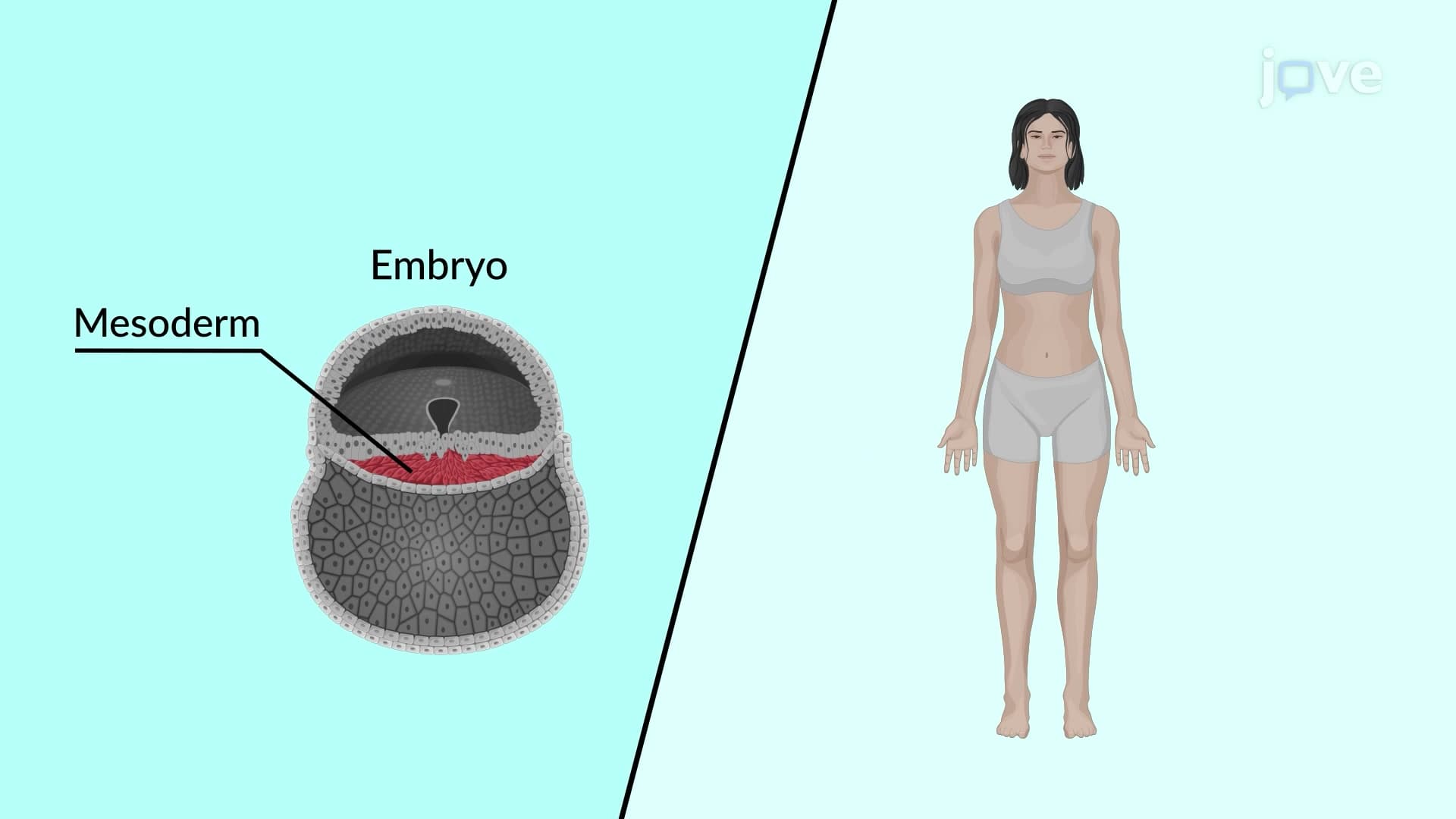
Body tissues biology forms the cornerstone of human anatomy, representing the organizational level between individual cells and complete organs. Think of tissues as specialized work crews in a construction project-each group has specific skills and tools, but they must coordinate to build a functional structure. This concept appears frequently on AP Biology exams and forms essential background knowledge for pre-med students preparing for the MCAT.
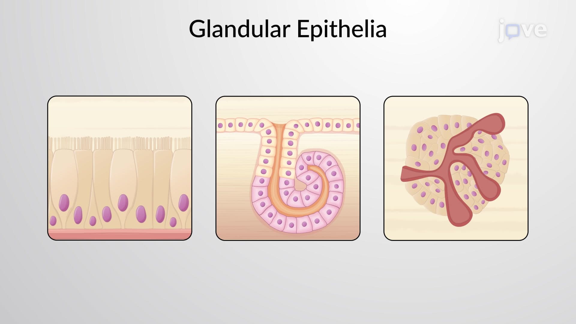
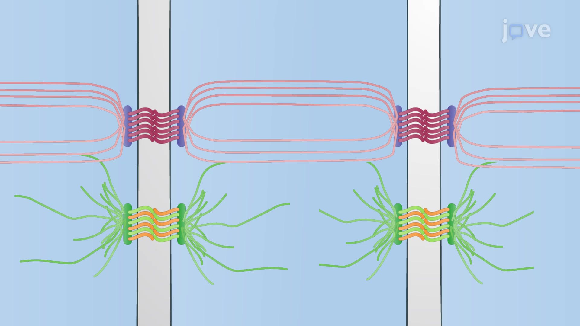
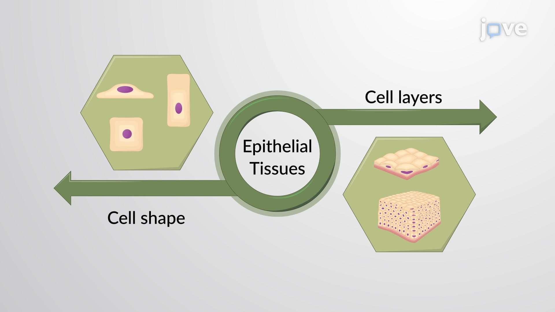
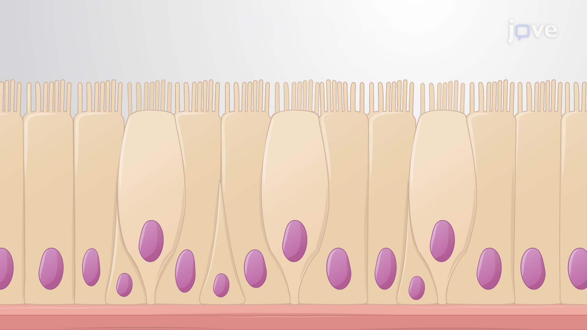
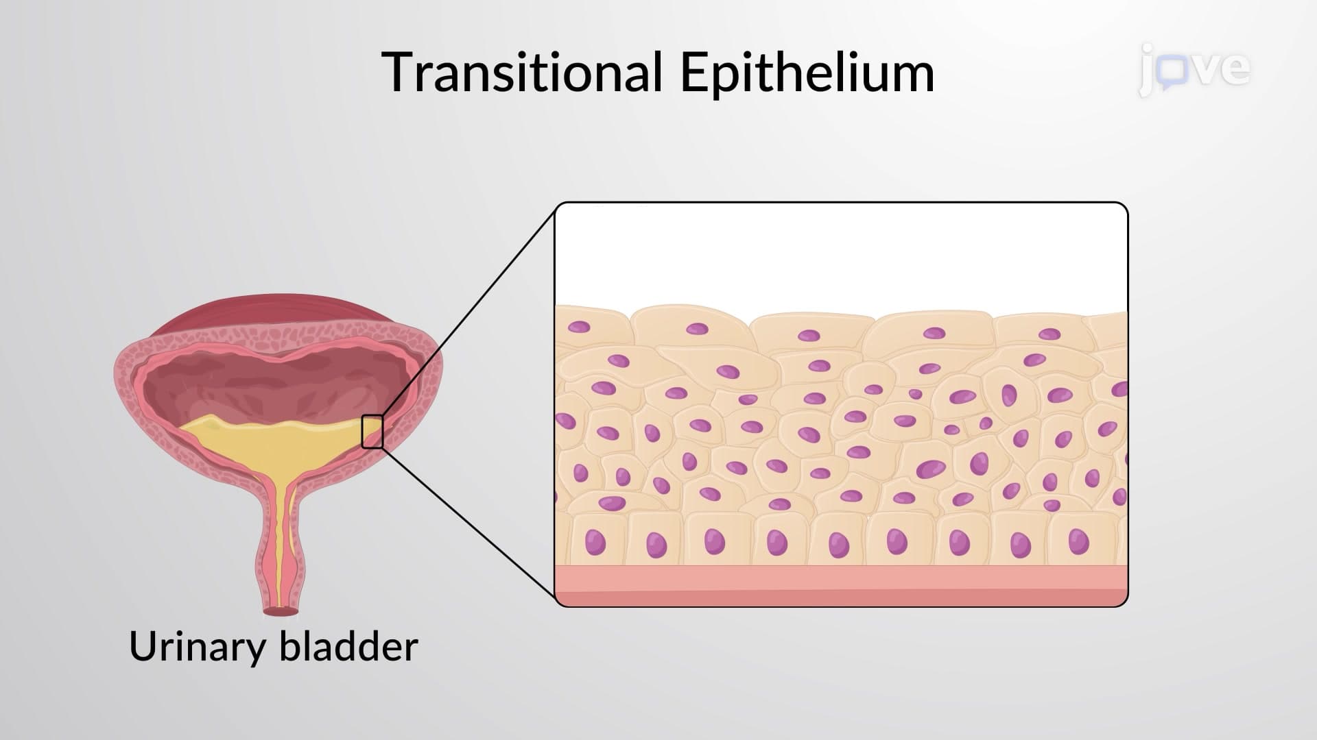
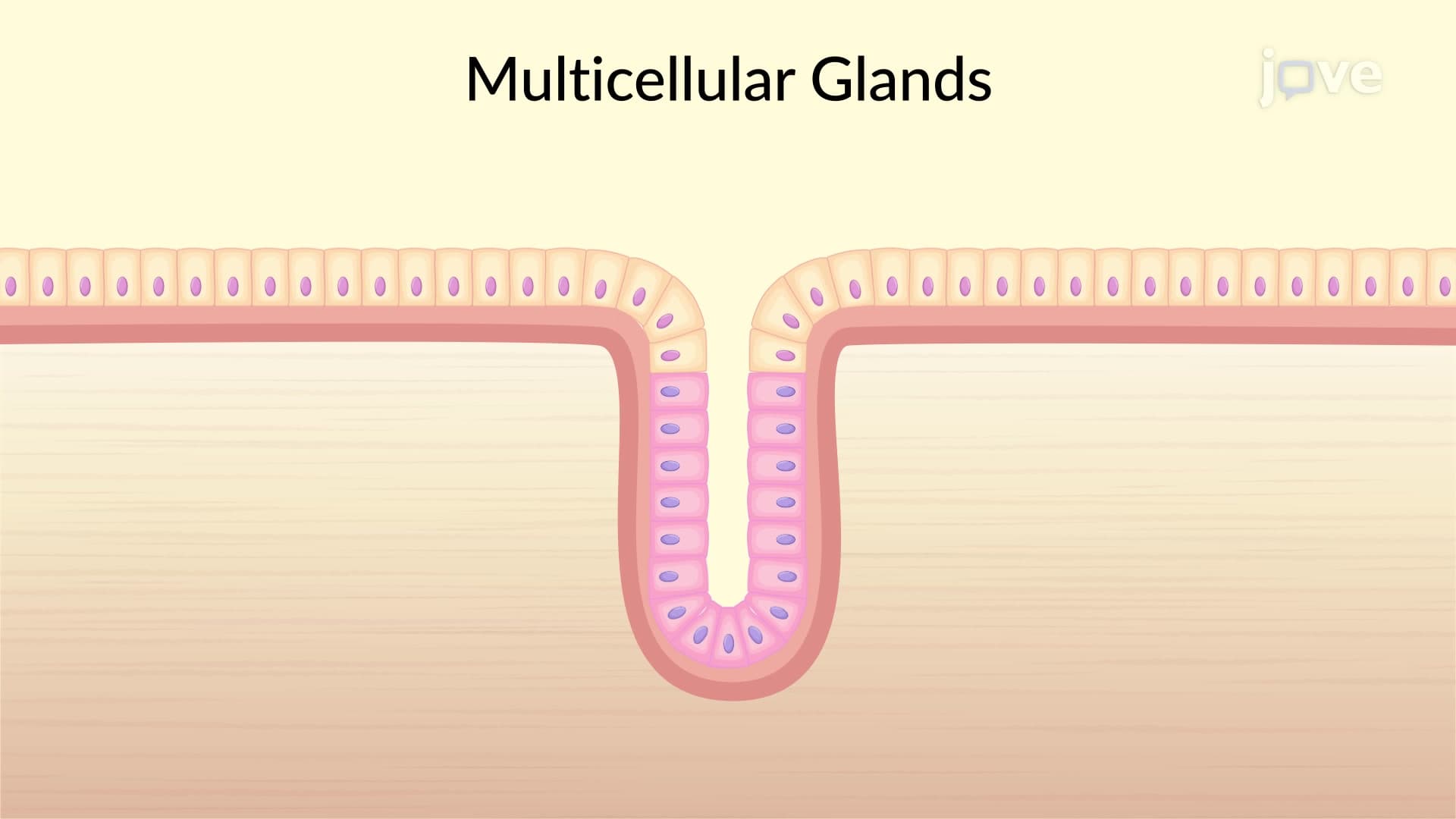
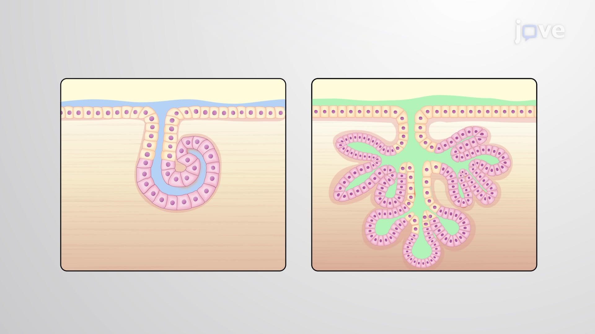
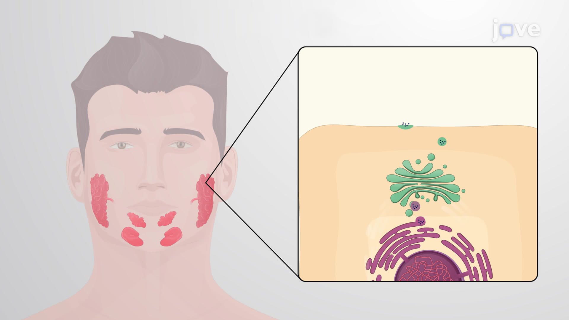
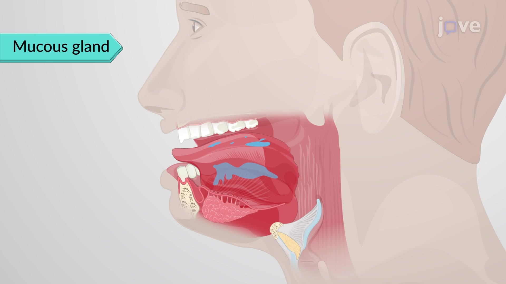
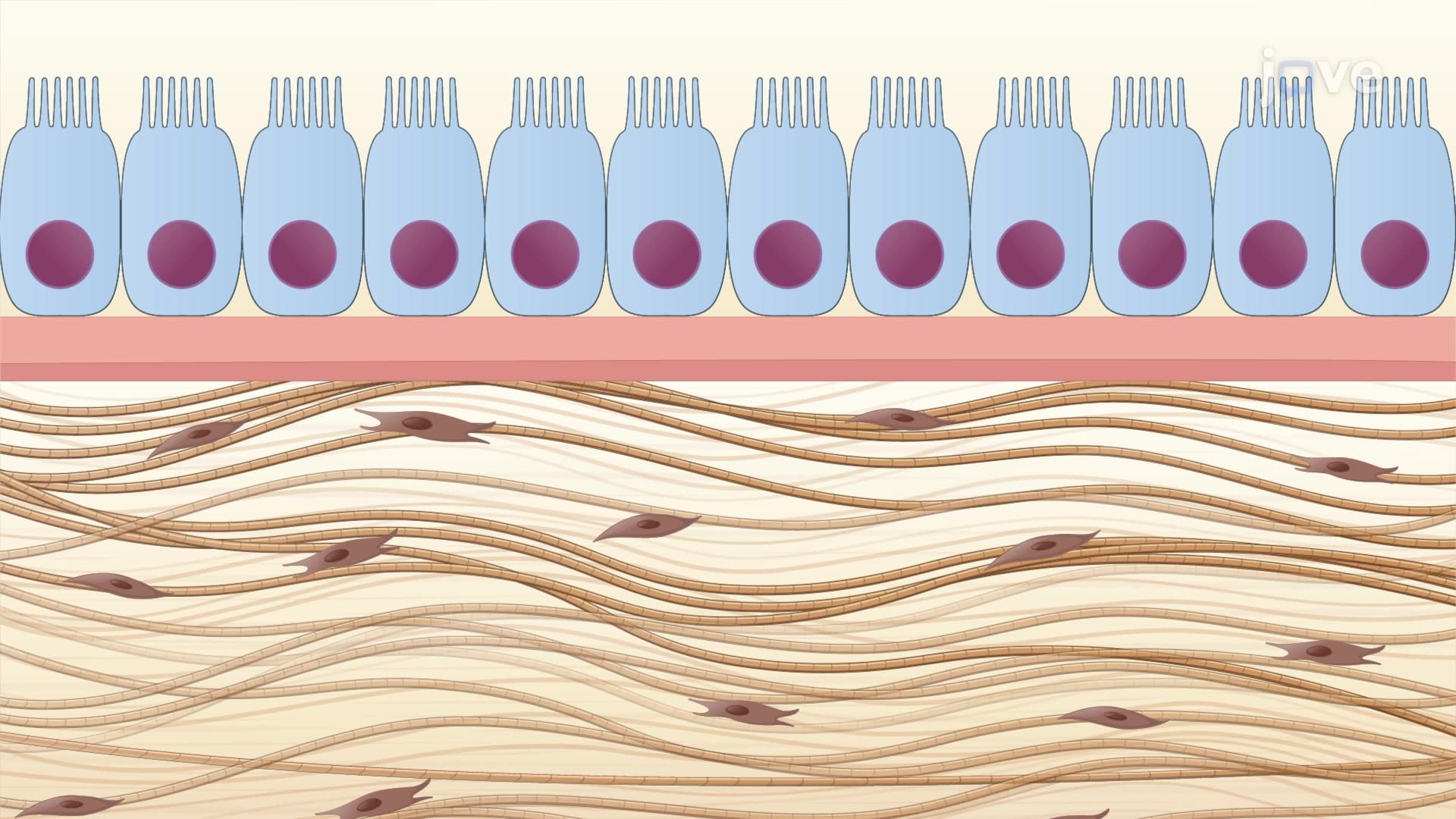
Epithelial tissues serve as the body's protective barriers and secretory specialists. These tightly packed cells form continuous sheets that line every body surface, from your skin's outer layer to your intestinal tract's inner walls. At institutions like Mayo Clinic, pathologists examine epithelial tissue samples to diagnose cancers, since many malignancies originate in these rapidly dividing cell populations.
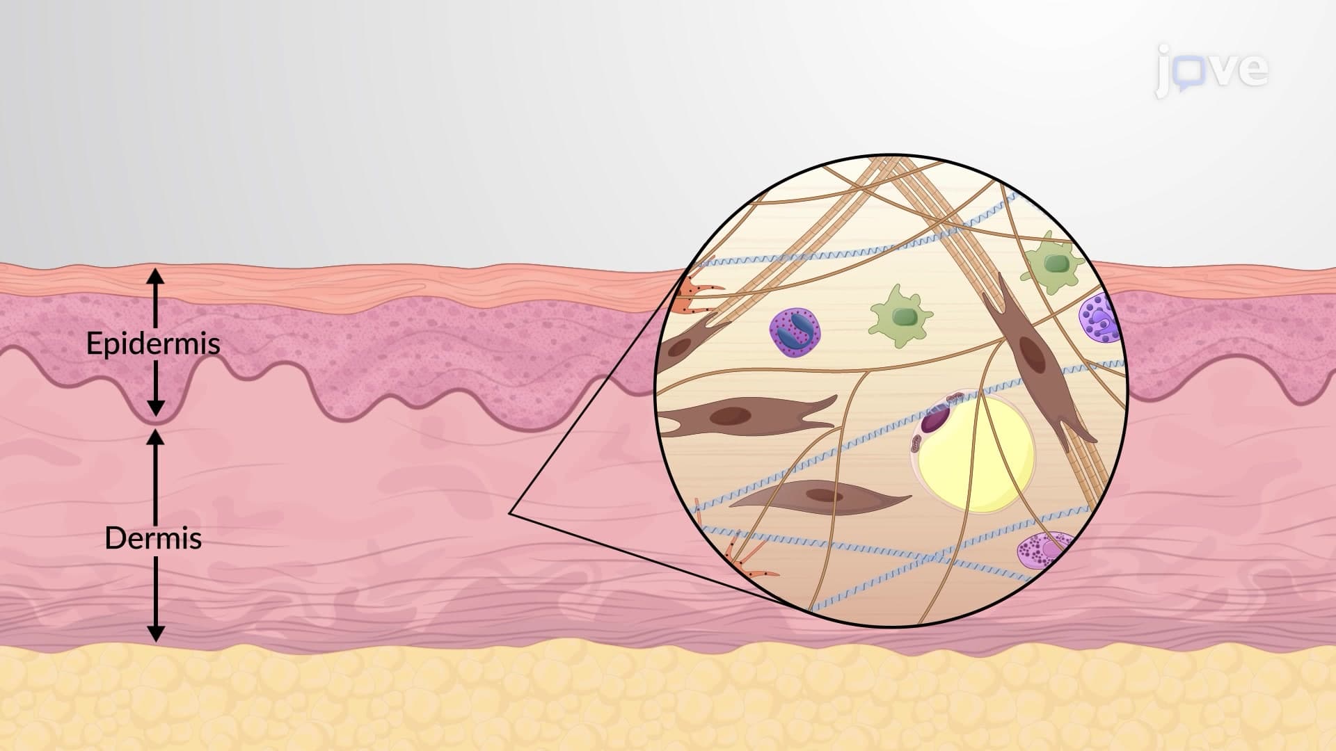
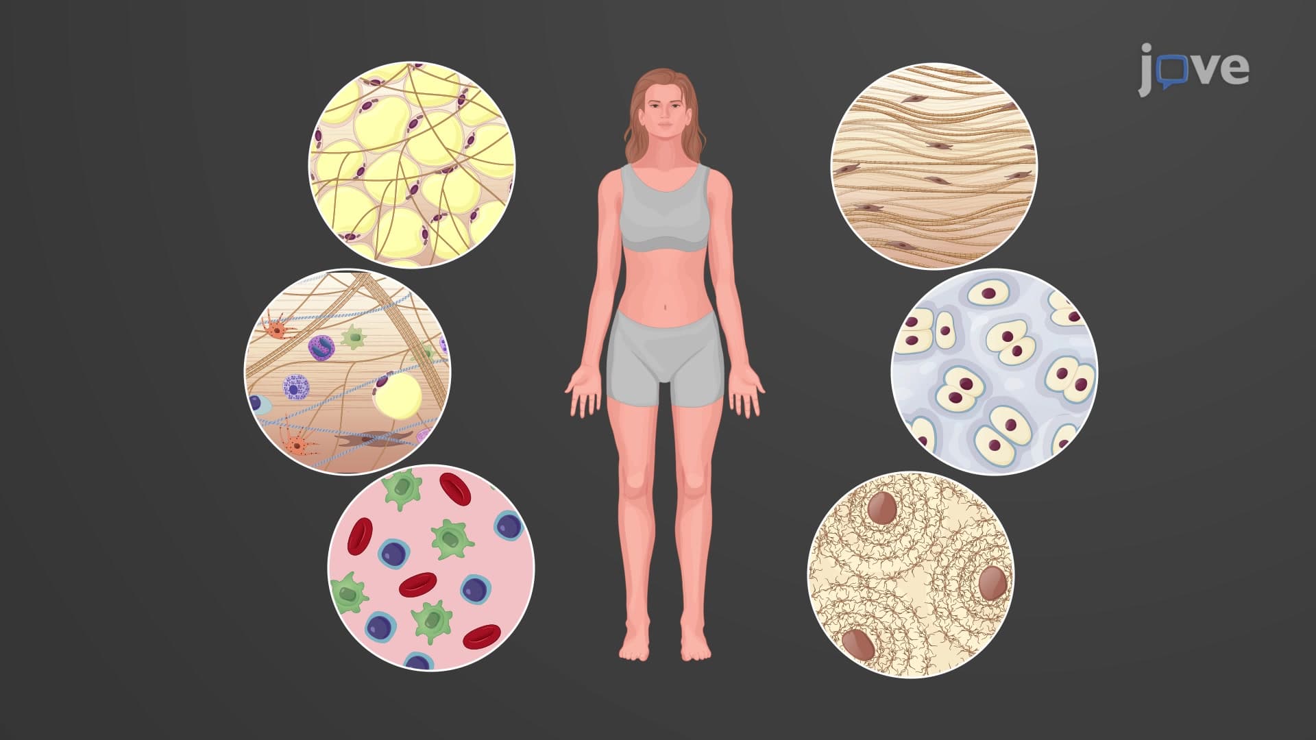
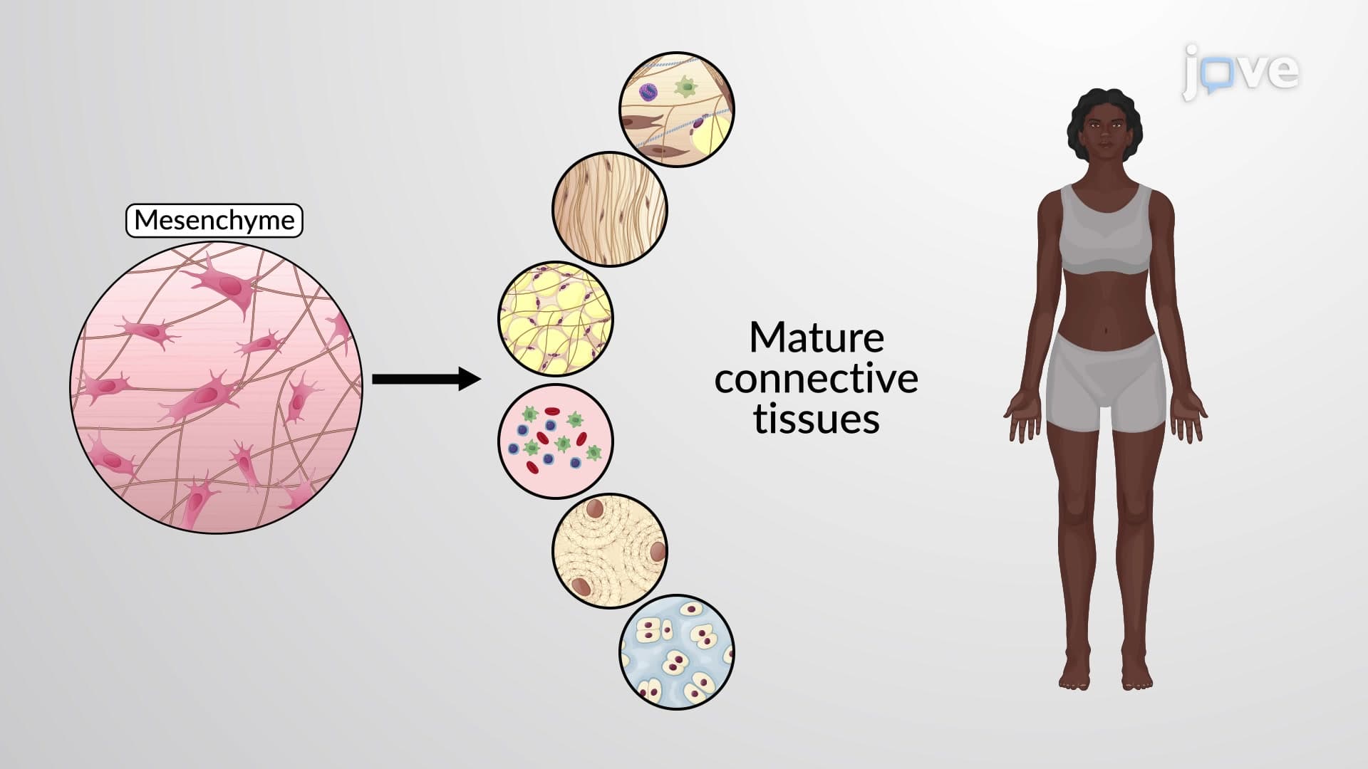
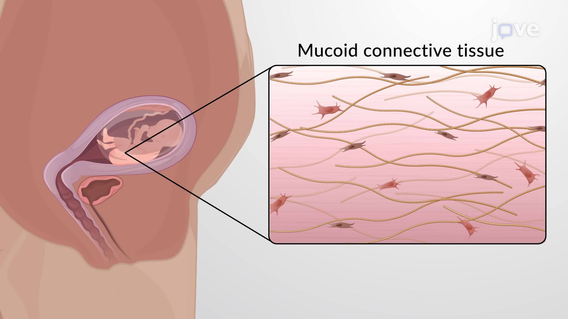
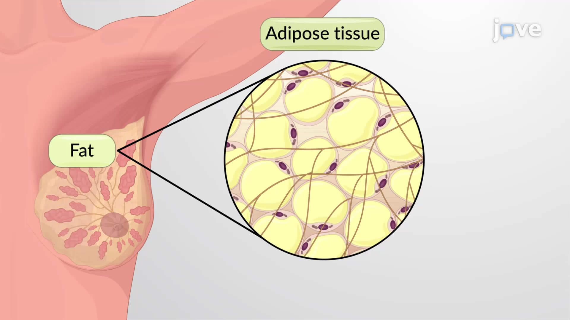
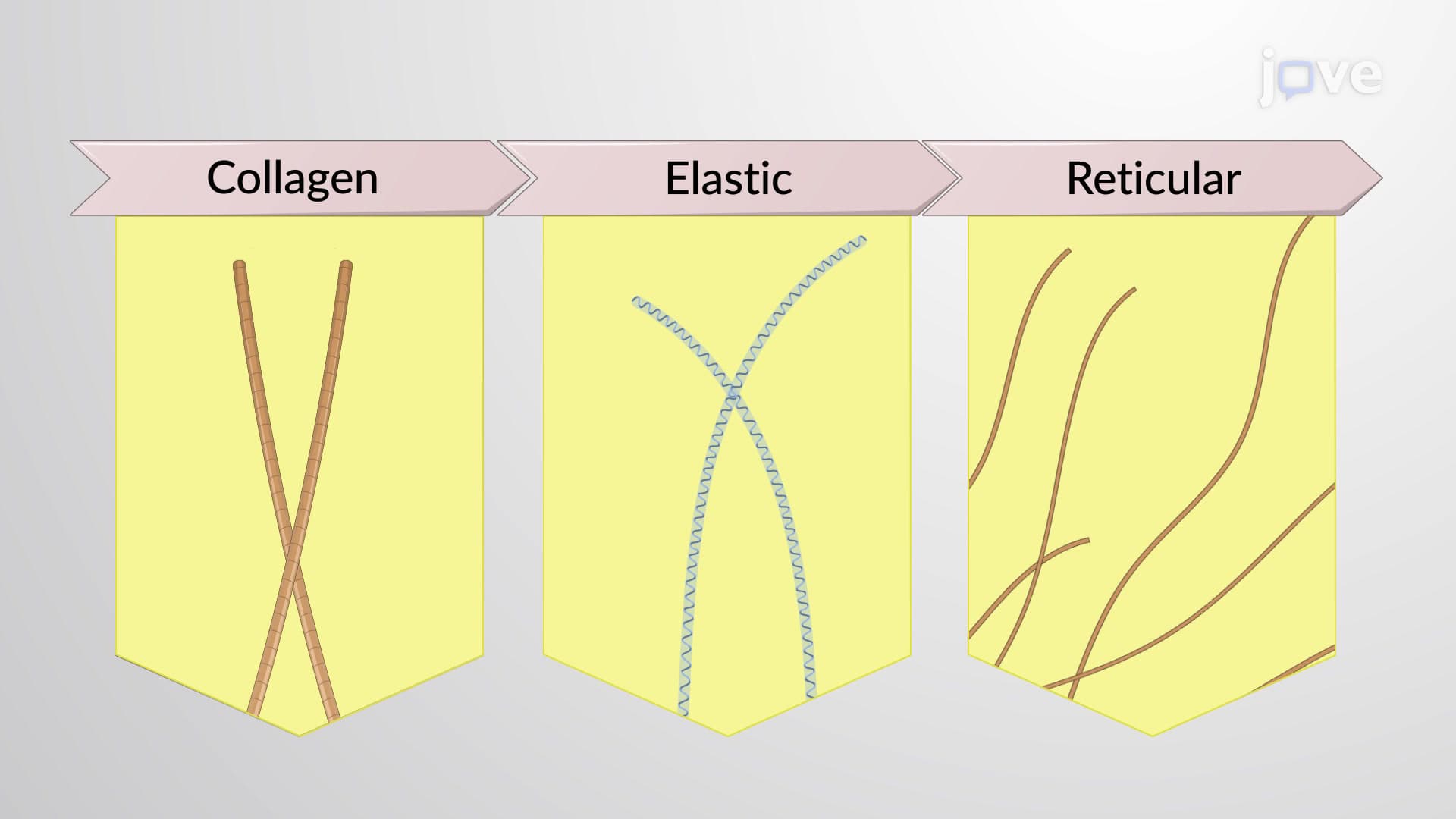
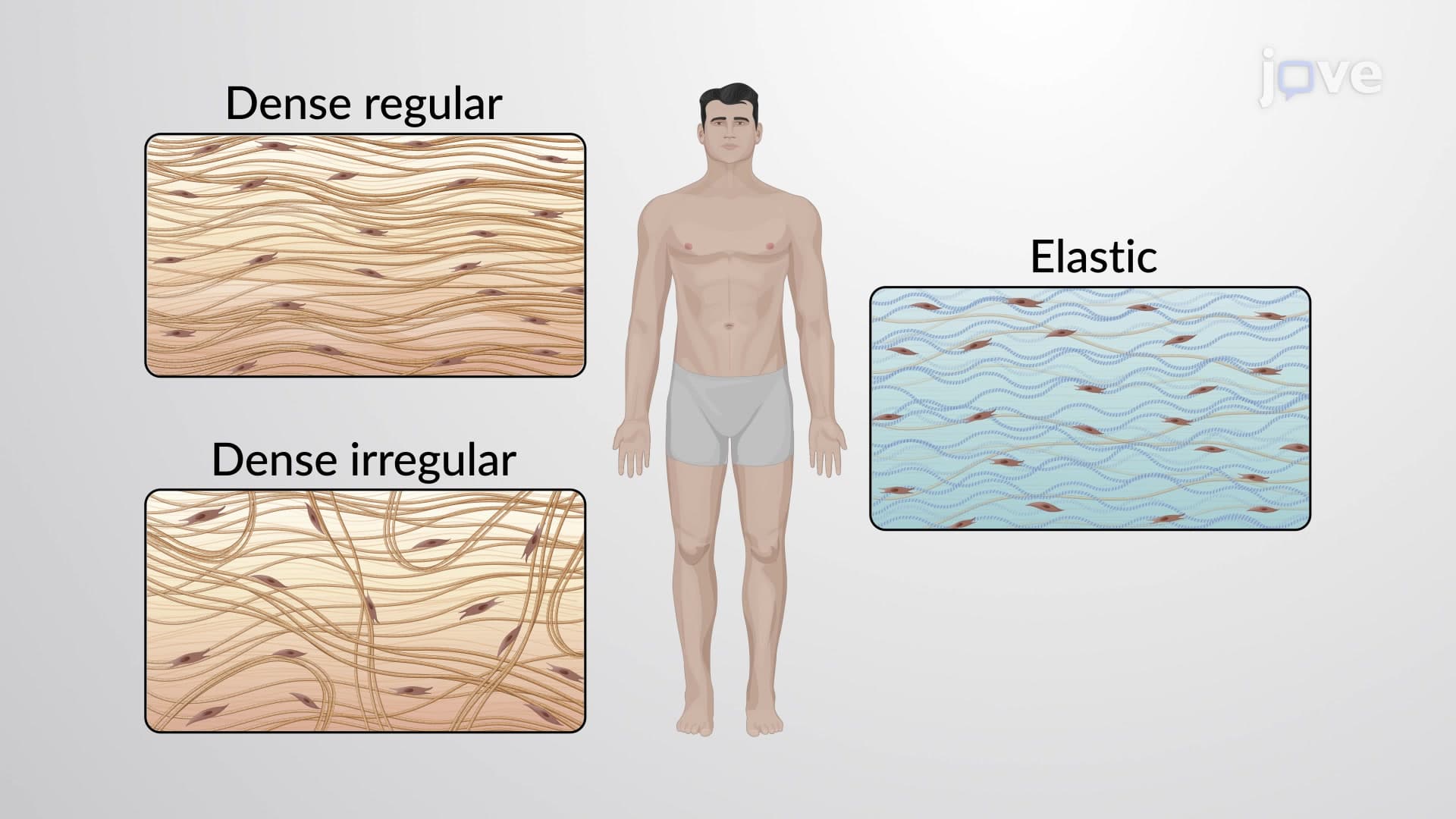
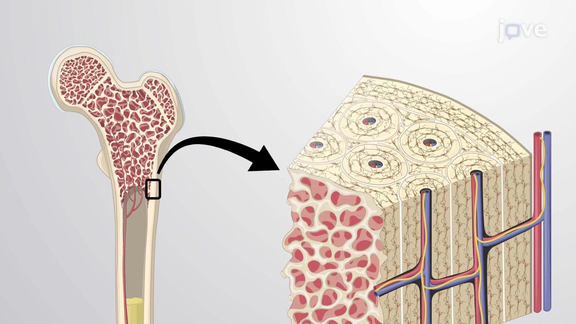
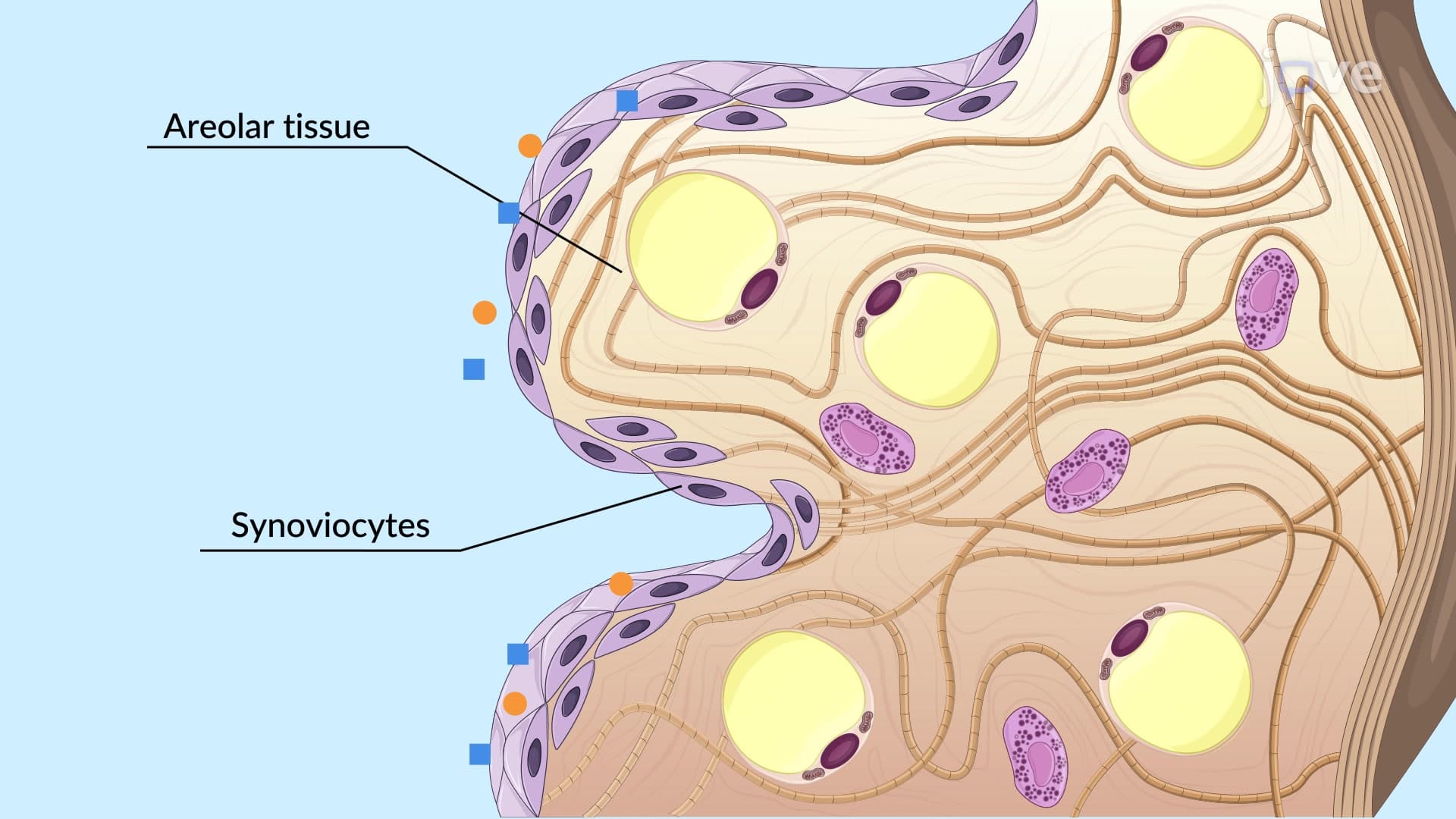
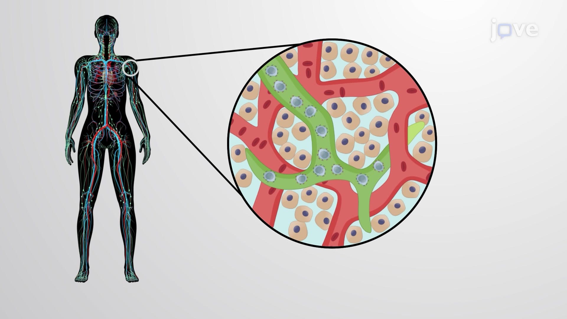
Connective tissues provide the body's structural framework and transportation network. Unlike epithelial tissues, connective tissue cells are scattered within an extensive extracellular matrix containing fibers and ground substance. Blood, bone, cartilage, and fat all represent different connective tissue types. During orthopedic surgeries at Cleveland Clinic, surgeons must understand how different connective tissues heal at varying rates-bone repairs faster than cartilage due to better blood supply.
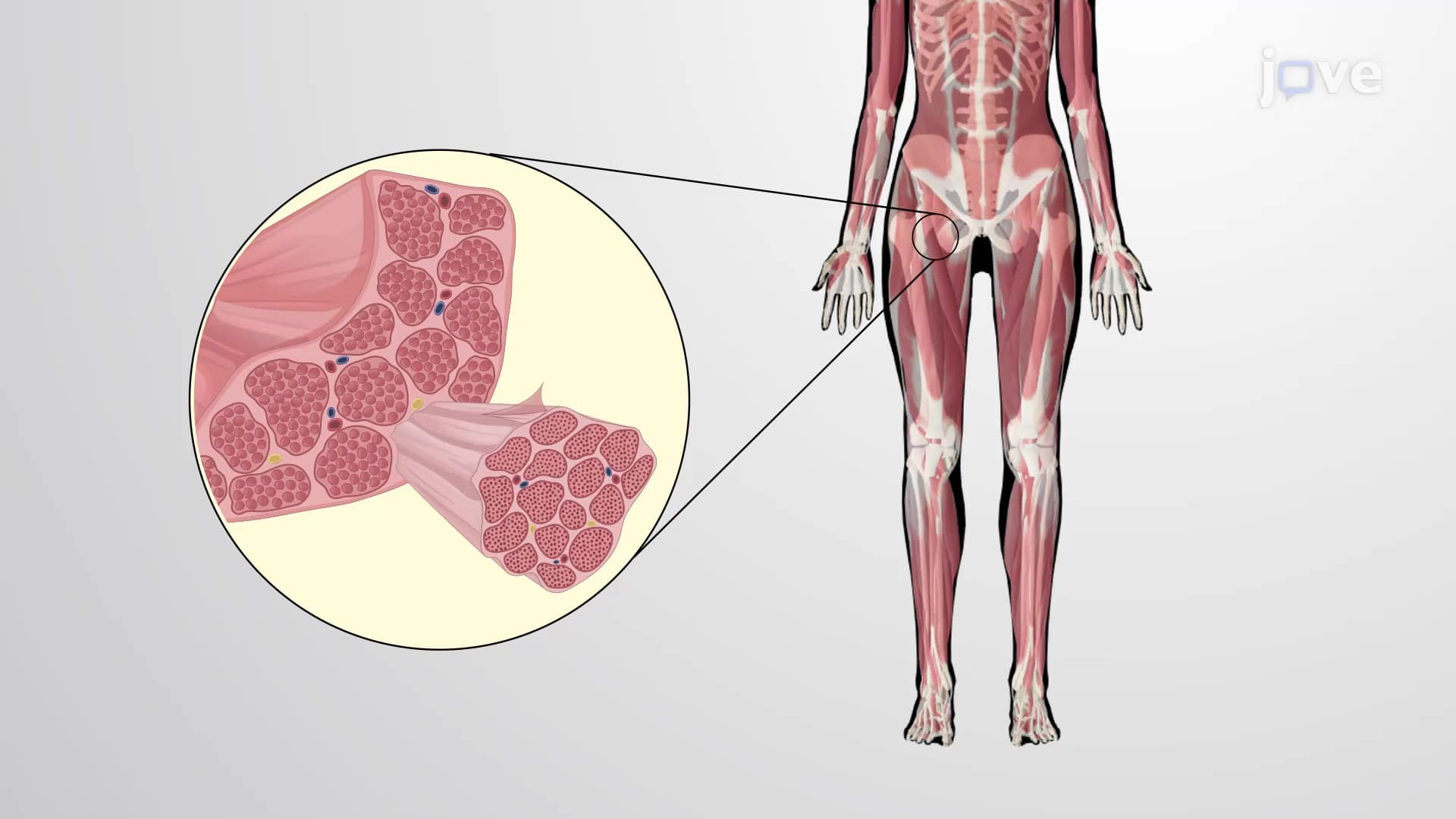
Muscle tissues generate the mechanical forces necessary for movement and organ function. The three muscle types-skeletal, cardiac, and smooth-each serve distinct purposes. Cardiac muscle's unique branched structure allows coordinated heart contractions, while smooth muscle in digestive organs creates the peristaltic waves that move food through your system.
Nervous tissues coordinate body activities through rapid electrical communication. Neurons transmit signals at speeds up to 120 meters per second, allowing split-second responses. This speed proves crucial in emergency medicine-neurologists at Massachusetts General Hospital rely on understanding nervous tissue function when treating stroke patients, where minutes can determine recovery outcomes.
The integration of these four tissue types creates organs capable of complex functions. Consider how your stomach demonstrates this coordination: nervous tissue receives hunger signals, epithelial tissue secretes digestive enzymes, smooth muscle tissue churns food mechanically, and connective tissue provides structural support while housing blood vessels that transport absorbed nutrients.
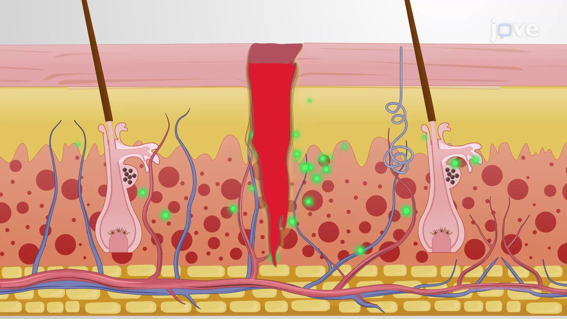
College anatomy courses emphasize this histology tissue biology because understanding tissue-level organization helps explain disease processes. When students encounter concepts like inflammation or wound healing on USMLE Step 1, they're applying these fundamental tissue principles to clinical scenarios.
Related Micro-courses