Loading video…


 26 Concepts
 323.1K views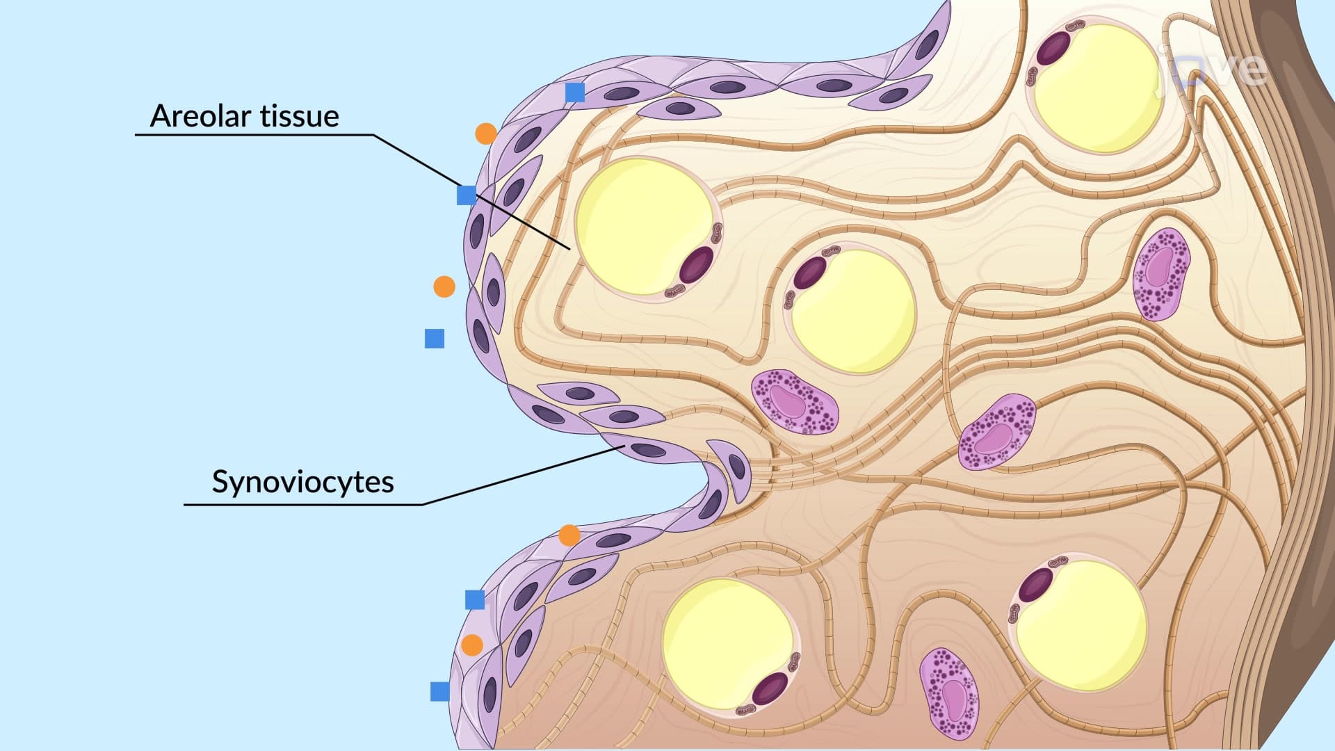

Video Summary: What are Tissue Membranes
Ever wonder why your mouth stays moist while your skin stays dry? Tissue membranes biology reveals how your body uses four specialized membrane types to protect and maintain different environments. From the mucous membranes lining your respiratory tract during a typical Texas allergy season to the serous membranes protecting your heart during exercise, understanding what are tissue membranes helps explain how your body maintains distinct internal conditions. These thin cellular sheets-mucous, serous, cutaneous, and synovial-each serve unique protective and regulatory functions throughout human anatomy. Watch the full video on JoVE Coach to master this concept with expert-led visuals and step-by-step explanations.
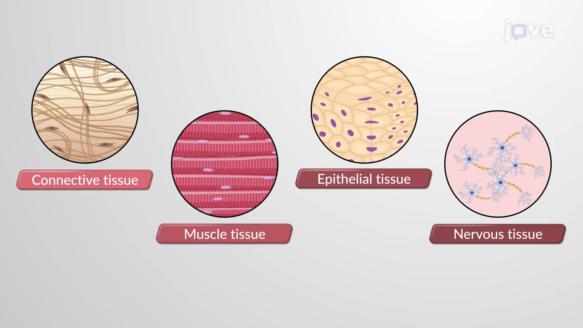
Tissue membranes biology forms a cornerstone of human anatomy, representing one of nature's most elegant solutions to compartmentalization and protection. These specialized structures consist of epithelial tissue bonded to underlying connective tissue, creating functional barriers that maintain distinct environments throughout the body. For students preparing for the AP Biology exam or college anatomy courses, mastering types of tissue membranes explained provides essential groundwork for understanding physiological processes from respiration to joint movement.
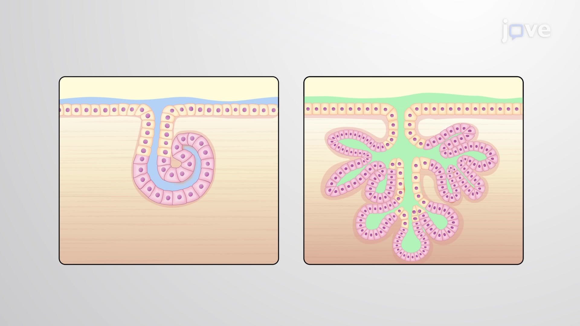
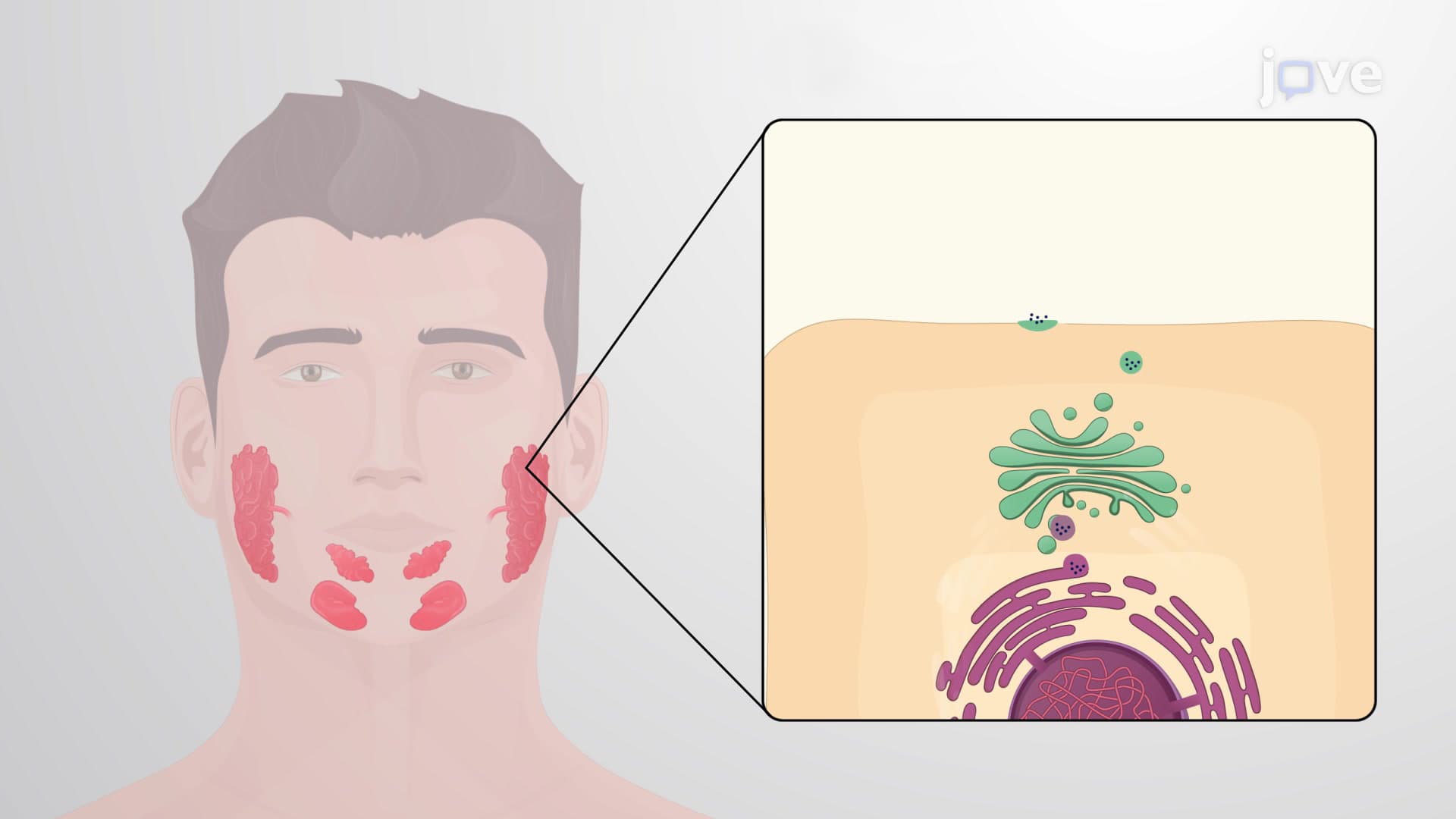
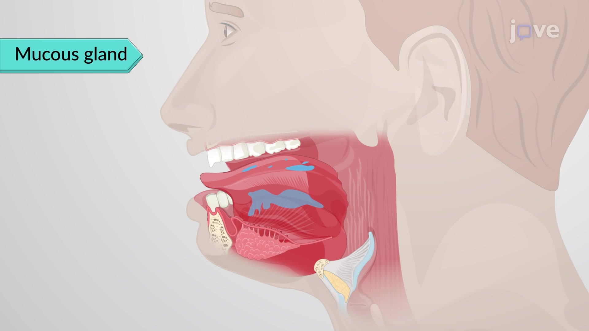
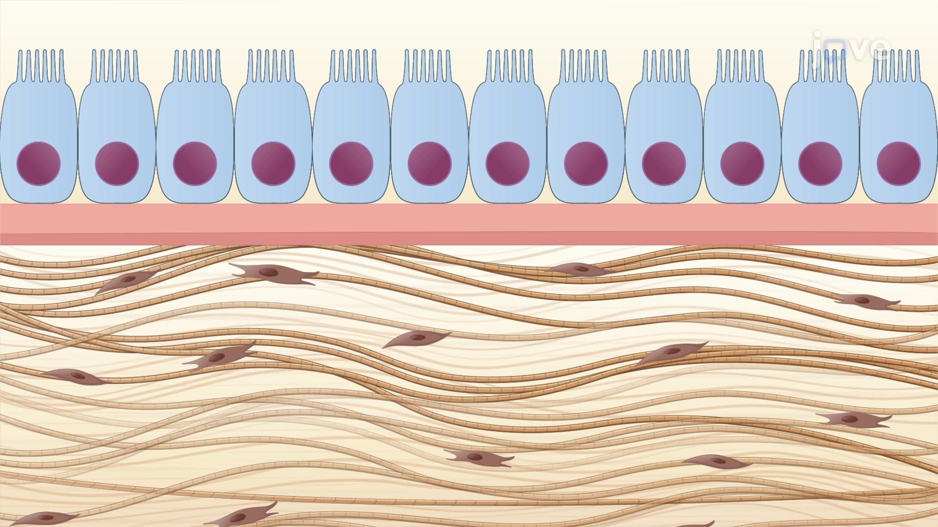
Mucous cutaneous membrane systems line all body cavities that communicate with the exterior environment. These membranes feature a characteristic two-layer structure: epithelial cells form the surface layer, while the lamina propria-composed of areolar connective tissue-provides structural support underneath. In the respiratory tract, mucous membranes trap particles and pathogens that enter during breathing, particularly relevant for understanding conditions like asthma or seasonal allergies common across the American Southeast. The digestive tract's mucous membranes secrete protective mucus that shields stomach lining from harsh digestive acids-a concept frequently tested on the MCAT's biological sciences section.
Epithelial serous membrane structures surround closed body cavities, creating friction-free environments for organ movement. The heart's pericardial serous membrane exemplifies this function perfectly: during exercise, your heart beats over 150 times per minute, requiring smooth movement within its protective sac. Serous membranes consist of mesothelium (simple squamous epithelium) supported by areolar connective tissue. These membranes produce serous fluid that lubricates between parietal (outer) and visceral (inner) layers, preventing dangerous friction during organ movement-a critical concept for USMLE Step 1 preparation.
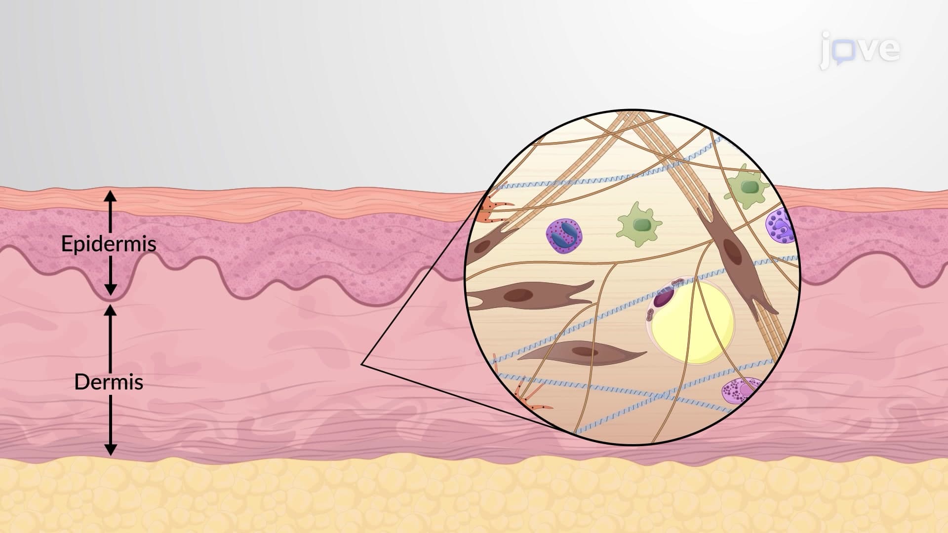
The cutaneous membrane (skin) represents the body's largest organ, featuring keratinized stratified squamous epithelium over dense connective tissue dermis. This tissue membrane classification creates waterproof protection essential for terrestrial life. Synovial connective membrane lines freely movable joints like the knee, secreting synovial fluid that lubricates articulating surfaces. Understanding synovial membrane function becomes crucial when studying joint injuries common in American high school athletics, from basketball knee injuries to football shoulder separations.
Related Micro-courses