Loading video…


 26 Concepts
 323.1K views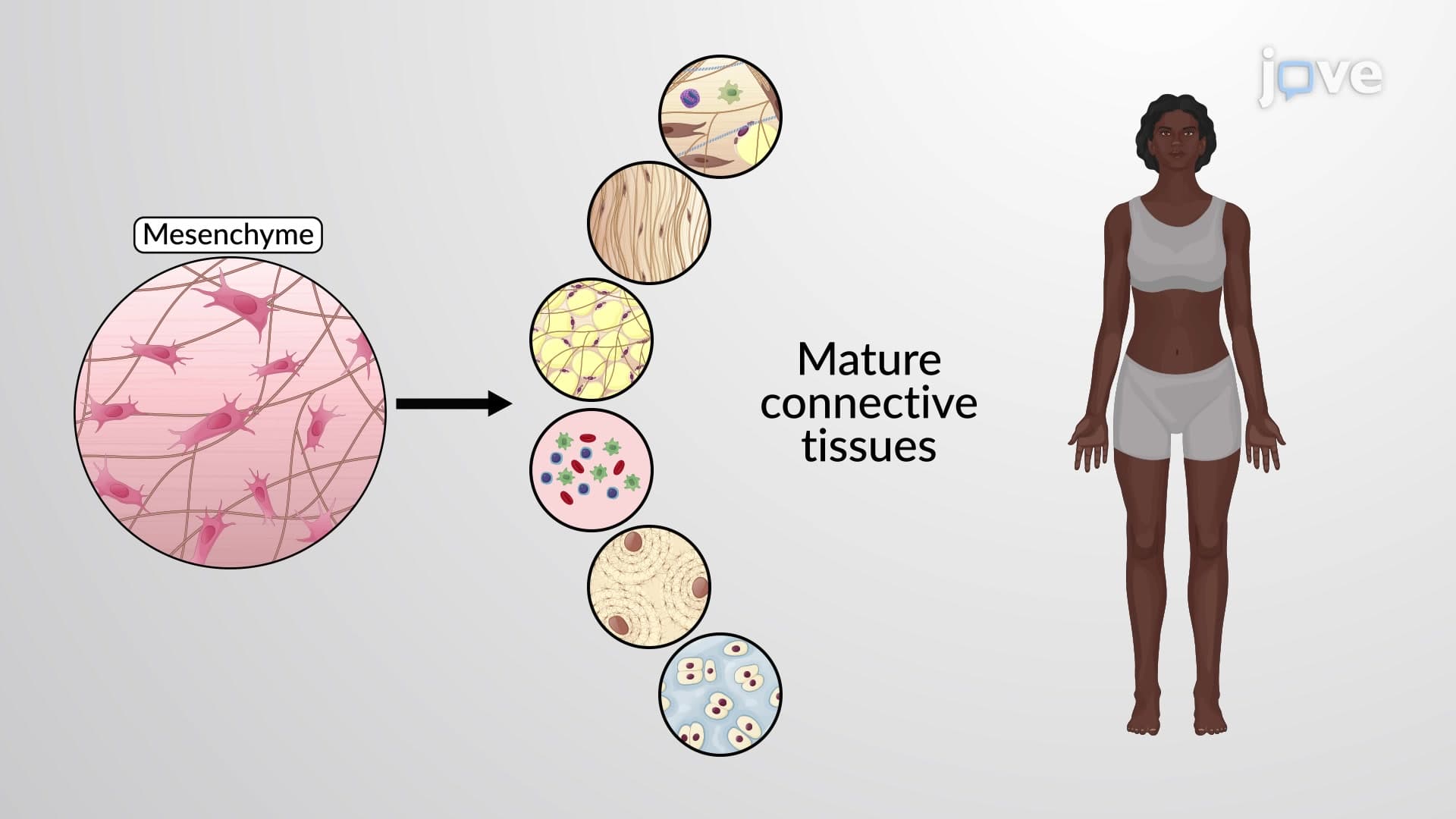

Video Summary: What are Classification of Connective Tissues
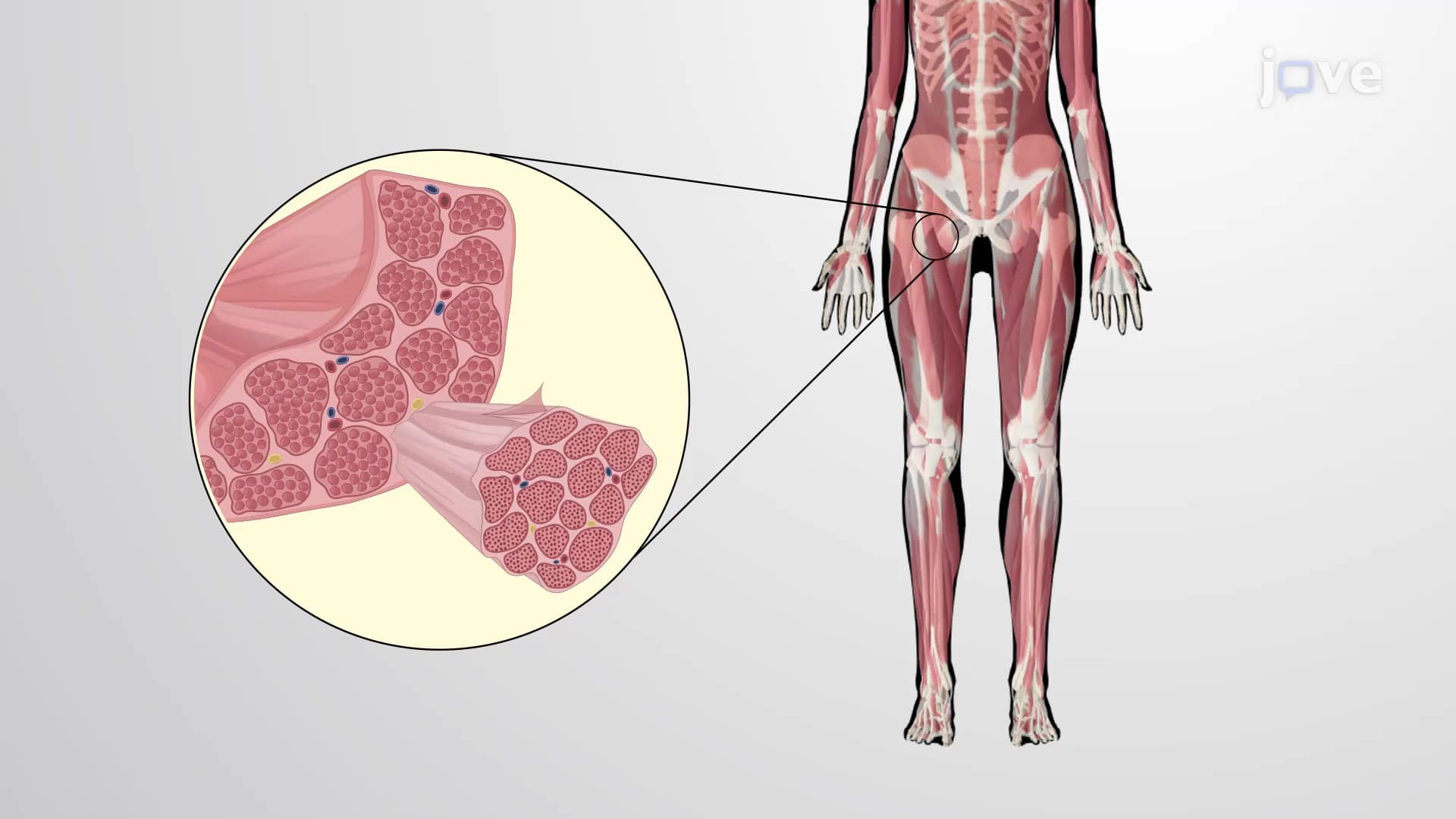
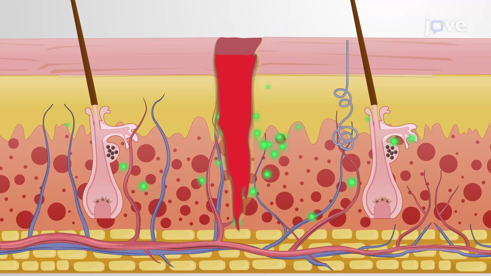
Why does a broken bone heal differently than a torn muscle? The answer lies in understanding connective tissue classification types and how different tissues respond to injury. From the dense collagen in your Achilles tendon that withstands the force of jumping to the fluid blood tissue that carries oxygen throughout your body, each connective tissue type serves unique functions. Watch the full video on JoVE Coach to master this concept with expert-led visuals and step-by-step explanations.
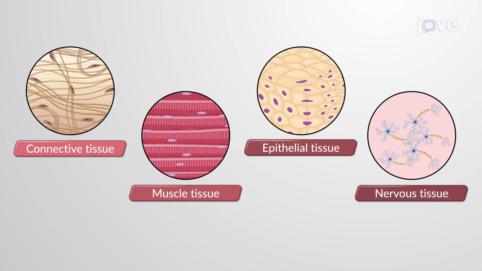
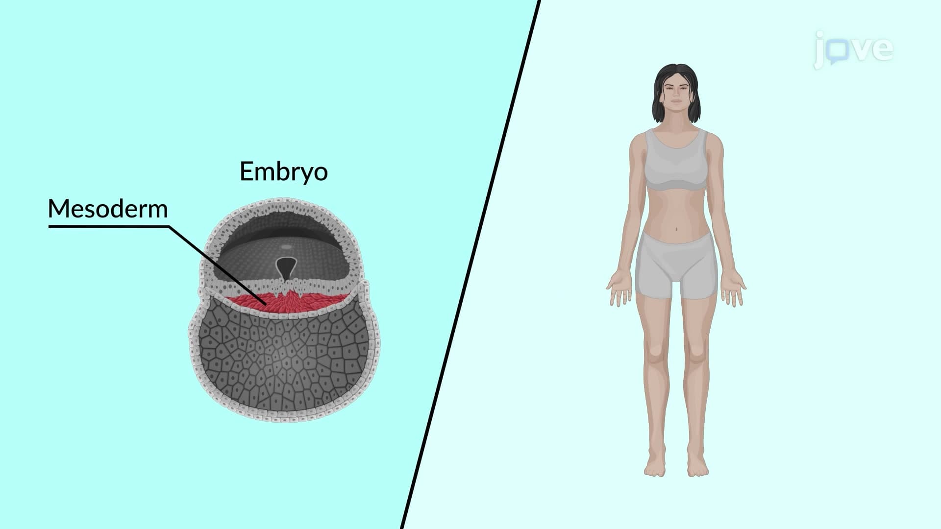
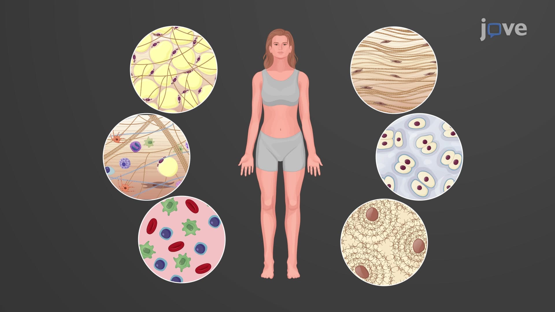
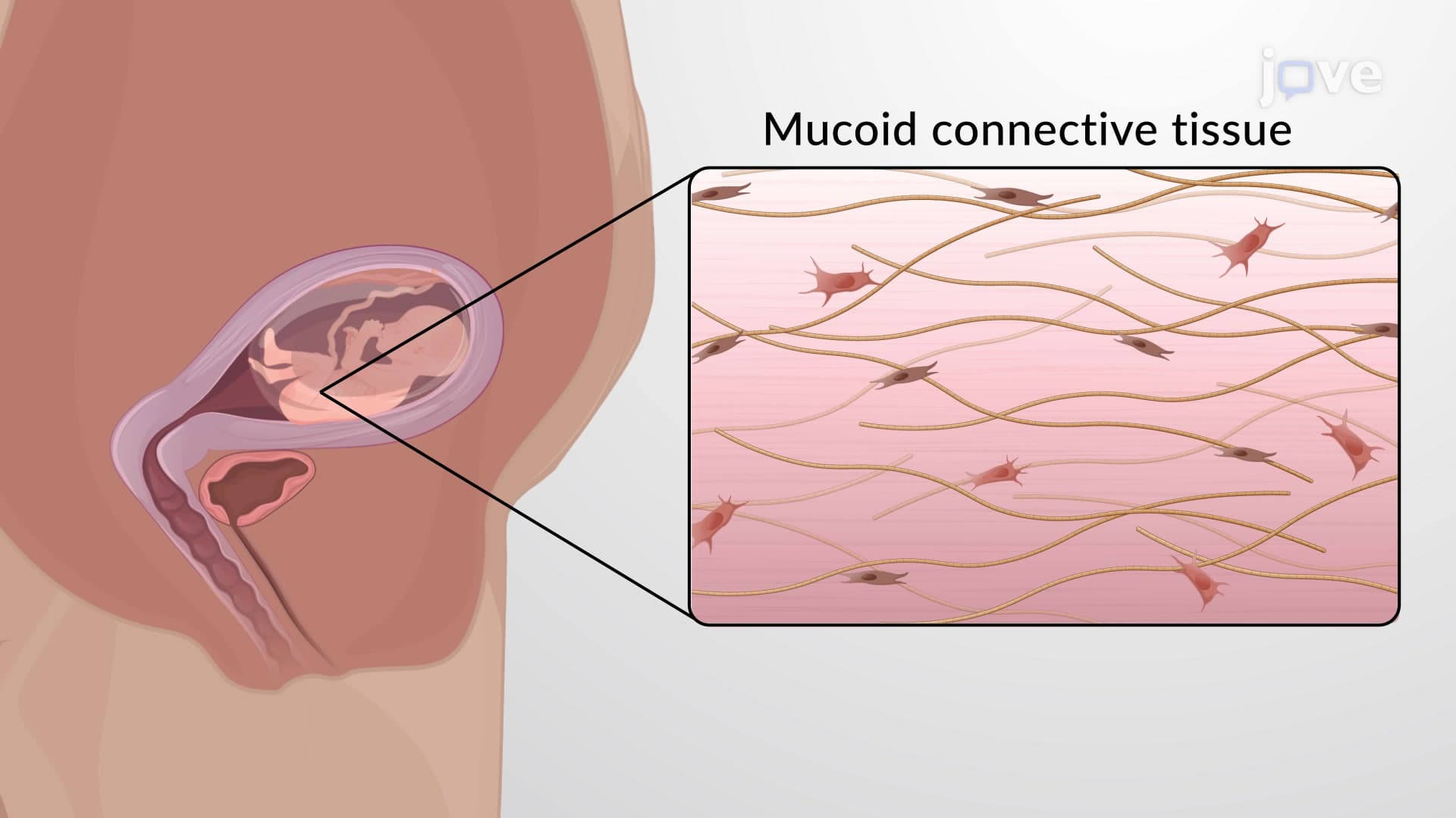
Connective tissue classification types form the foundation for understanding how our bodies maintain structural integrity and physiological function. All connective tissues originate from mesenchyme during embryonic development, but mature into distinctly different forms based on their extracellular matrix composition and cellular organization. This classification system helps medical professionals predict tissue behavior, healing patterns, and disease susceptibility.
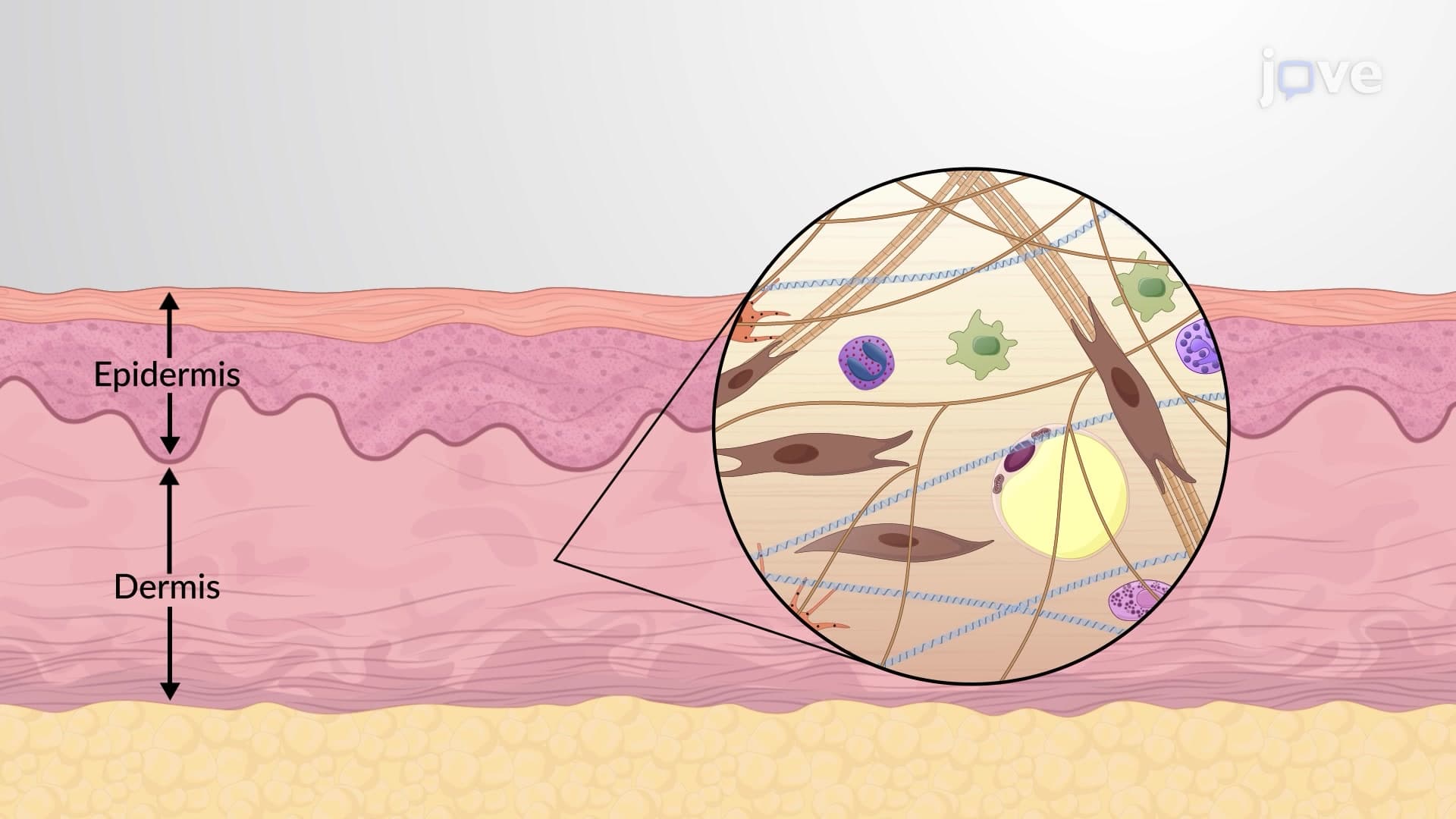
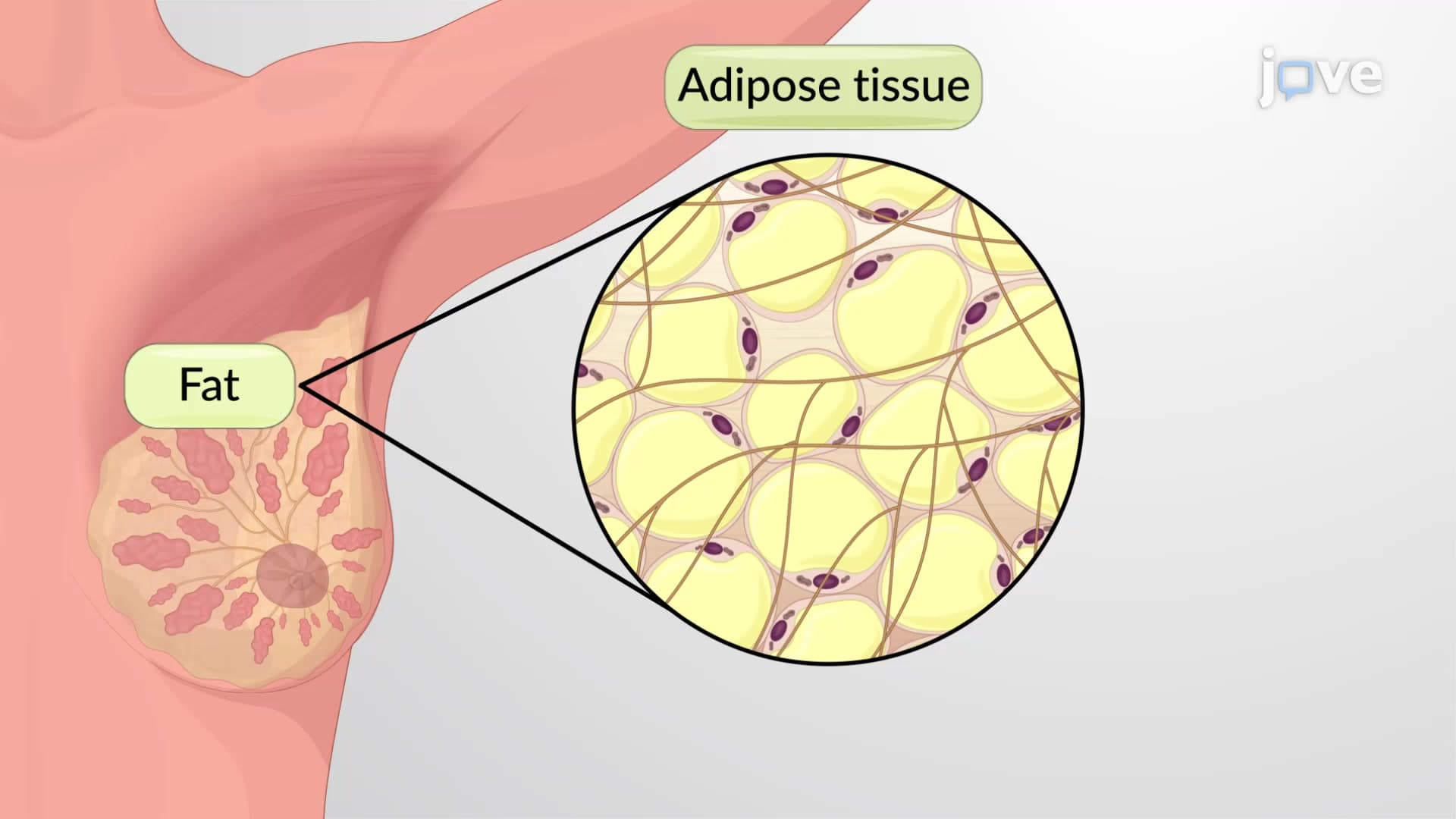
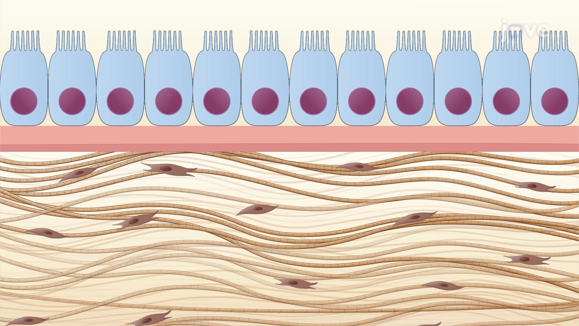
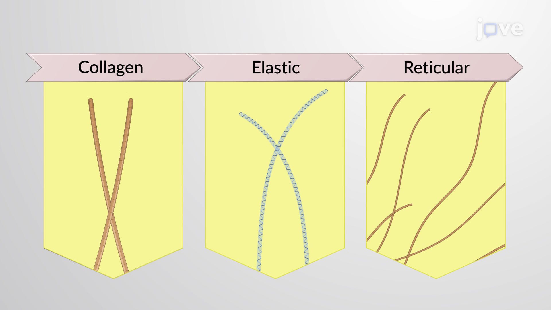
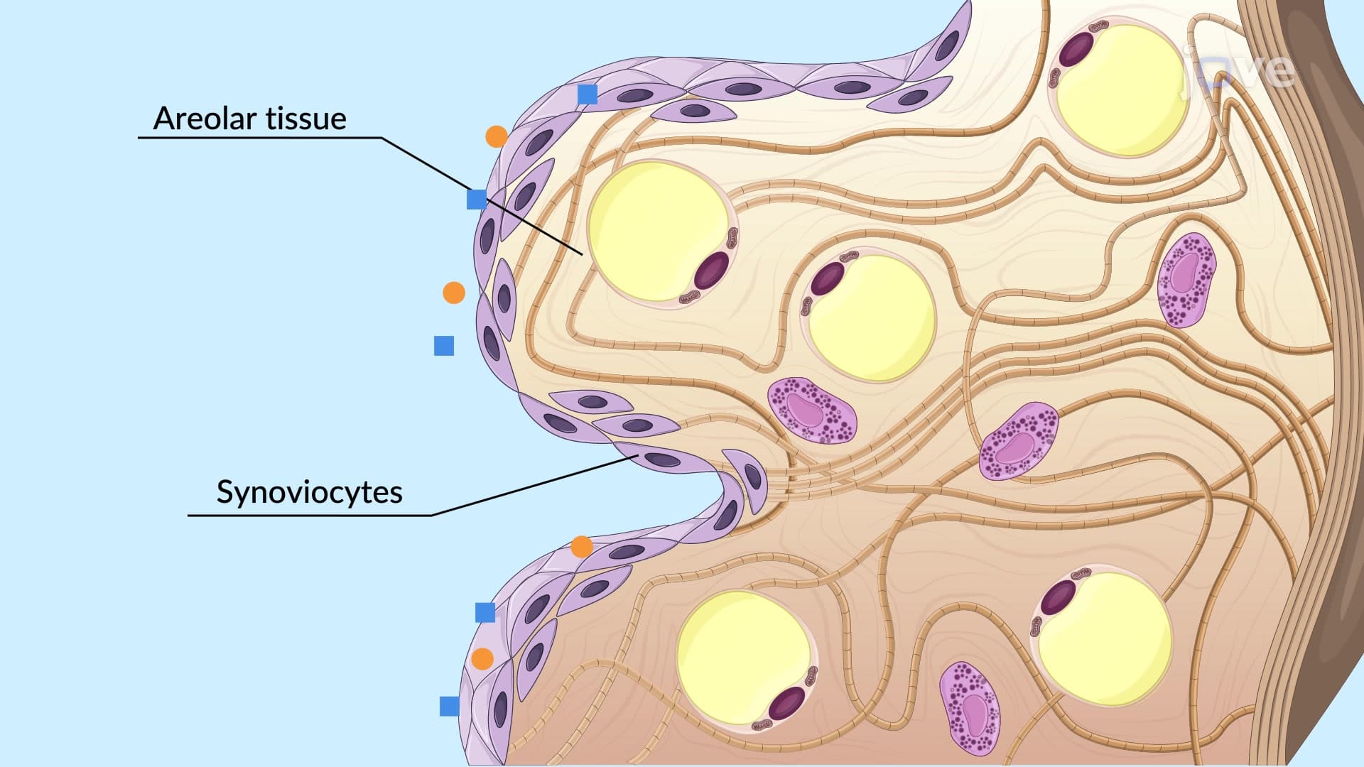
How connective tissues are classified begins with understanding connective tissue proper, which includes both loose and dense varieties. Loose connective tissue features widely spaced cells with abundant ground substance and loosely arranged protein fibers. Areolar tissue, found beneath skin and around organs, provides cushioning and immune cell pathways. Adipose tissue stores energy and insulates organs-the subcutaneous fat layer that varies among individuals exemplifies this tissue type. Reticular tissue forms the structural framework of lymphoid organs like the spleen and lymph nodes.
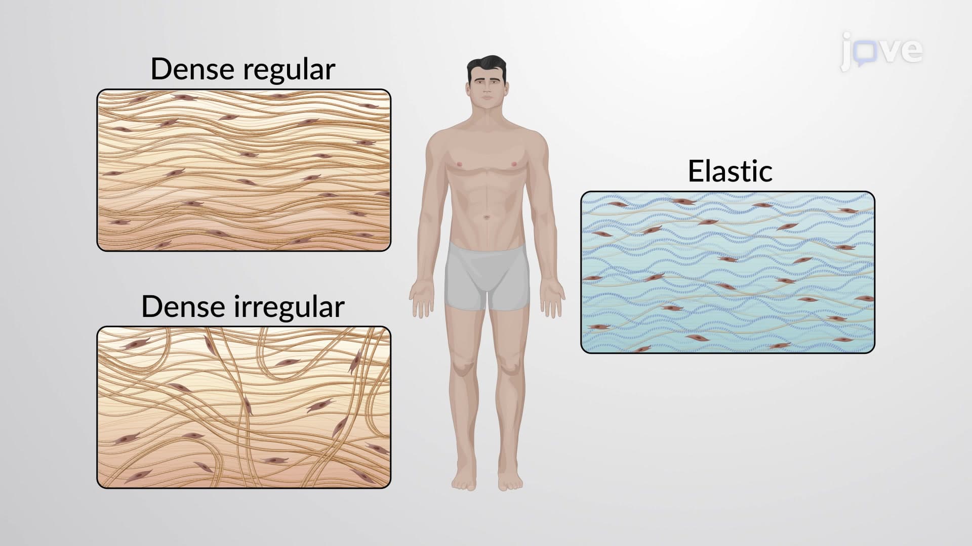
Dense connective tissue proper contains tightly packed collagen fibers with fewer cells. Regular dense tissue, like tendons and ligaments, aligns fibers in one direction for maximum tensile strength. The patellar tendon connecting your kneecap to shinbone demonstrates this organization. Irregular dense tissue, found in joint capsules and organ walls, arranges fibers in multiple directions for multi-directional strength. Elastic dense tissue in arterial walls and vocal cords contains elastin fibers for stretch and recoil.
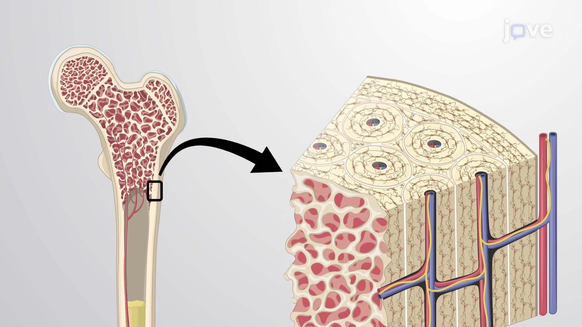
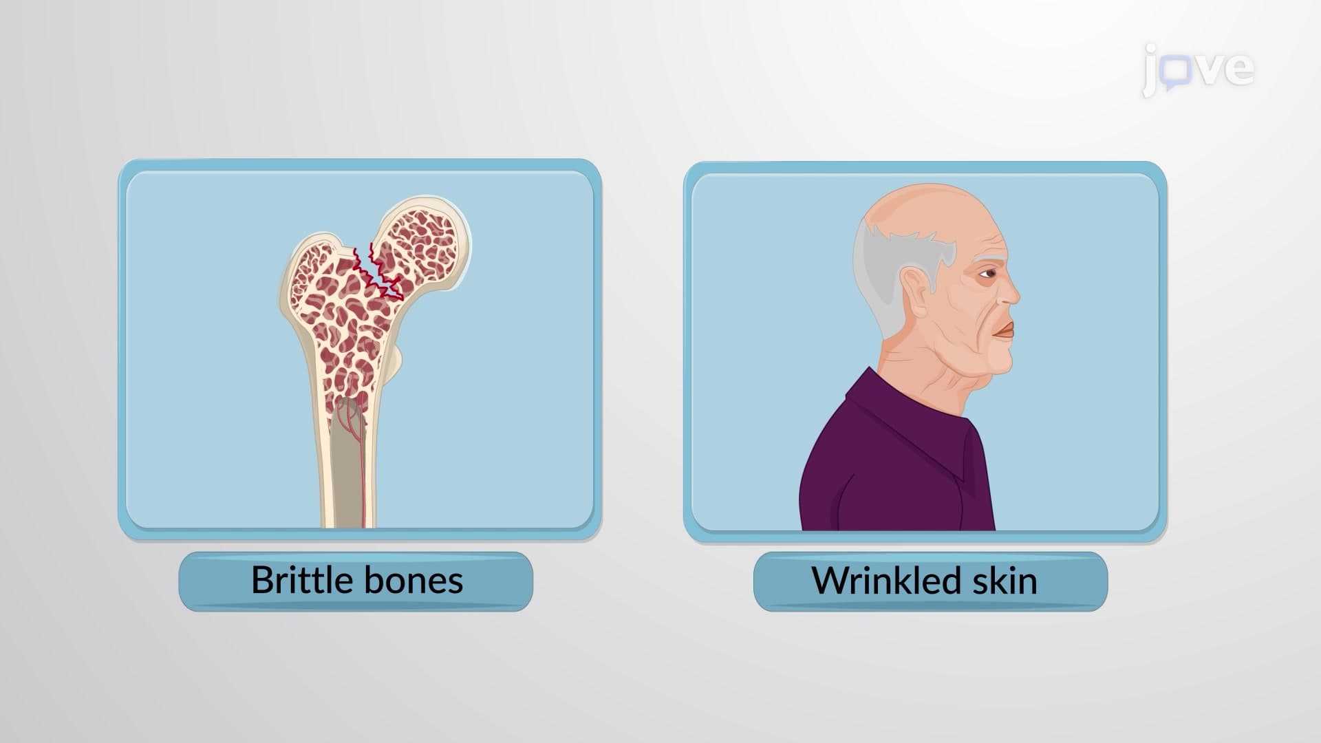
Supporting connective bone cartilage tissues provide structural framework and protection. Bone tissue features a calcified extracellular matrix containing calcium phosphate crystals. Compact bone forms dense outer layers, while spongy bone creates internal scaffolding that reduces weight while maintaining strength. This organization explains why osteoporosis primarily affects spongy bone regions like vertebrae and hip joints.
Cartilage provides firm yet flexible support with properties intermediate between dense connective tissue and bone. Hyaline cartilage covers joint surfaces and forms the fetal skeleton. Fibrocartilage in intervertebral discs combines strength with shock absorption. Elastic cartilage in ear and epiglottis maintains shape while allowing flexibility.
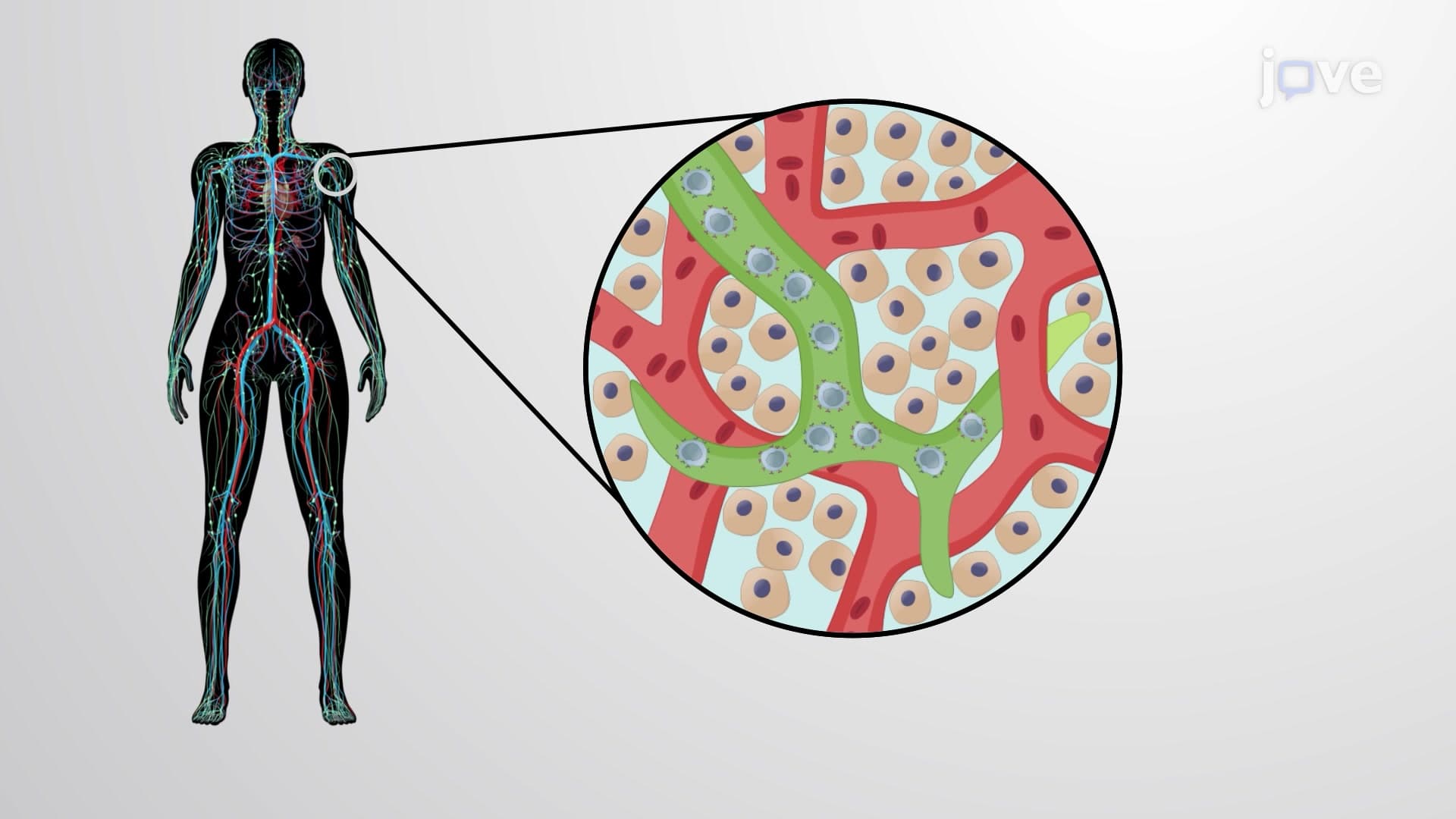
Fluid connective tissue includes blood and lymph, where cells float in liquid extracellular matrix rather than fibrous networks. Blood transports nutrients, gases, and waste products throughout the body. Lymph collects tissue fluid and returns it to circulation while filtering pathogens through lymph nodes.
Understanding these classifications proves essential for AP Biology, college anatomy courses, and pre-health professional exams like the MCAT. Students often encounter questions comparing tissue types or predicting healing outcomes based on tissue classification.
Related Micro-courses