Loading subject page…


323,101 views

Video Summary: Connective Tissue Fibers and Ground Substance Explained
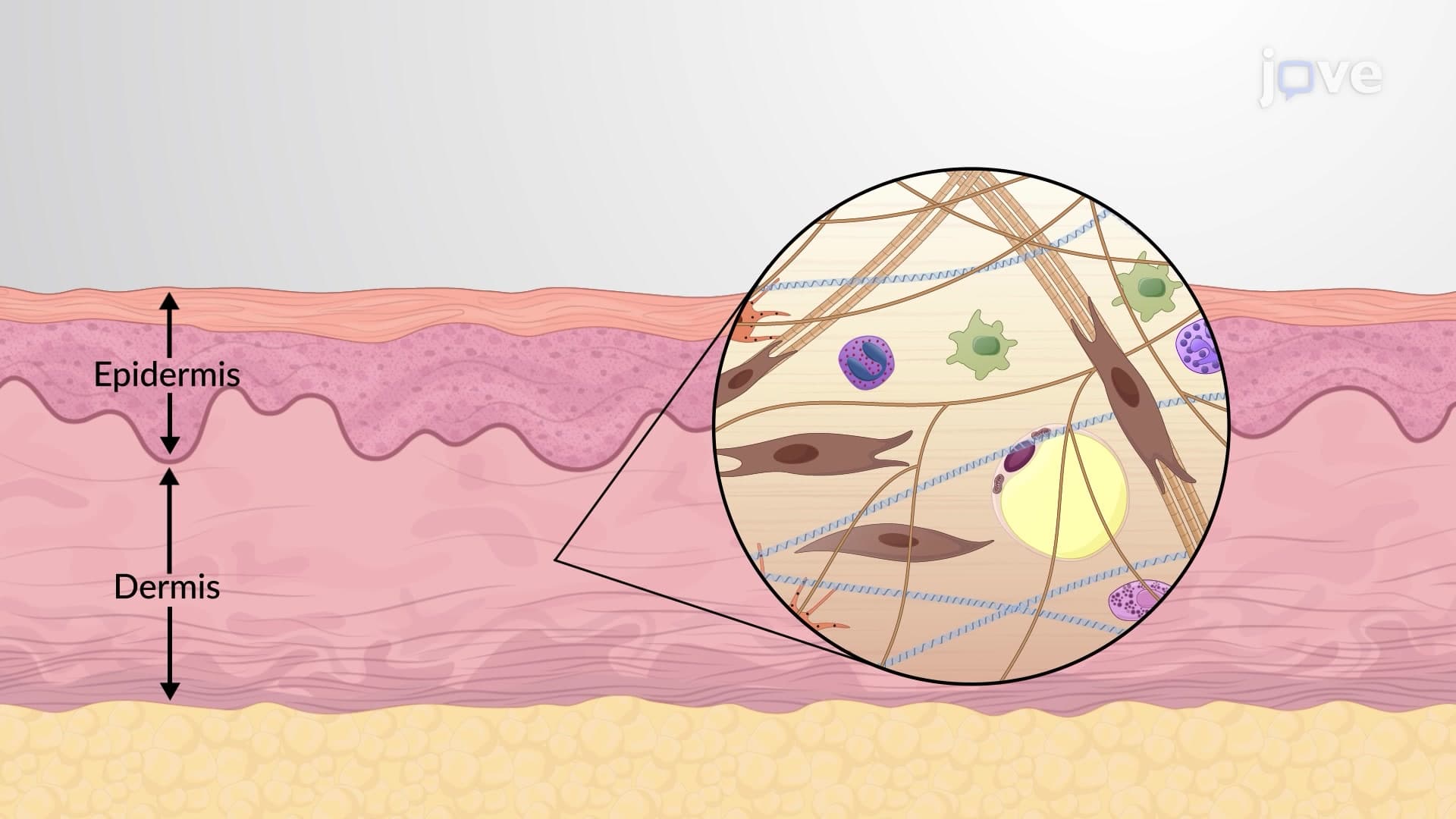
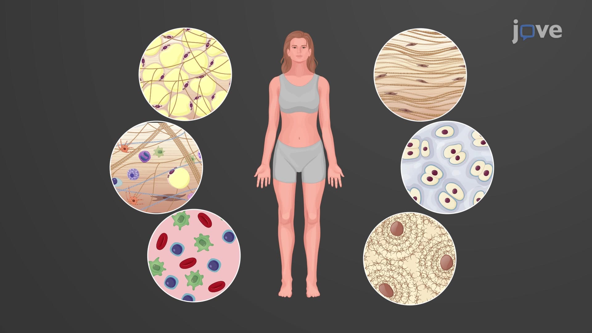
Did you know that your Achilles tendon can withstand forces equivalent to 12 times your body weight during a sprint? This incredible strength comes from the intricate organization of connective tissue fibers and ground substance that form the extracellular matrix. These specialized components work together like a biological scaffold, with collagen providing tensile strength while proteoglycans trap water to maintain tissue hydration. Connective Tissue Fibers And Ground Substance Explained reveals how this cellular architecture supports everything from bone formation to blood vessel elasticity. Watch the full video on JoVE Coach to master this concept with expert-led visuals and step-by-step explanations.
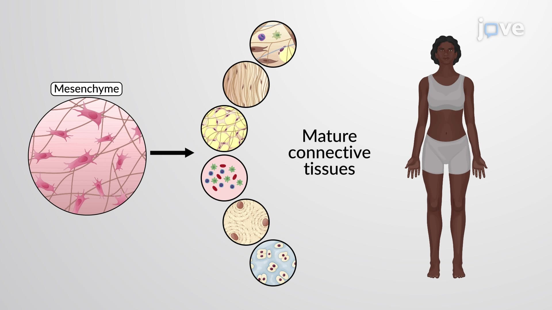
The extracellular matrix (ECM) represents one of the most sophisticated biological engineering systems in the human body. This complex network provides structural support, facilitates cellular communication, and maintains tissue integrity across diverse anatomical locations. For students preparing for the AP Biology exam or college-level anatomy courses, mastering these concepts is essential for understanding how tissues maintain their functional properties under mechanical stress.
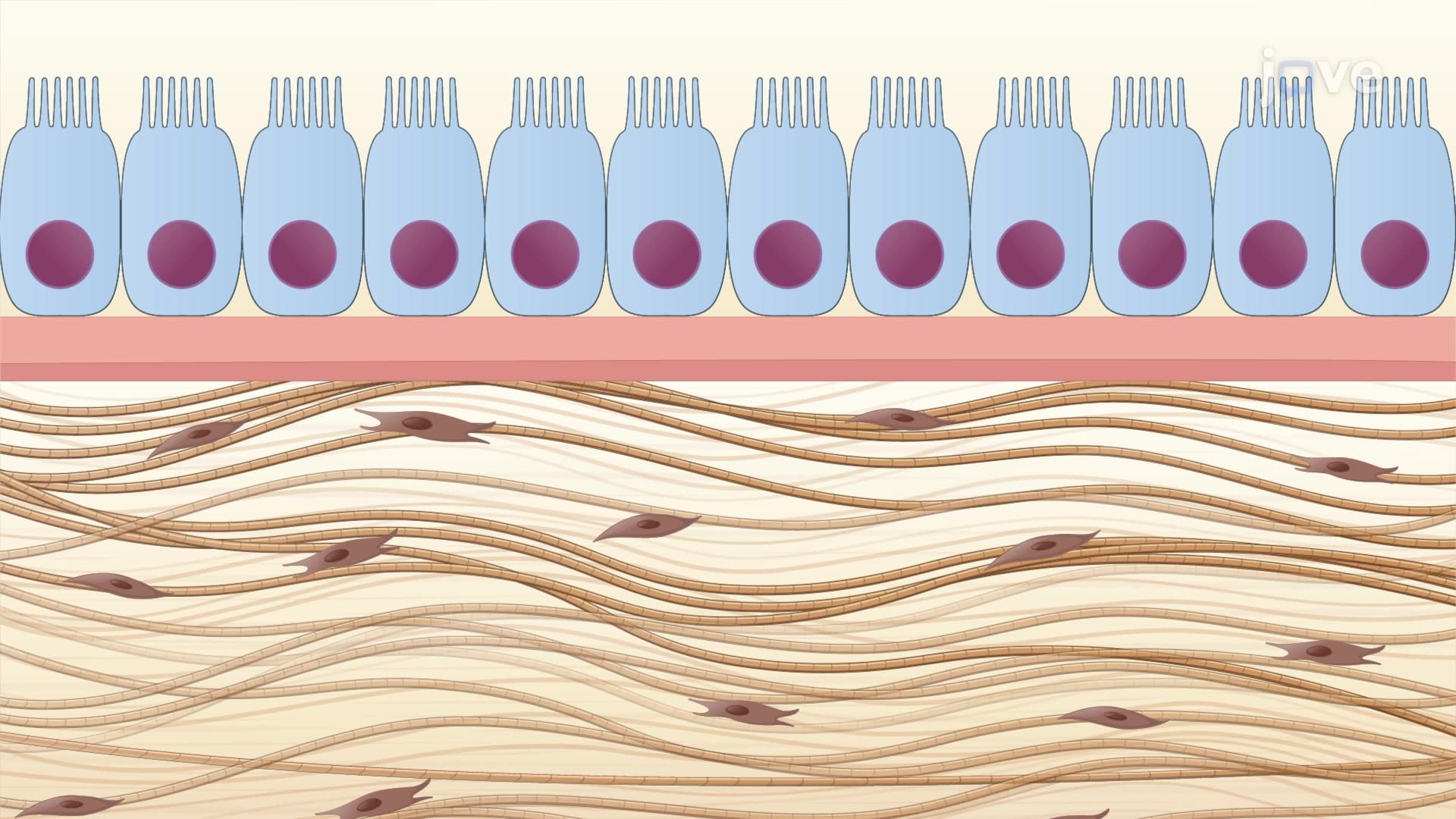
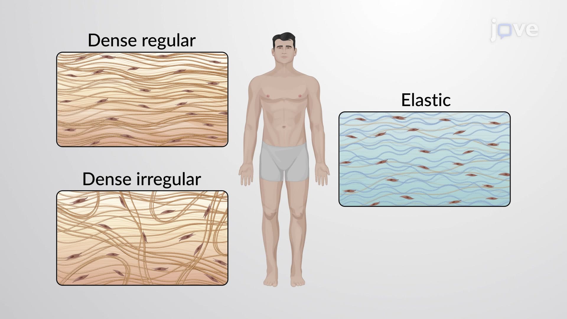
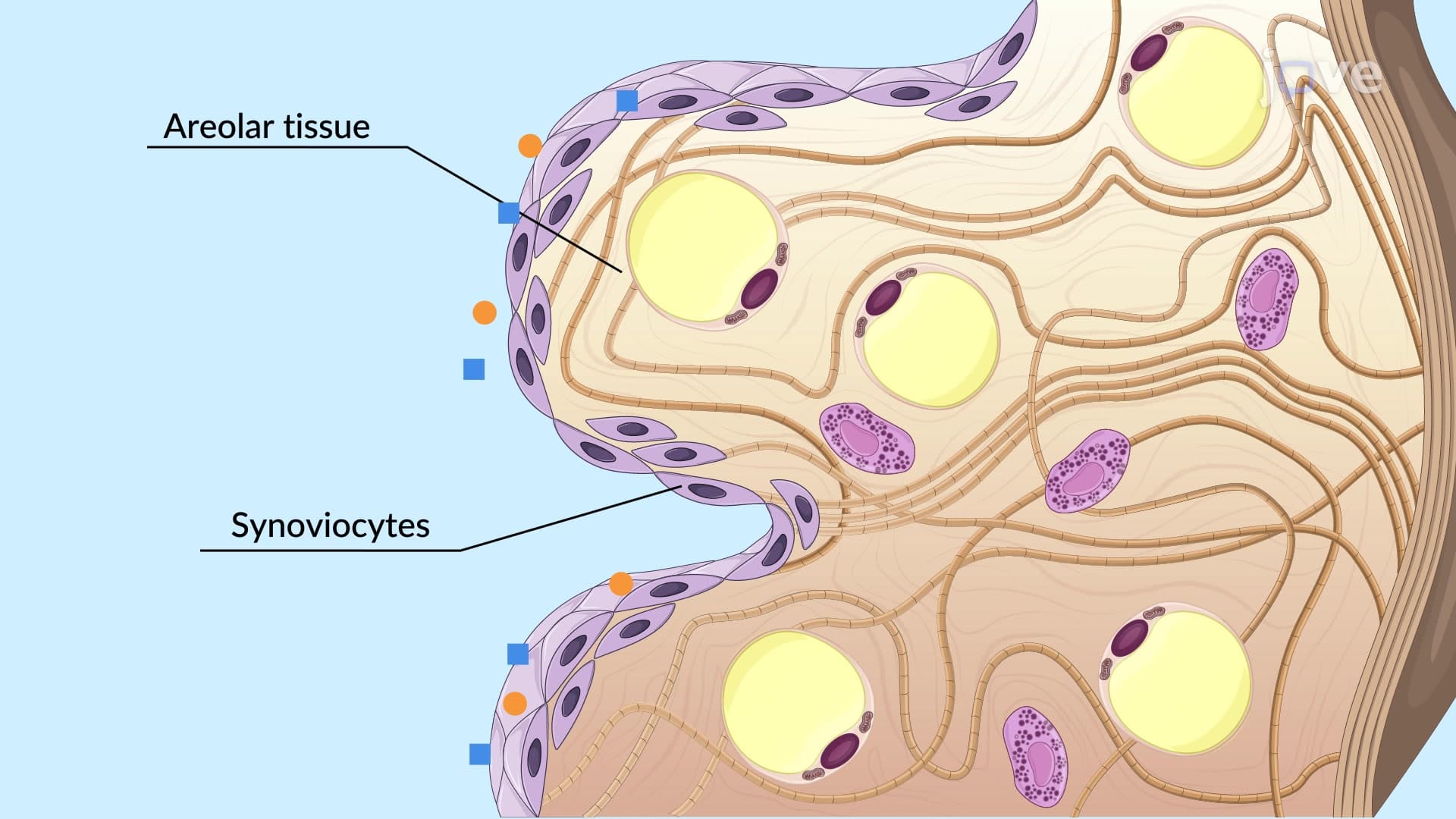
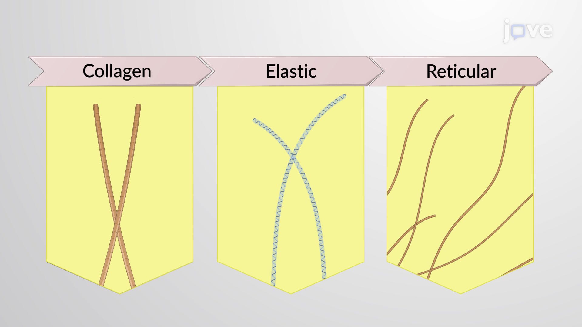
Collagen fibers dominate the connective tissue landscape, with Type I collagen being the most abundant protein in the human body. These unbranched bundles create rope-like structures that resist pulling forces-imagine the cables supporting the Golden Gate Bridge, but at a microscopic scale. In clinical settings, collagen disorders like Ehlers-Danlos syndrome demonstrate the critical importance of proper fiber formation, as patients experience joint hypermobility and skin fragility due to defective collagen synthesis.
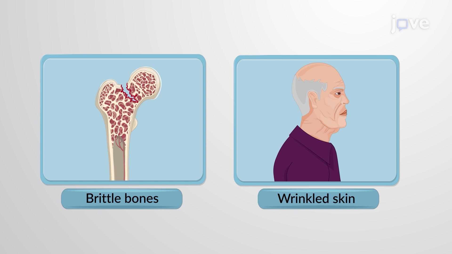
Elastic fibers provide the rebound capacity essential for tissues that stretch repeatedly. The combination of tropoelastin and fibrillin creates branched networks resembling a three-dimensional spider web. Consider how your aorta expands with each heartbeat-this elasticity prevents vessel rupture while maintaining continuous blood flow. Medical students studying for the MCAT often encounter questions about elastin degradation in aging, where reduced elastic fiber integrity contributes to arterial stiffening and hypertension.
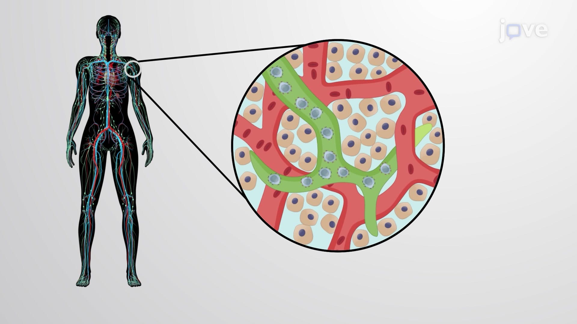
Reticular fibers form delicate meshworks that support cellular structures, particularly in lymphoid organs and blood vessel walls. These Type III collagen networks act like biological scaffolding, providing framework without overwhelming smaller structures. Pathology students recognize reticular fiber patterns as diagnostic markers for certain tissue types under silver staining techniques.
The ground substance creates a hydrated gel environment that facilitates molecular transport and cellular interactions. Proteoglycans serve as molecular sponges, with their glycosaminoglycan (GAG) chains trapping water molecules through ionic interactions. This mechanism explains why cartilage can compress under load yet spring back to original shape-a principle tested frequently on USMLE Step 1 examinations.
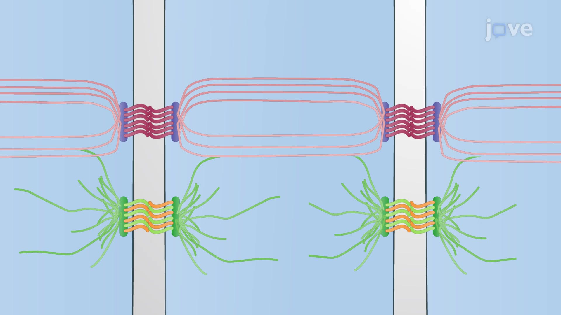
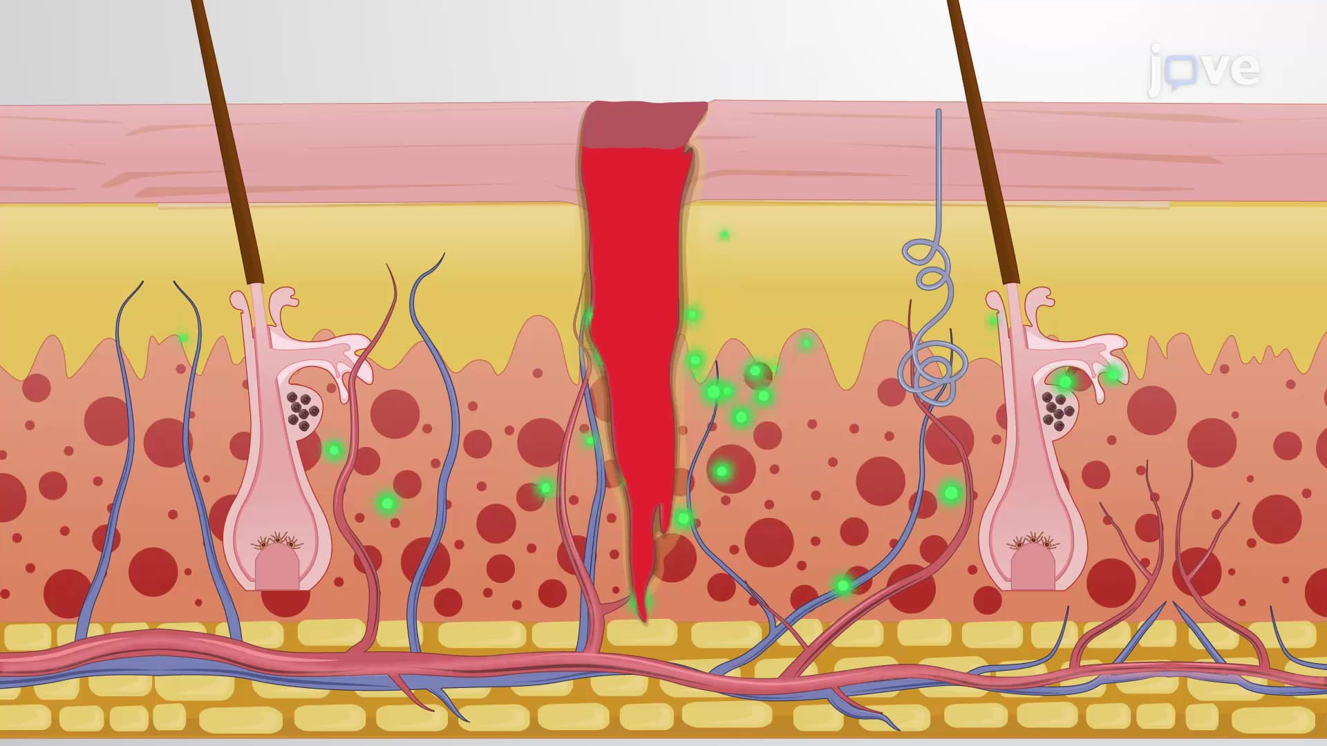
Fibronectin and other cell adhesion molecules act as cellular GPS systems, guiding cell migration during development and tissue repair. Emergency medicine residents observe this process during wound healing, where fibronectin helps organize clot formation and guides inflammatory cell recruitment to injury sites.
Related Micro-courses