Loading subject page…


323,100 views
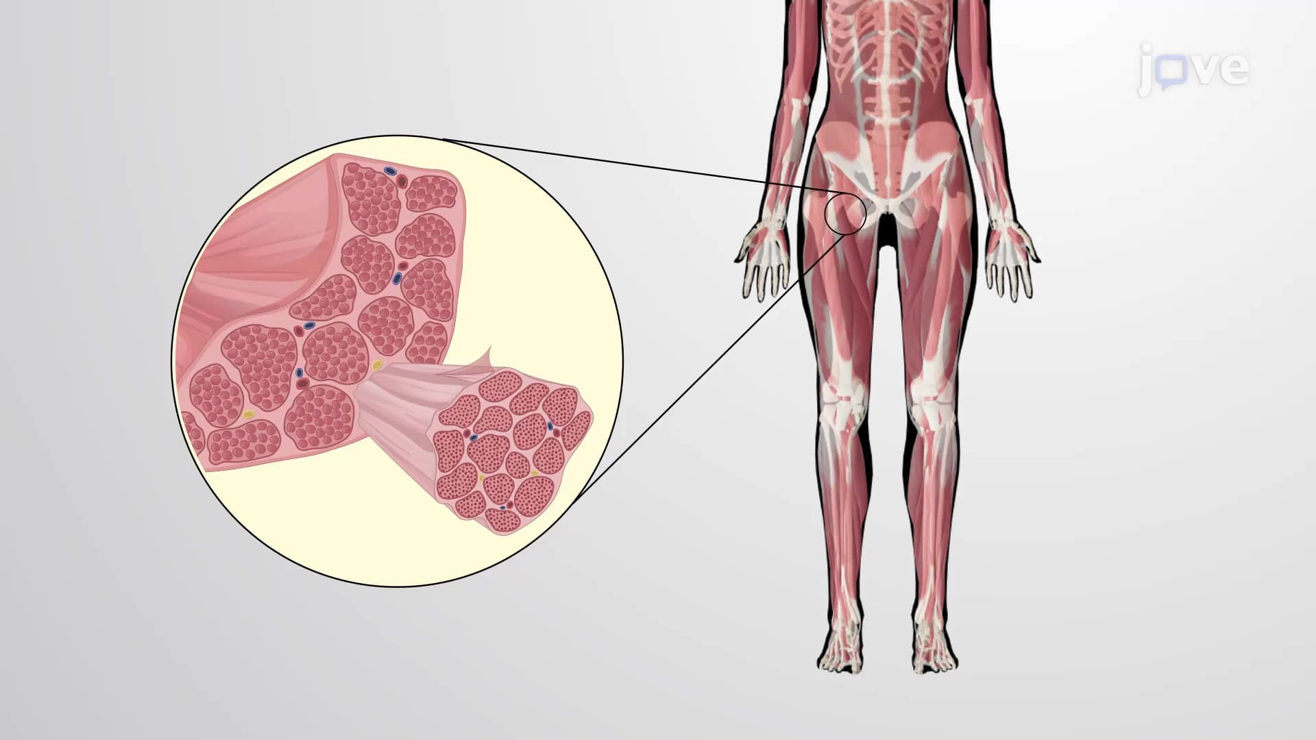

Video Summary: Layers of Connective Tissue Proper Explained
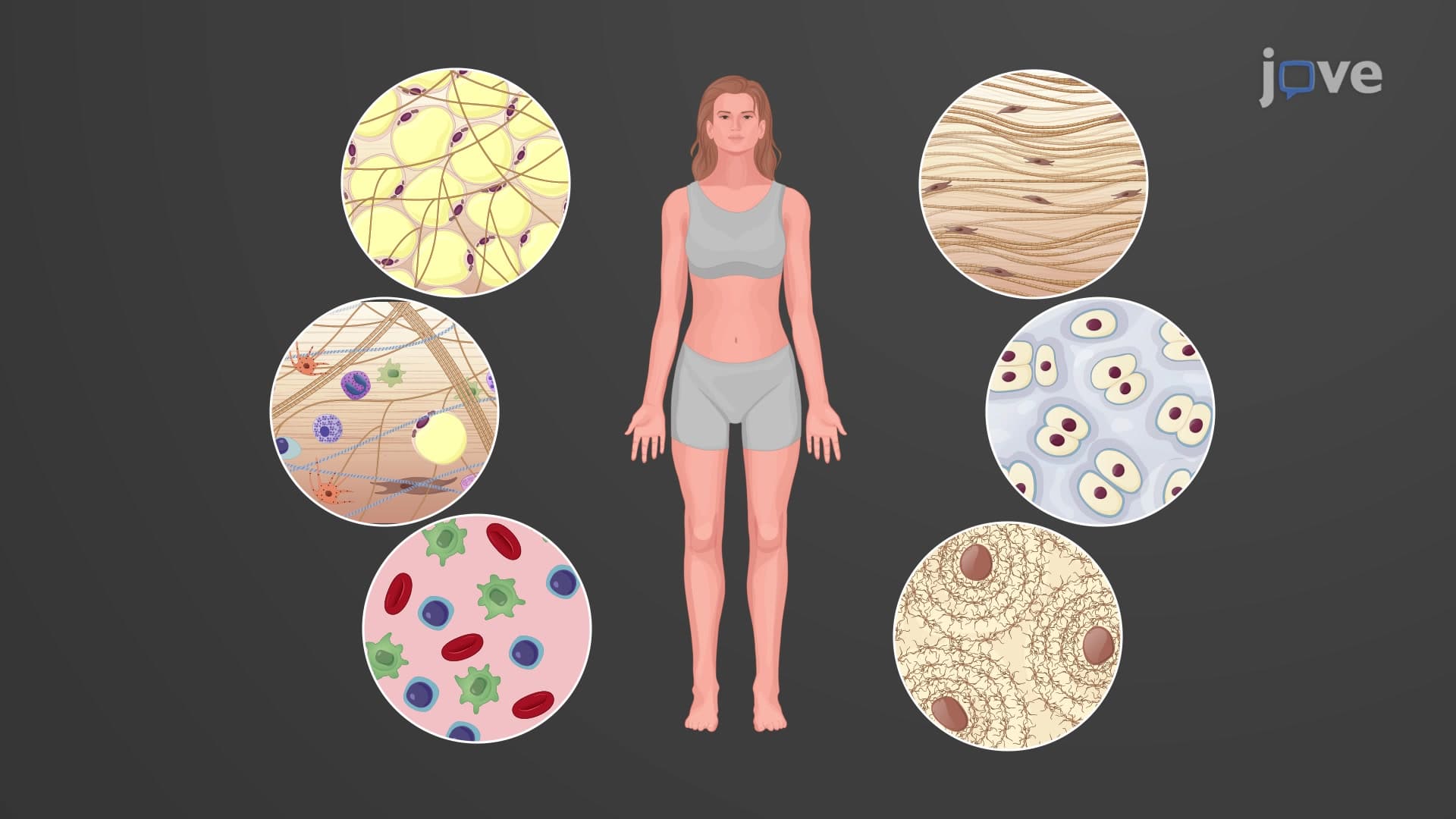
Did you know that the connective tissue proper layers beneath your skin are what prevent your muscles from sliding around when you move? These specialized layers of connective tissue proper anatomy work like invisible scaffolding throughout your body. When a surgeon at Johns Hopkins Hospital makes an incision, they must carefully navigate through these distinct fascial planes to reach internal organs safely. Understanding the Layers of Connective Tissue Proper Explained becomes crucial for anyone studying human anatomy. Watch the full video on JoVE Coach to master this concept with expert-led visuals and step-by-step explanations.
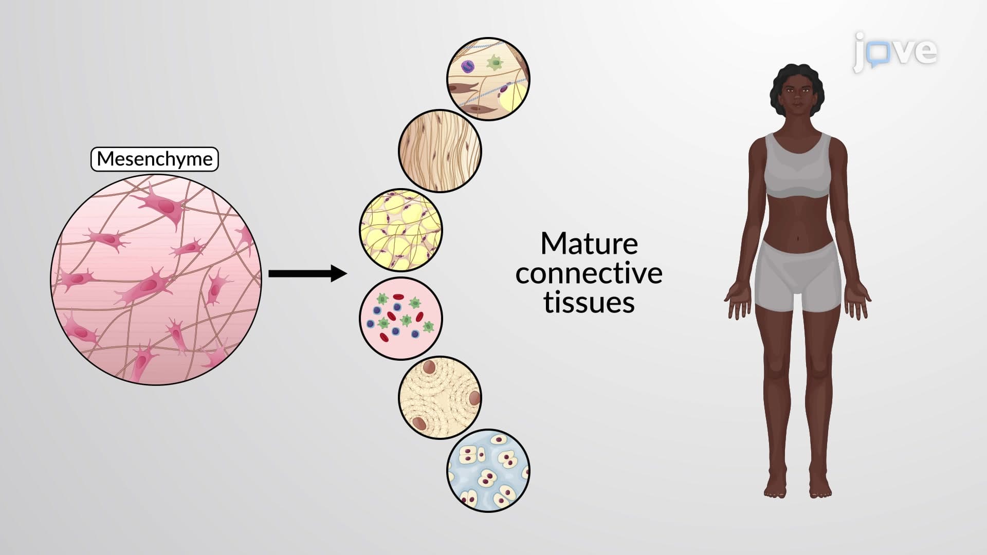
The human body's structural integrity depends on a sophisticated network of connective tissue proper layers that function as biological scaffolding. These layers, collectively known as fascia, represent one of anatomy's most elegant organizational systems. Unlike the simple categorization often taught in introductory courses, these layers connective tissue proper anatomy form a complex, interconnected web that surgeons and clinicians must navigate with precision.
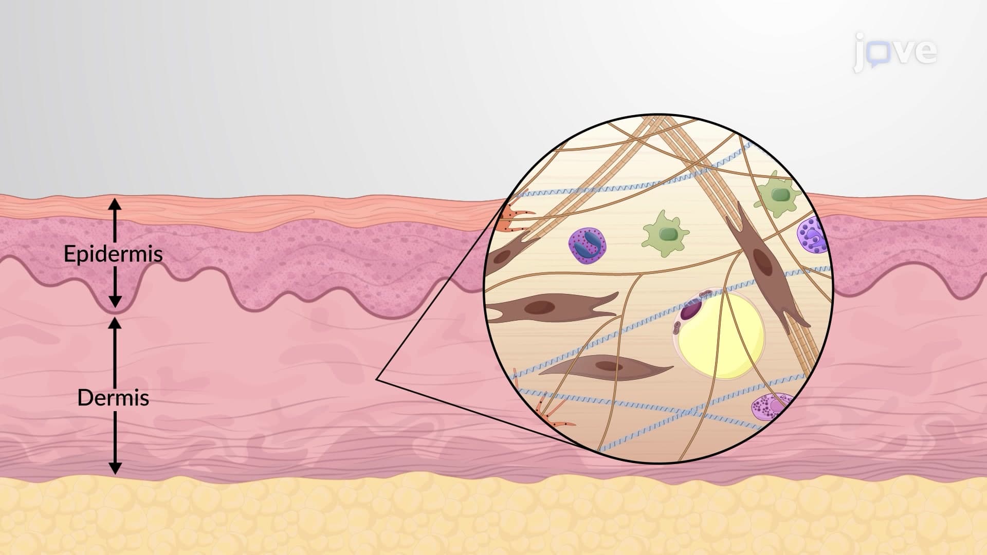
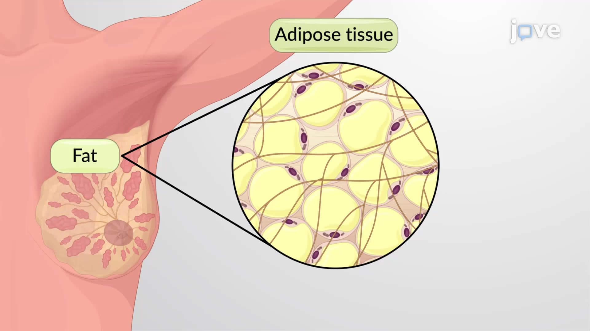
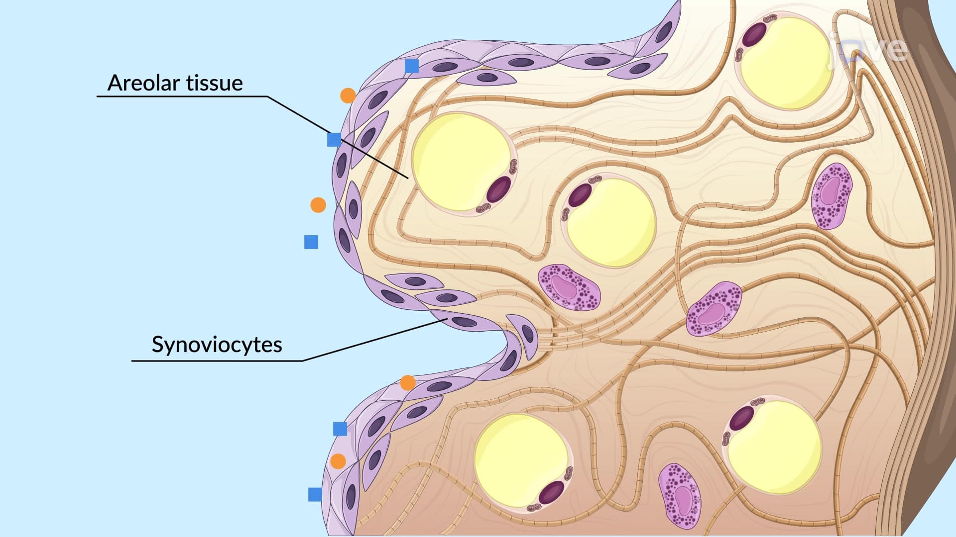
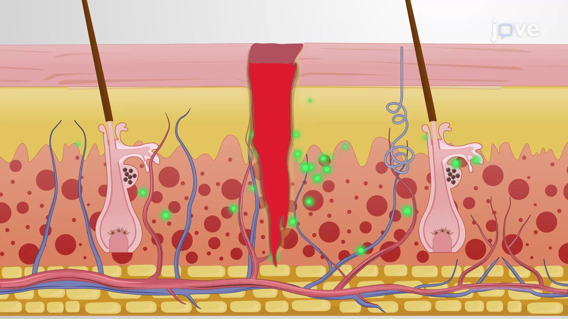
The superficial fascia, or hypodermis, represents the most accessible connective tissue body layer in human anatomy. Located directly beneath the dermis, this layer combines areolar connective tissue with varying amounts of adipose tissue. At Massachusetts General Hospital, plastic surgeons regularly work within this layer during cosmetic procedures, understanding that its thickness varies dramatically-from thin coverage over the shins to substantial padding across the abdomen and buttocks.
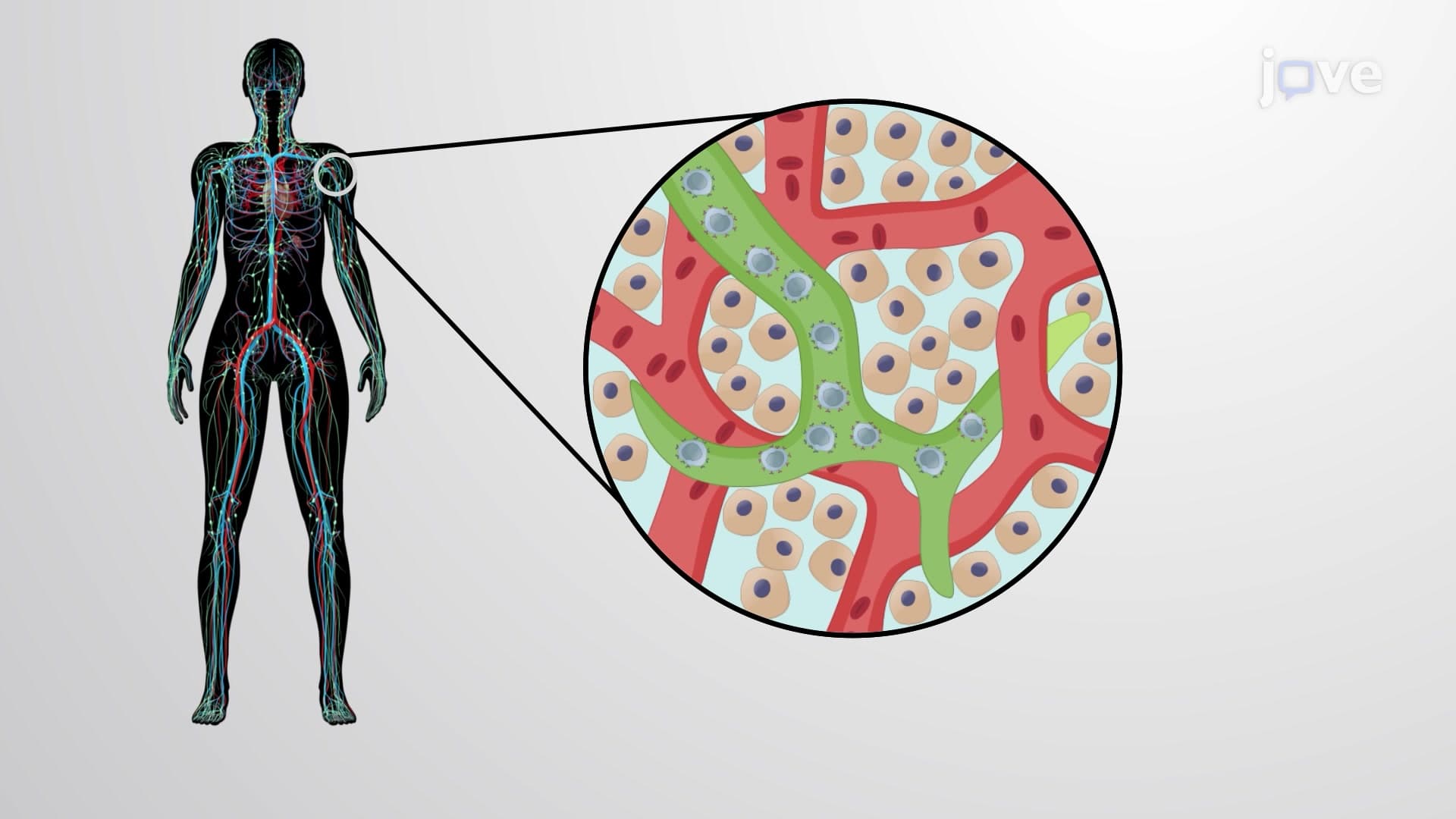
This fascial layer anatomy serves multiple critical functions beyond simple padding. The areolar tissue creates pathways for nerves, blood vessels, and lymphatic vessels, while the adipose component provides thermal insulation and energy storage. For students preparing for the MCAT or AP Biology exams, remember that this layer's composition directly influences body temperature regulation and drug absorption rates in clinical settings.
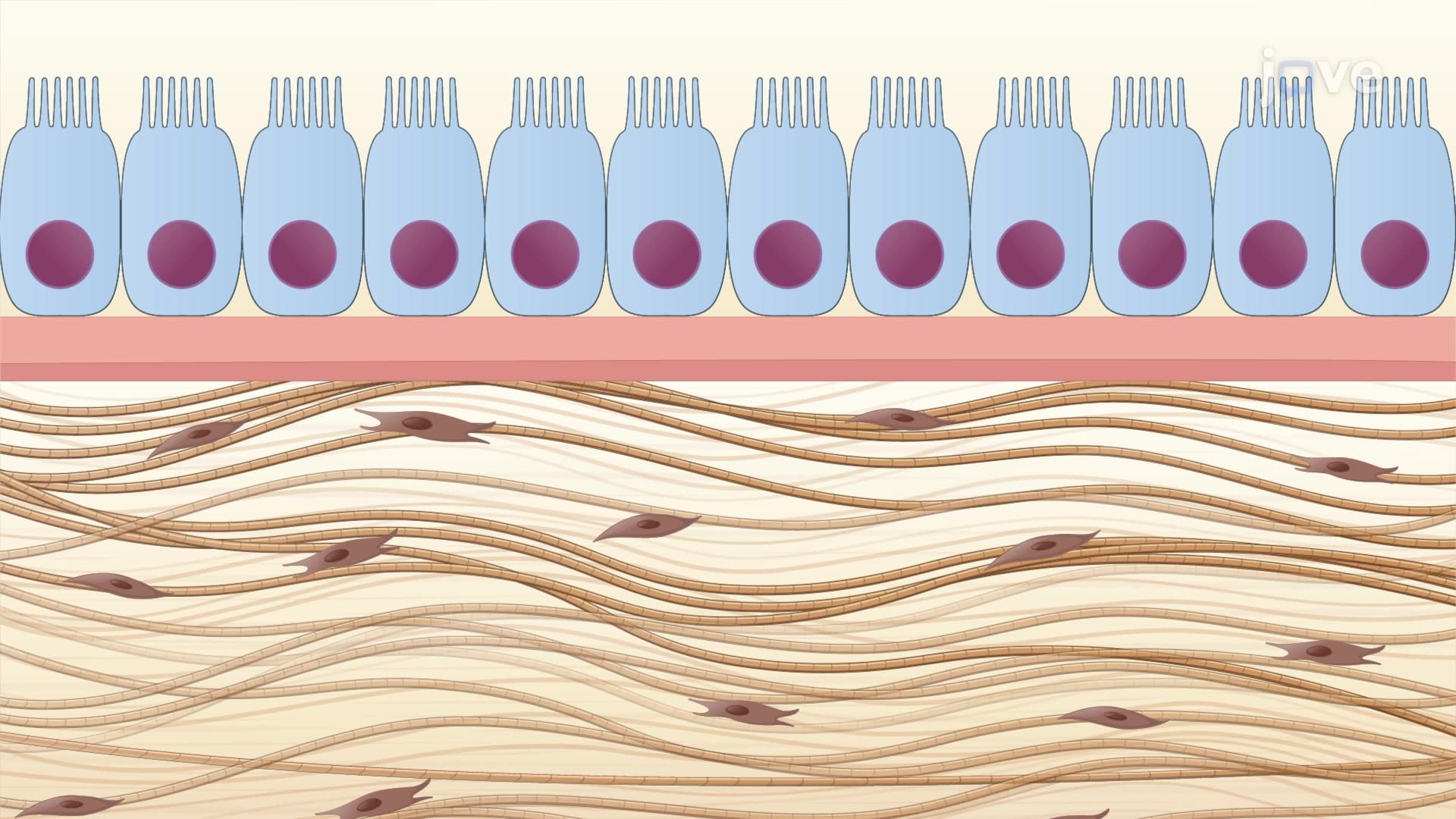
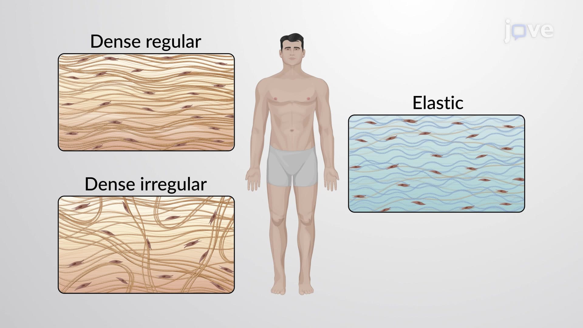
Deep fascia represents the most structurally sophisticated dermis connective tissue layer extension in the body. Composed primarily of dense regular connective tissue enriched with hyaluronic acid, this layer creates the epimysium surrounding skeletal muscles. Orthopedic surgeons at the Cleveland Clinic rely on understanding these fascial planes when treating compartment syndrome, a condition where pressure builds within fascial compartments.
The connective tissue proper organization becomes particularly evident in the thigh, where deep fascia creates distinct anterior, medial, and posterior compartments. Each compartment houses specific muscle groups with shared functions and nerve supplies. This organization proves essential for students studying kinesiology or preparing for NCLEX examinations, as dysfunction in one compartment can affect entire movement patterns.
The deepest superficial deep fascia layers system includes subserous fascia, which provides the final protective barrier before reaching serous membranes. This areolar tissue layer cushions vital organs like the heart and lungs, allowing them to move freely within their cavities during breathing and cardiac cycles. Thoracic surgeons at Johns Hopkins understand that this layer's flexibility prevents organ adhesion while maintaining protective cushioning.
Related Micro-courses