Loading subject page…


323,100 views

Video Summary: Overview of Connective Tissues Proper Explained
Did you know that the tendons connecting your bicep muscle to your arm bones can withstand forces up to 4,000 pounds per square inch? This incredible strength comes from understanding connective tissue proper overview, which reveals how different arrangements of protein fibers create tissues with vastly different functions. From the cushioning fat under your skin to the tough ligaments in your knee, connective tissue proper types explained show us how structure determines function in the human body. Watch the full video on JoVE Coach to master this concept with expert-led visuals and step-by-step explanations.
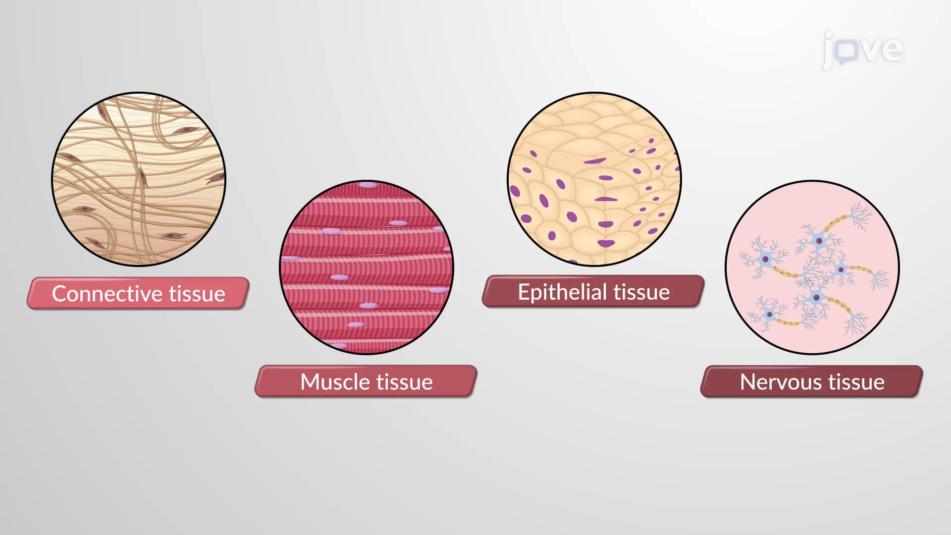
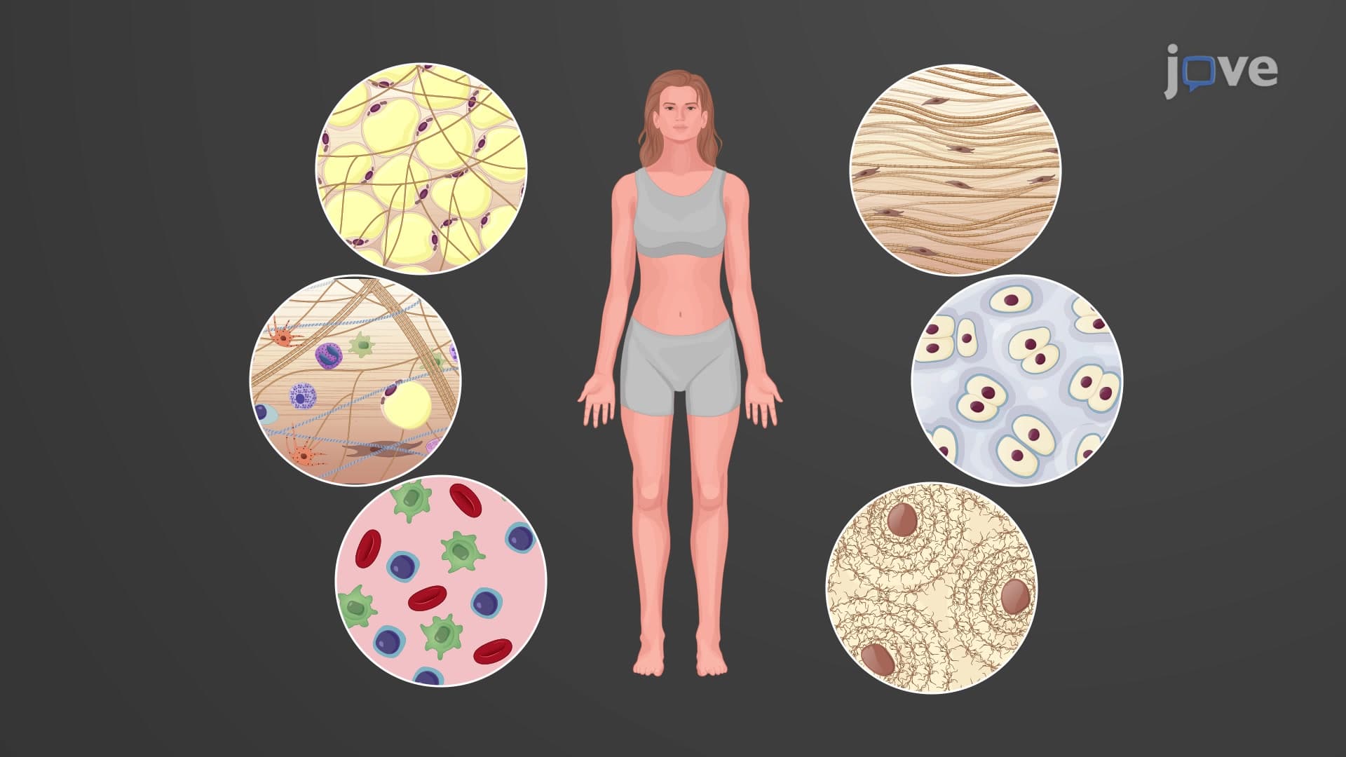
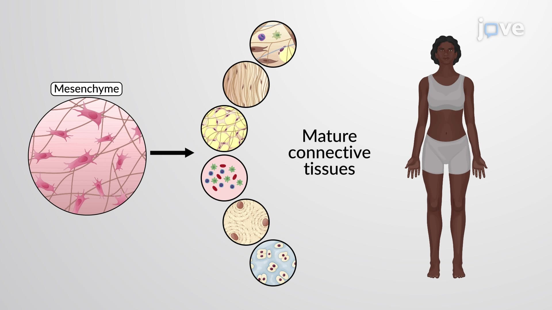
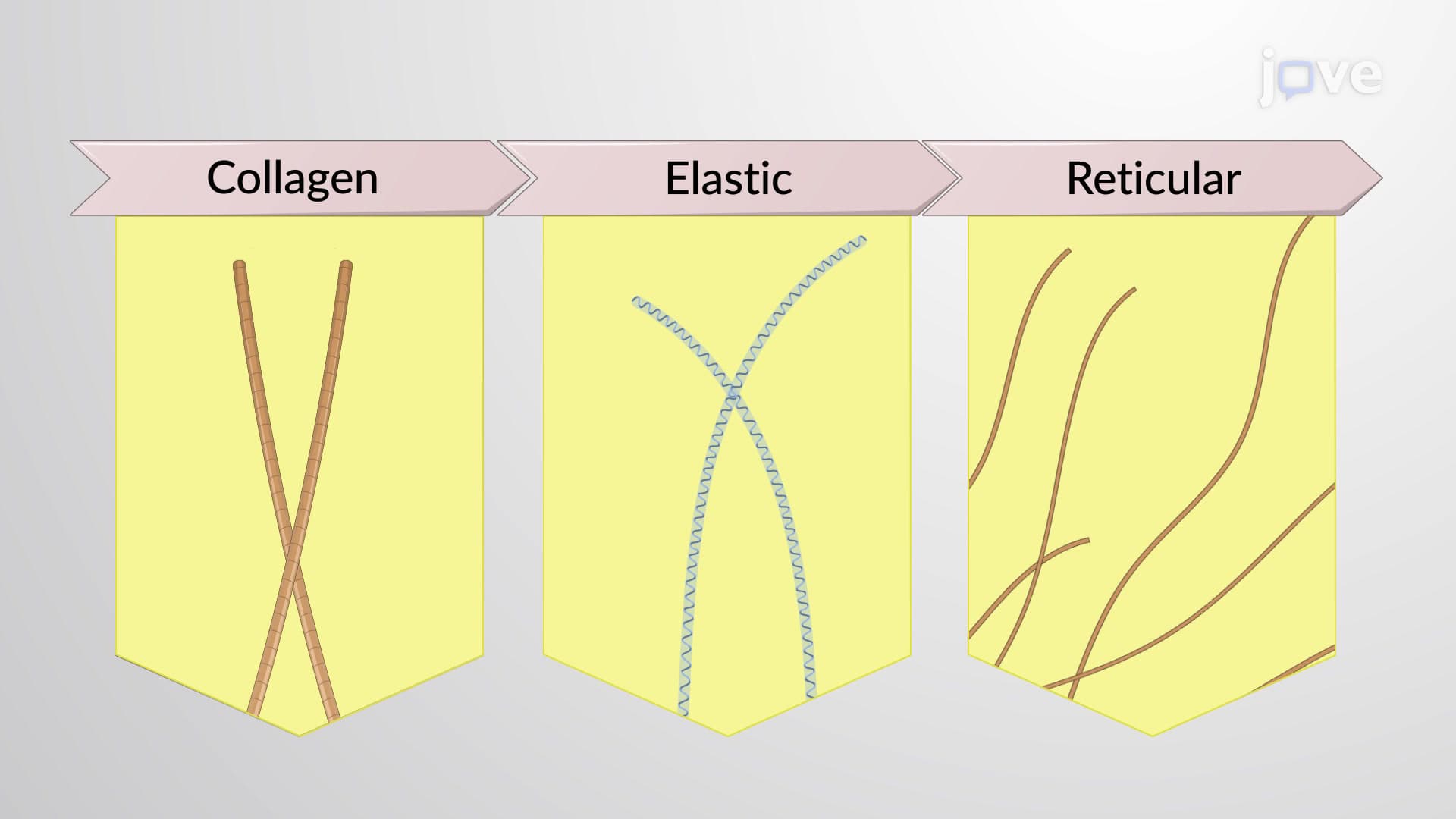
Connective tissue proper represents one of the most diverse and functionally important tissue categories in the human body. Unlike epithelial tissues that form barriers, or muscle tissues that generate force, connective tissue proper specializes in providing structural support, protection, and connection between different organs and systems. The key to understanding these tissues lies in recognizing how the arrangement of protein fibers within the extracellular matrix determines their mechanical properties and biological functions.
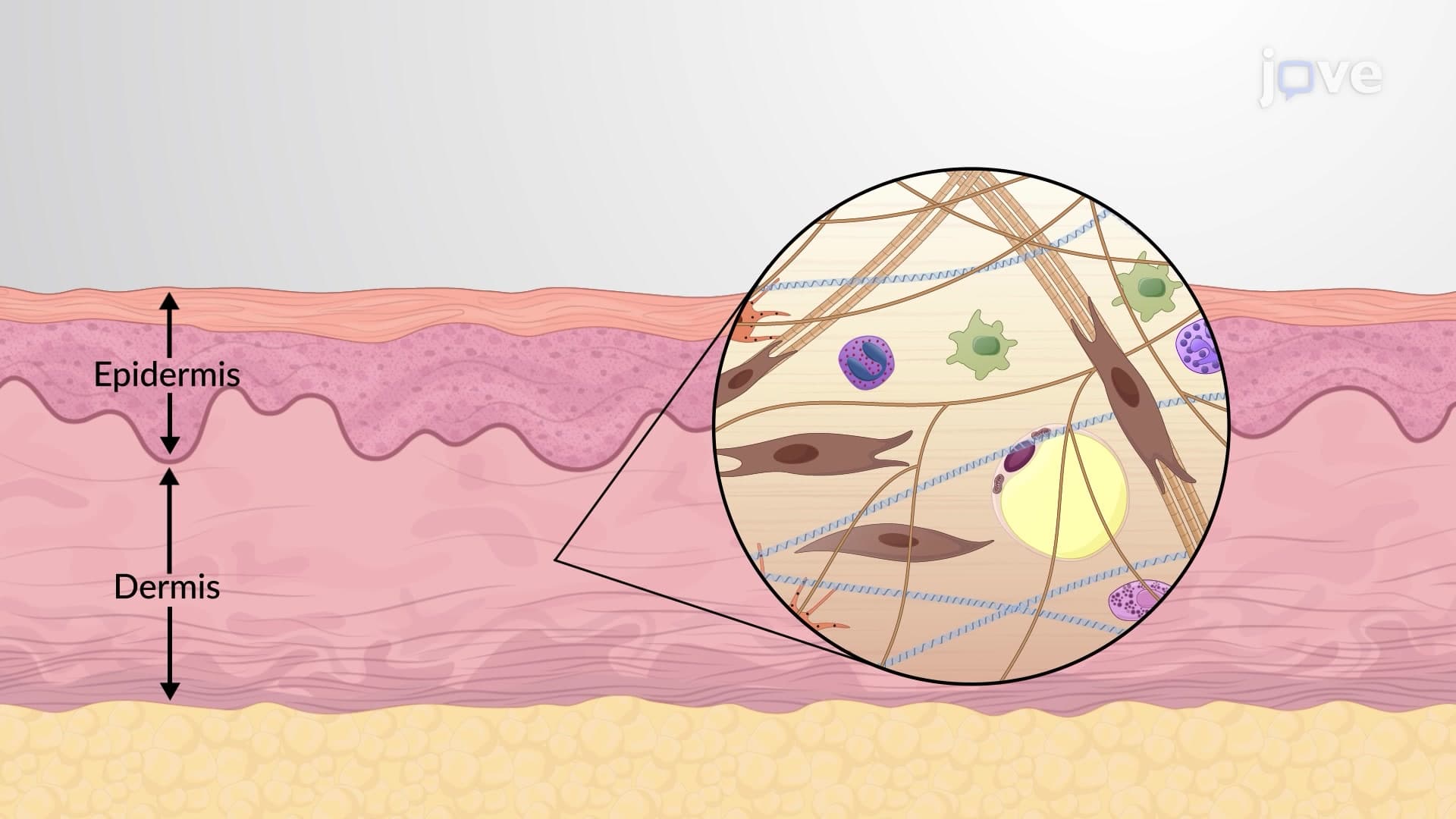
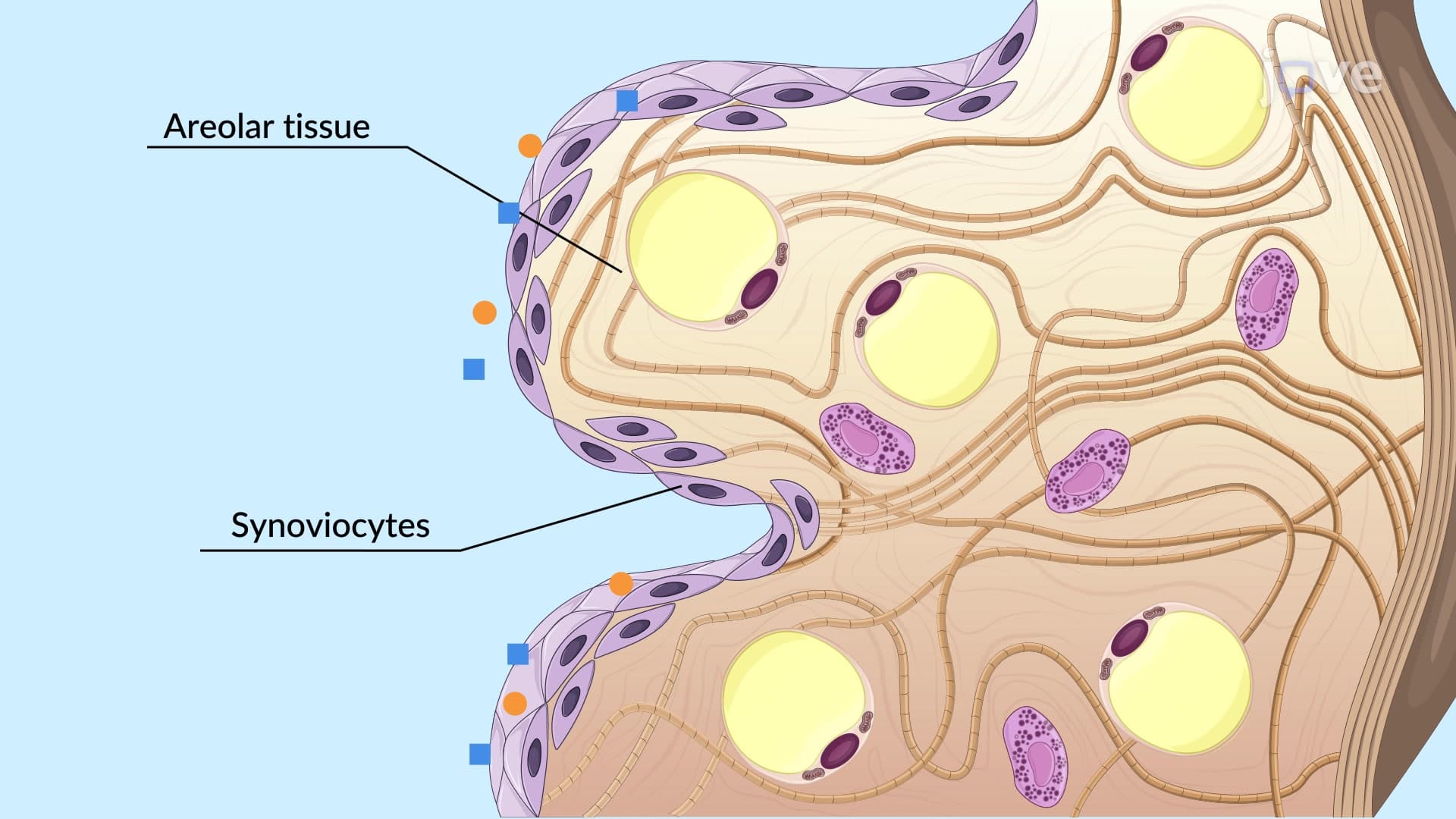
Loose connective tissues are characterized by widely spaced protein fibers embedded in an abundant ground substance, creating a gel-like matrix that provides flexibility while maintaining structural integrity. Areolar loose connective tissue serves as the body's universal packing material, found beneath the skin and surrounding organs like the heart and lungs. This tissue's loose fiber arrangement allows for easy movement and provides pathways for blood vessels and nerves.
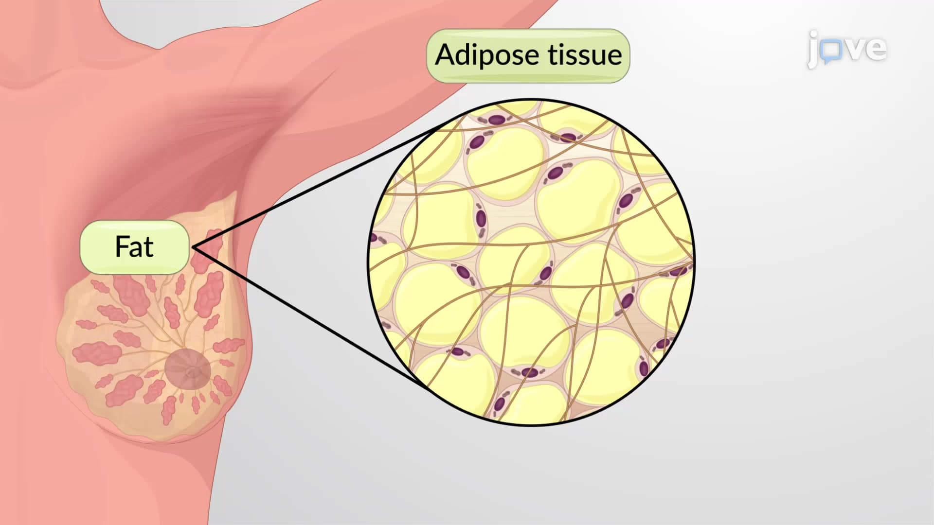
Adipose reticular connective tissue includes two distinct but related types. Adipose tissue, concentrated in areas like the subcutaneous layer and around organs such as the kidneys, serves dual functions of energy storage and thermal insulation. A typical adult American carries 15-20% of their body weight as adipose tissue. Reticular tissue, found in lymph nodes, the spleen, and bone marrow, creates a three-dimensional scaffold that supports immune cells and blood cell production.
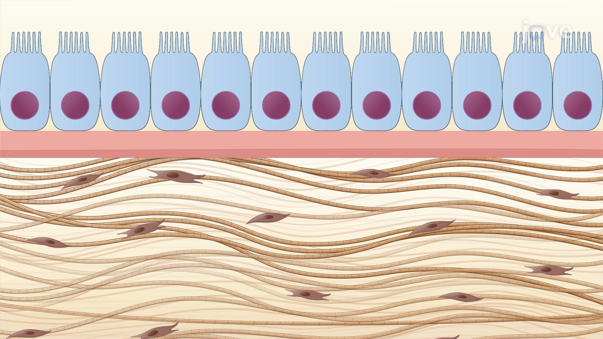
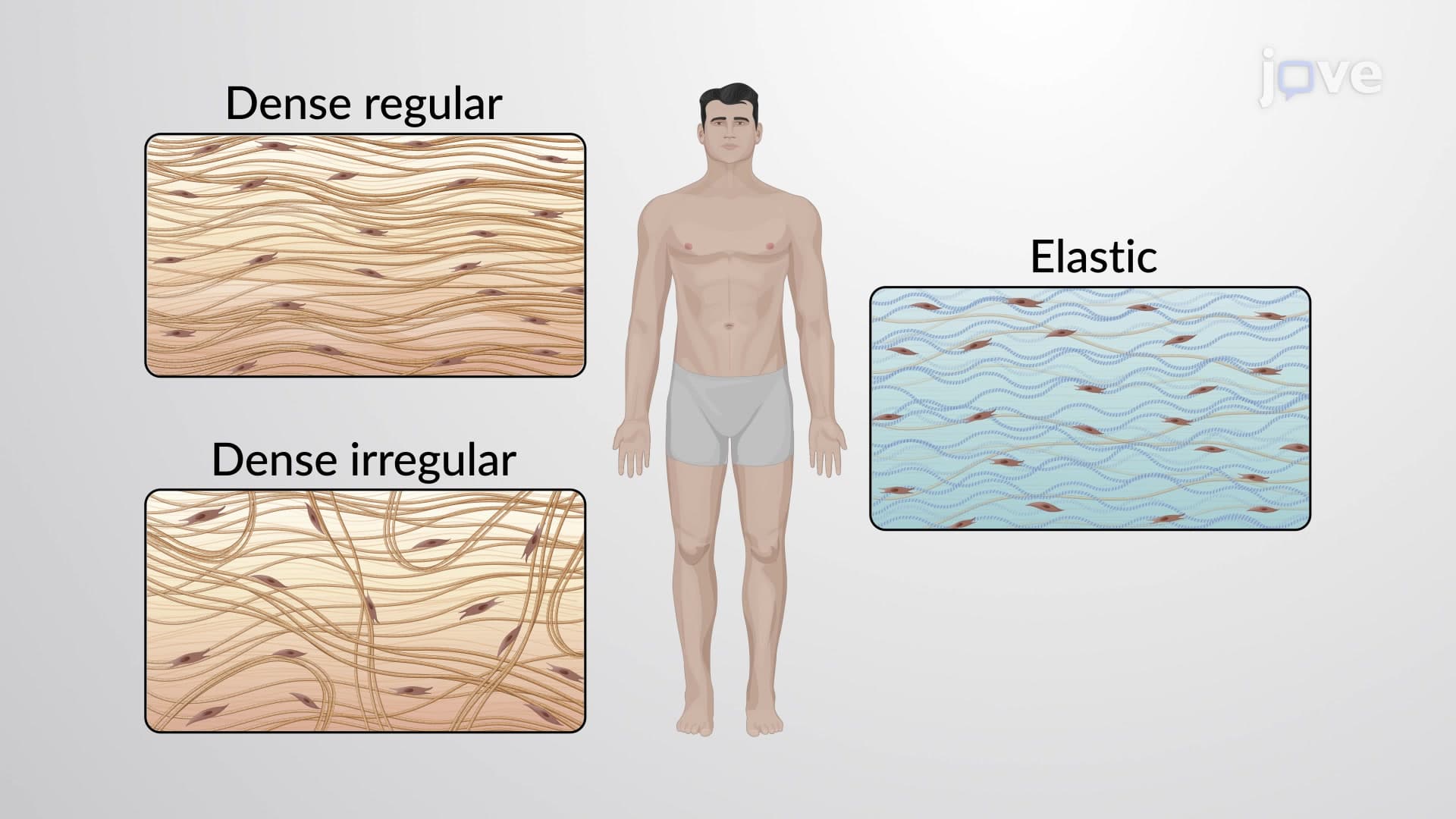
Dense connective tissues contain tightly packed collagen fibers with minimal ground substance, creating structures capable of withstanding tremendous mechanical stress. Dense regular connective tissue, exemplified by tendons and ligaments, arranges collagen fibers in parallel bundles that can resist forces in one primary direction. The Achilles tendon, connecting your calf muscle to your heel bone, demonstrates this principle perfectly-it can withstand forces equivalent to lifting a small car.
Dense irregular connective tissue, found in joint capsules and organ coverings, weaves collagen fibers in multiple directions to resist stress from various angles. This arrangement makes the tough outer covering of your liver or the fibrous capsule around your shoulder joint incredibly durable against multi-directional forces.
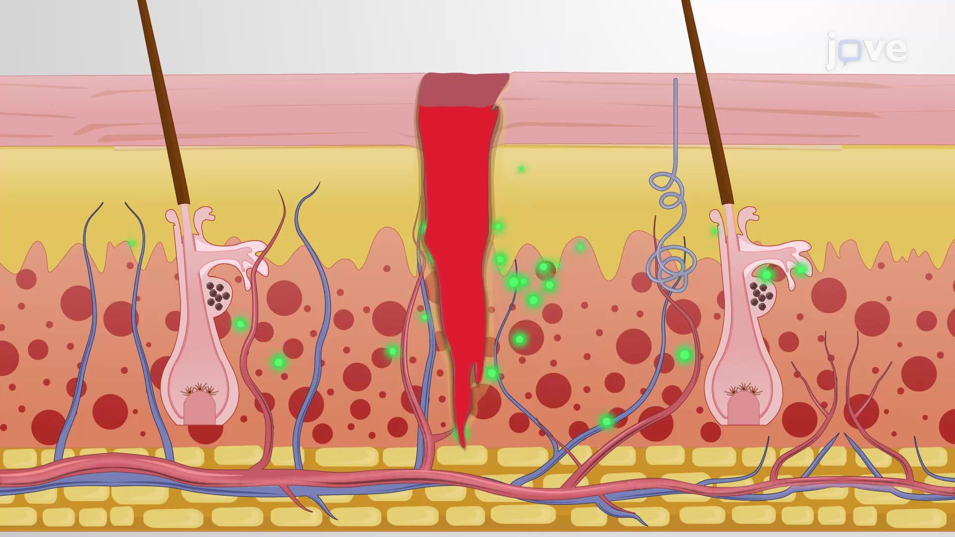
Understanding connective tissue proper proves essential for success on standardized exams like the MCAT, AP Biology, and college anatomy courses. Students frequently encounter questions about structure-function relationships, asking them to predict tissue locations based on mechanical properties or explain why certain injuries heal more slowly than others. For pre-med students, this knowledge becomes foundational for understanding conditions like Marfan syndrome, where defective connective tissue affects multiple organ systems, or why surgical repairs of tendons require extensive rehabilitation compared to muscle injuries.
Related Micro-courses