Loading subject page…


323,106 views

Video Summary: Tissue Injury Inflammation and Repair Explained
When you get a paper cut, why does your finger turn red and swell within minutes? This immediate response demonstrates tissue injury inflammation repair in action-your body's sophisticated defense system that protects against infection and promotes healing. Understanding how tissue injury inflammation and repair occur is crucial for pre-med students studying for the MCAT and anyone interested in human biology. From emergency rooms treating trauma patients at Johns Hopkins to understanding why athletes ice injuries, these biological processes impact healthcare decisions daily. Watch the full video on JoVE Coach to master this concept with expert-led visuals and step-by-step explanations.
Tissue injury inflammation repair represents one of biology's most essential survival mechanisms. When tissues suffer damage-whether from a surgical incision at Mayo Clinic, a sports injury during high school football, or cellular damage from infection-the body initiates a precisely coordinated response. This process has evolved over millions of years to protect organisms from further harm while restoring normal tissue function.
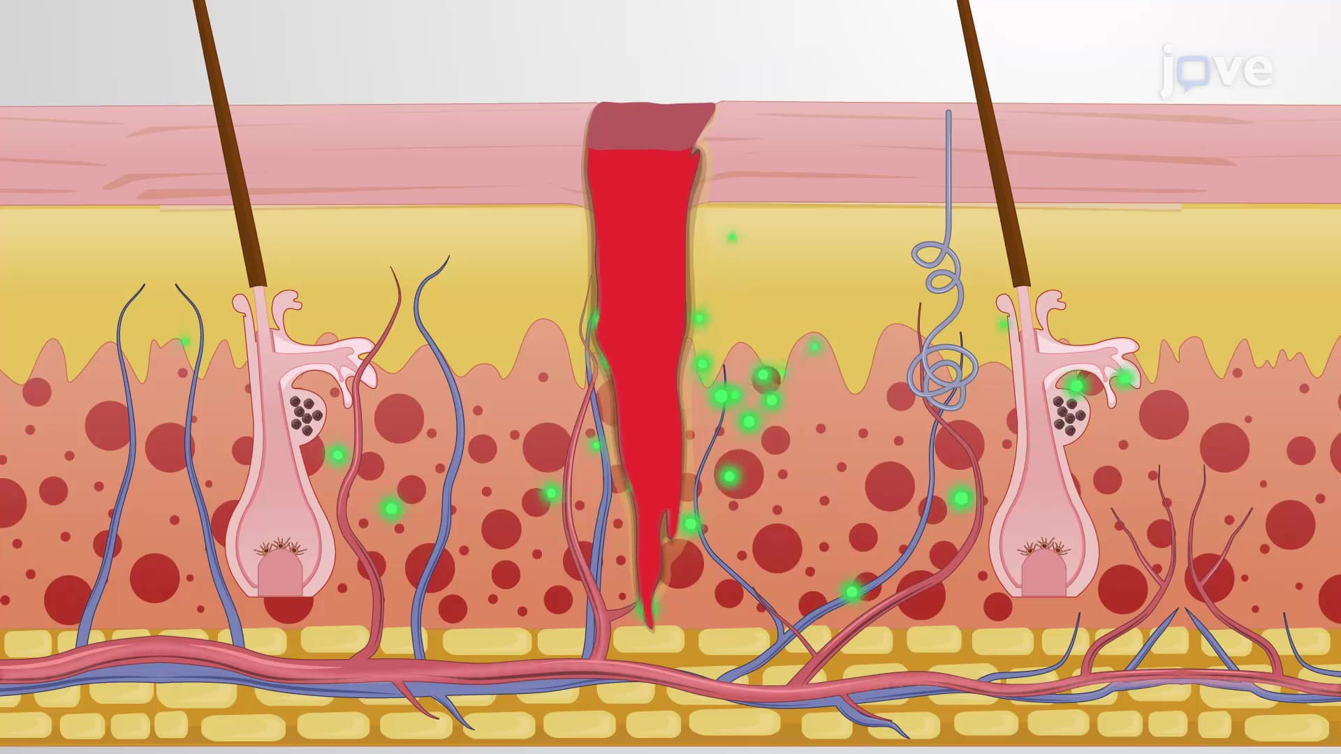
The acute inflammatory response begins within seconds of tissue injury. Blood vessels at the injury site immediately constrict (vasoconstriction) for 5-10 seconds, then dramatically dilate (vasodilation). This increased blood flow creates the characteristic redness and heat. Simultaneously, vessel walls become more permeable, allowing plasma proteins and immune cells to enter tissues, causing swelling (edema).
Inflammatory mediators tissue components orchestrate this response. Histamine, released from mast cells, causes immediate vasodilation. Prostaglandins amplify the inflammatory response and sensitize pain receptors. Complement proteins create membrane attack complexes that destroy pathogens. For AP Biology students, understanding these molecular signals is crucial for exam success.
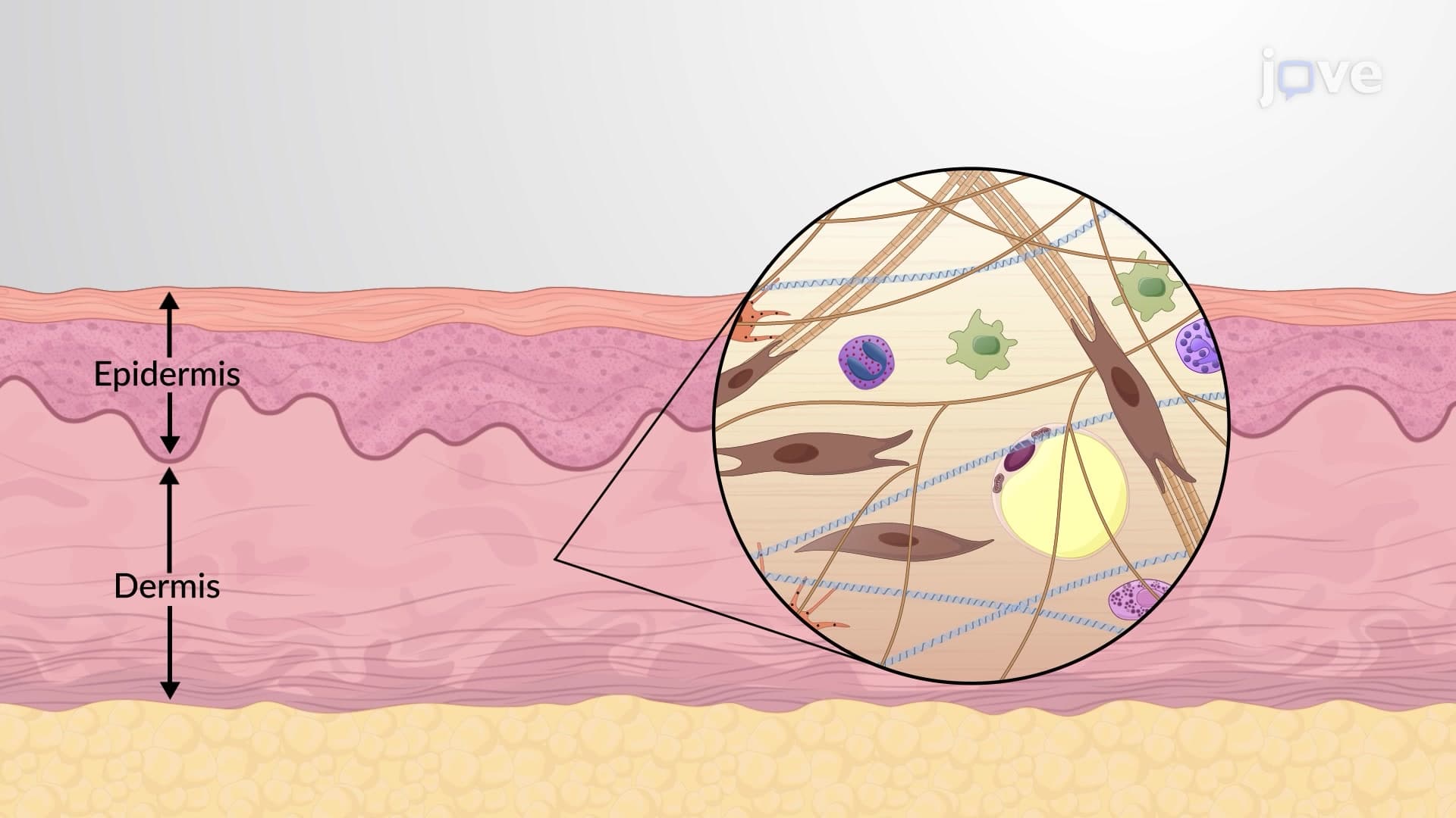
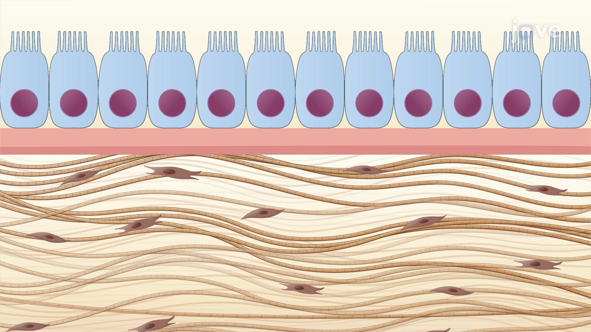
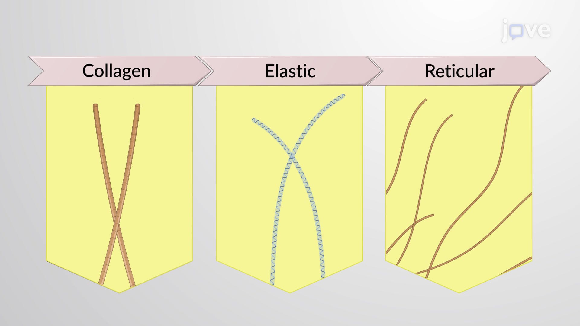
Following inflammation, tissue repair regeneration occurs through two pathways. Regeneration replaces damaged cells with identical functional cells-common in epithelial tissues like skin and intestinal lining. However, when regenerative capacity is exceeded, fibrosis scar tissue formation occurs. Fibroblasts proliferate and secrete collagen, creating strong but functionally different replacement tissue.
Healthcare professionals at institutions like Cleveland Clinic rely on understanding wound healing phases to optimize patient outcomes. The hemostasis phase stops bleeding through platelet aggregation and clot formation. The inflammatory phase (days 1-3) brings immune cells to clear debris. The proliferation phase (days 3-21) rebuilds tissue through angiogenesis and collagen synthesis. Finally, the remodeling phase (months to years) reorganizes collagen fibers for maximum strength.
This knowledge proves essential for MCAT preparation, particularly in understanding how diabetes impairs healing by affecting neutrophil function and collagen synthesis-concepts frequently tested in medical school admission exams.
Related Micro-courses