Loading subject page…


323,101 views

Video Summary: What are Classification of Epithelial Tissues
Ever wonder why your skin can withstand daily wear while your lungs allow oxygen to pass through effortlessly? The secret lies in epithelial tissue classification, which determines how these protective barriers function throughout your body. From the tough, multi-layered skin protecting a construction worker's hands to the delicate single-cell lining in hospital patients' lung alveoli, understanding what are classification of epithelial tissues reveals how form follows function in human anatomy. Watch the full video on JoVE Coach to master this concept with expert-led visuals and step-by-step explanations.
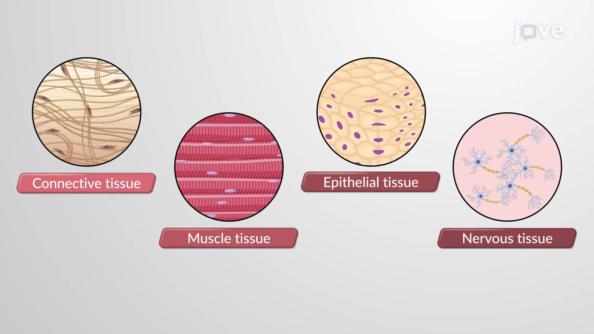
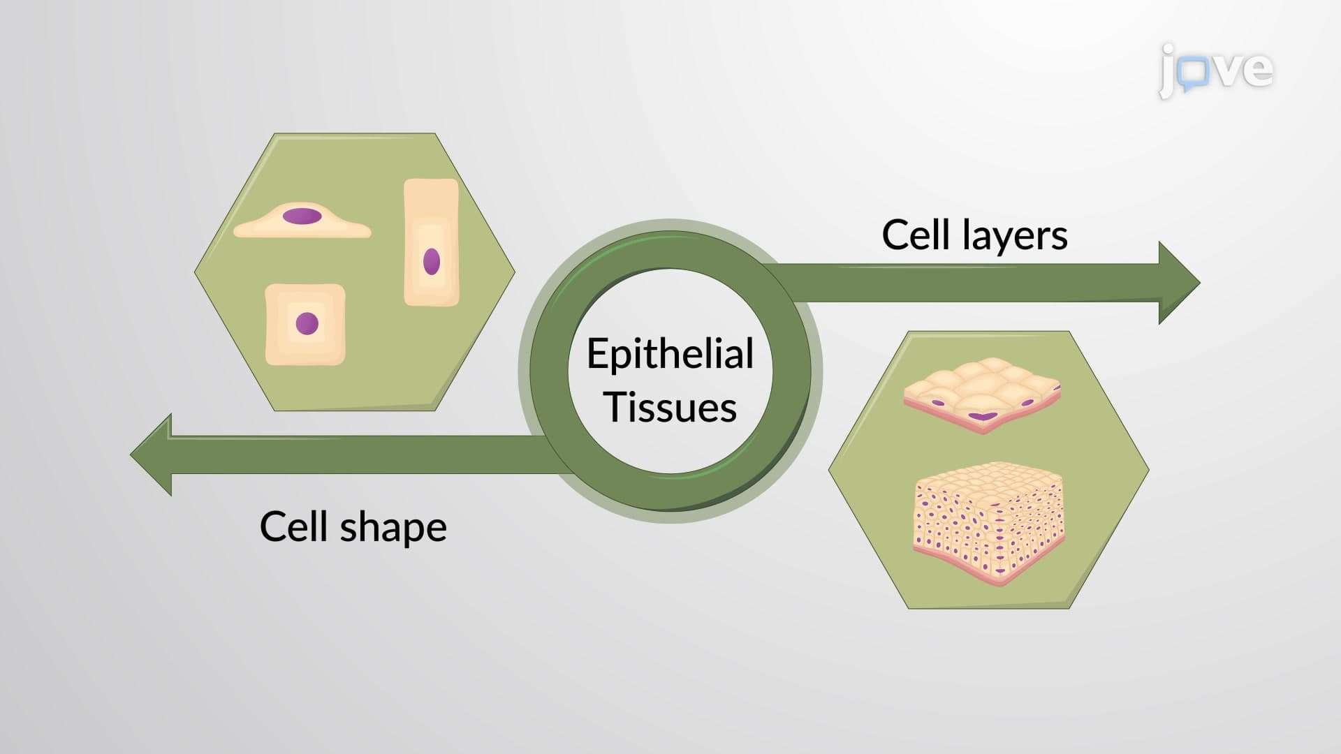
Epithelial tissue classification forms the foundation of histology and human anatomy studies. This systematic approach categorizes tissues based on two critical characteristics: cell shape and the number of cellular layers. This dual classification system enables healthcare professionals and students to predict tissue function simply by identifying structural features under a microscope.
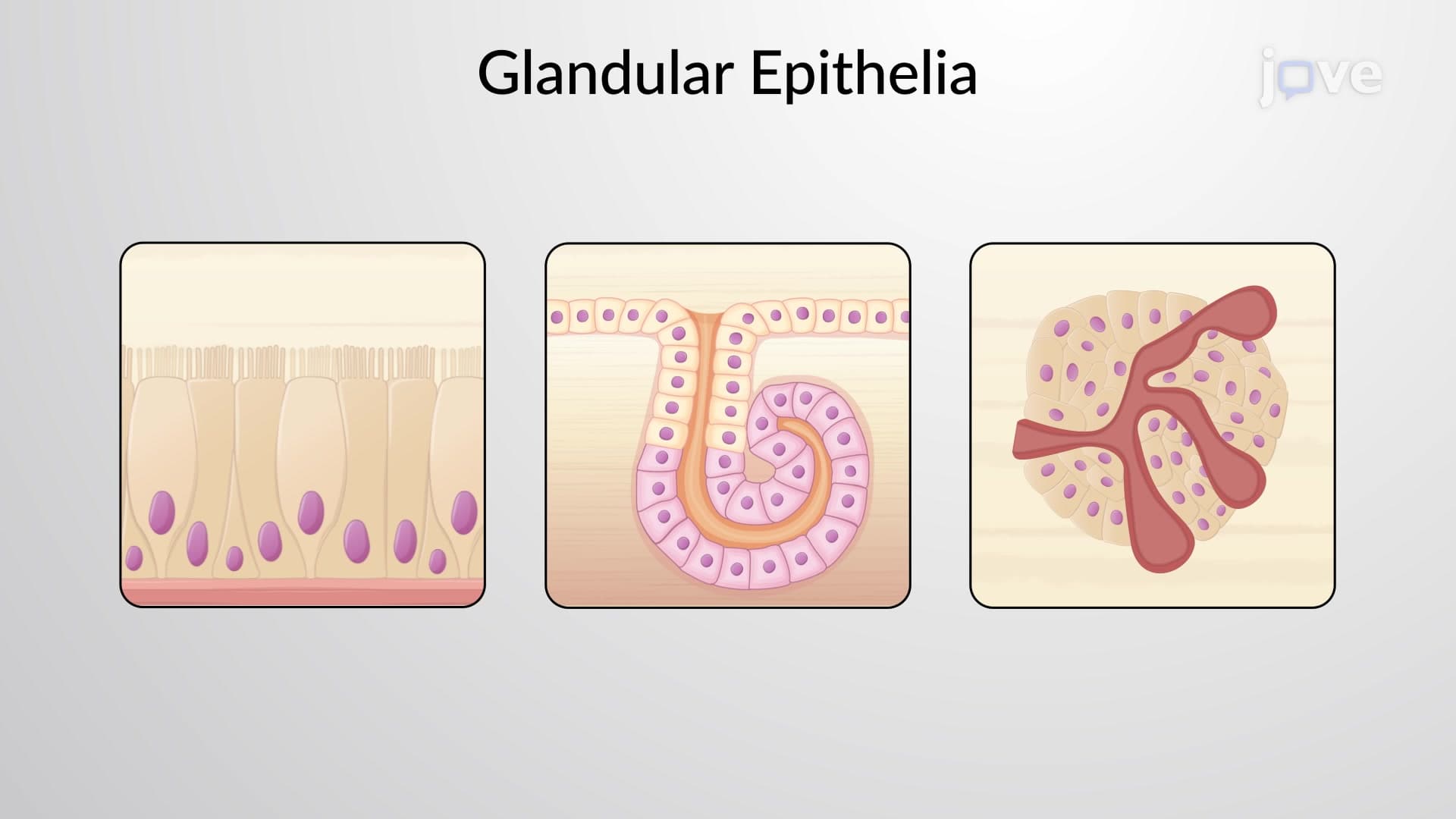
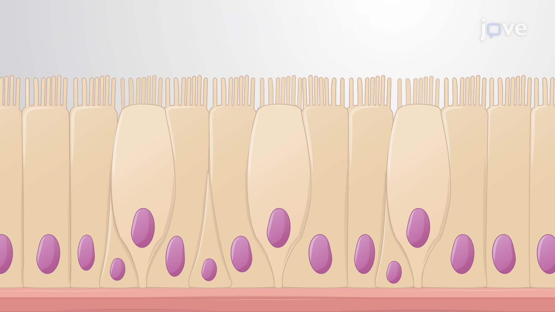
The epithelial tissue classification system recognizes three distinct cell shapes, each optimized for specific functions. Squamous cells appear flattened like floor tiles, with their thin profile facilitating rapid diffusion. These cells line blood vessels and lung alveoli, where materials must pass quickly across minimal distances. Cuboidal cells resemble perfect squares in cross-section, featuring centrally positioned nuclei that indicate active metabolic functions. These cells predominate in kidney tubules and glandular tissues where secretion and absorption occur. Columnar cells stand tall like skyscrapers, with elongated nuclei positioned near their base. These cells line the digestive tract, where their height accommodates specialized structures like microvilli for enhanced absorption.
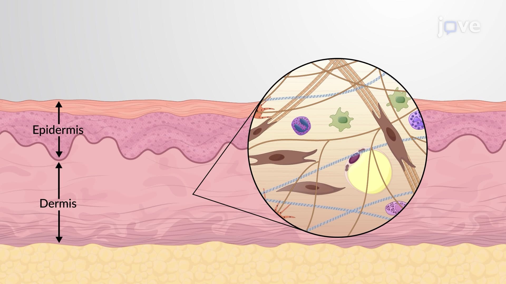
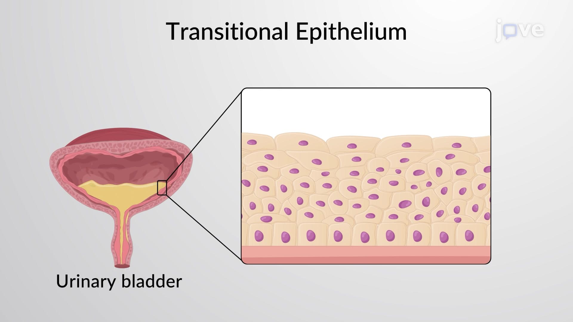
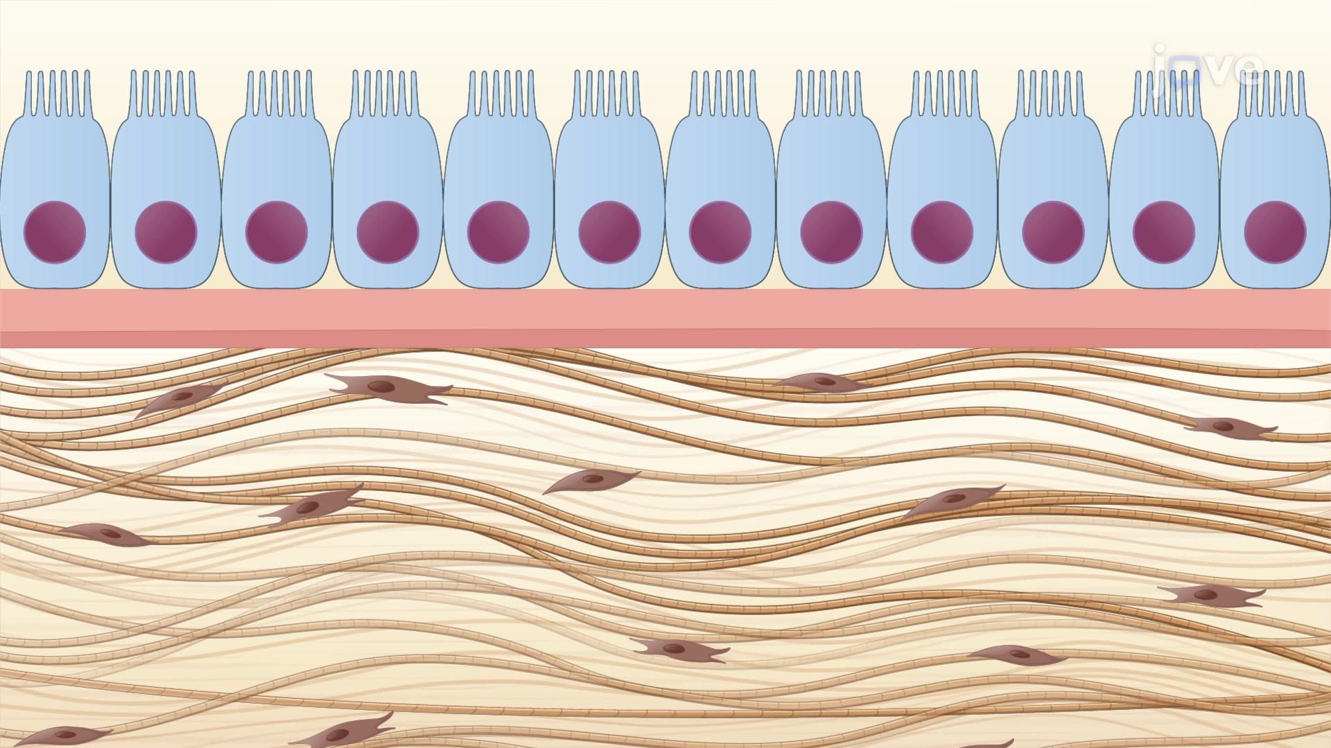
The layer classification distinguishes between simple and stratified arrangements. Simple epithelium consists of a single cell layer directly contacting the basement membrane. This thin barrier appears in locations requiring efficient material exchange, such as the air sacs of lungs where oxygen and carbon dioxide must transfer rapidly, or kidney glomeruli where blood filtration occurs. Stratified epithelium features multiple cell layers stacked like a brick wall, providing robust protection against mechanical stress and chemical damage. The skin's epidermis exemplifies stratified squamous epithelium, protecting against environmental hazards from UV radiation to physical abrasion.
Understanding epithelial tissue classification proves essential for success in AP Biology, college anatomy courses, and pre-health professional exams like the MCAT. Medical students studying for USMLE Step 1 frequently encounter questions linking epithelial structure to pathological changes. For instance, Barrett's esophagus involves the replacement of normal stratified squamous epithelium with columnar epithelium, demonstrating how classification knowledge applies to disease diagnosis. Nursing students preparing for NCLEX examinations must understand these concepts to comprehend wound healing, where epithelial regeneration follows specific patterns based on tissue type.
Related Micro-courses