Loading subject page…


323,100 views

Video Summary: What are Embryonic Connective Tissues
Did you know that every bone, cartilage, and blood vessel in your body traces back to the same embryonic tissue type? Embryonic connective tissue types serve as the foundation for all adult connective structures, originating from the mesoderm germ layer. From the protective Wharton's jelly cushioning umbilical cord blood vessels to the stem cells that repair a broken bone at Johns Hopkins Hospital, these early tissues shape our entire structural framework. Watch the full video on JoVE Coach to master this concept with expert-led visuals and step-by-step explanations.
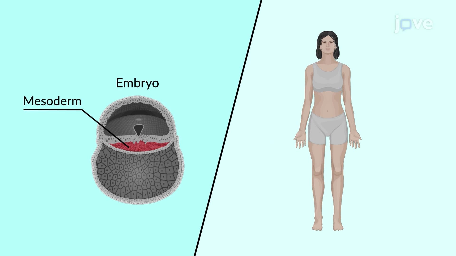
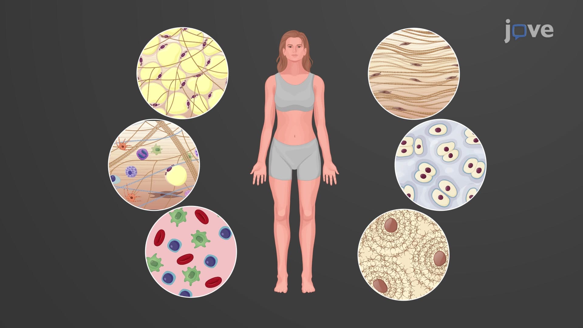
Embryonic connective tissue types represent the foundational structural elements that emerge during the earliest stages of human development. These specialized tissues originate exclusively from the mesoderm, one of three primary germ layers established during gastrulation around the third week of human embryonic development. Unlike their mature counterparts, embryonic connective tissues maintain remarkable plasticity and differentiation potential, making them crucial for proper organ formation and lifelong tissue maintenance.
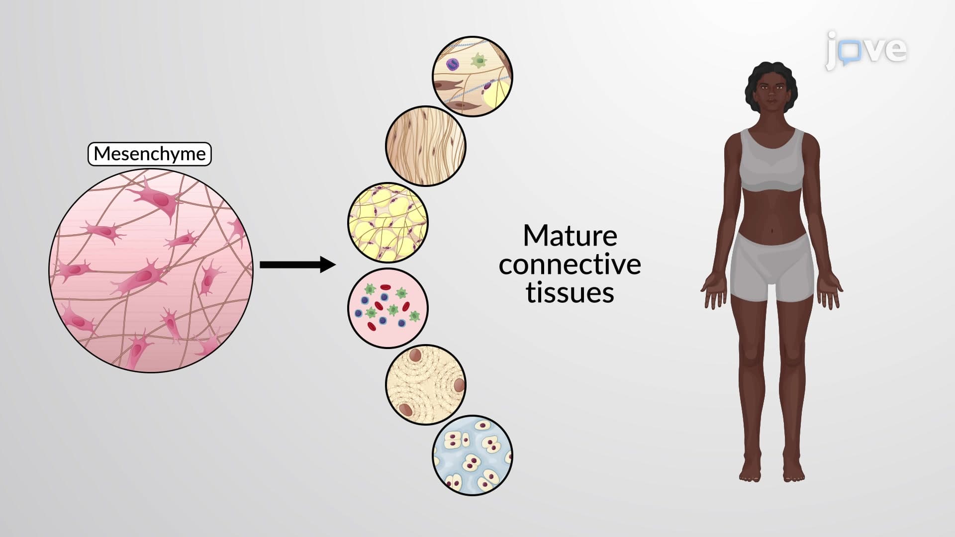
Mesenchyme embryonic tissue stands as the first connective tissue formed during human development, typically appearing by the fourth week of gestation. This primitive tissue consists of distinctive star-shaped mesenchymal stem cells suspended within a fluid-rich extracellular matrix containing sparse reticular fibers. The unique stellate morphology of these cells, with their branching cytoplasmic extensions, allows for extensive cell-to-cell communication essential for coordinated development.
Fetal tissue mesenchyme serves as the master pool for all future connective tissues. These multipotent stem cells possess the remarkable ability to differentiate into bone-forming osteoblasts, cartilage-producing chondroblasts, blood-forming hematopoietic cells, and numerous other specialized cell types. Students preparing for the MCAT or AP Biology exams should note that mesenchymal stem cells retain this differentiation capacity even in adult tissues, explaining how broken bones heal and wounds repair themselves.
Research at institutions like the Mayo Clinic and Stanford University has demonstrated that small clusters of mesenchymal stem cells persist throughout adult life, strategically positioned near blood vessels in what scientists call "perivascular niches." These cellular reserves activate during injury or disease, migrating to damaged sites and differentiating into the specific cell types needed for repair.
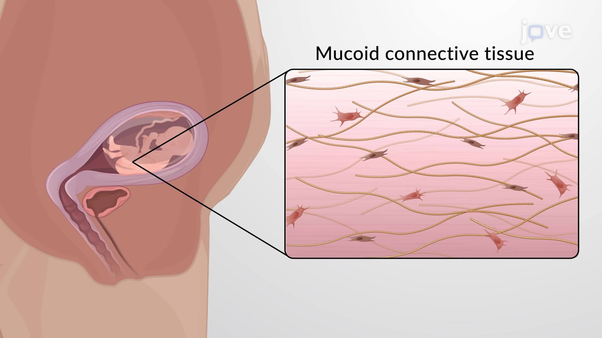
The second embryonic connective tissue type, mucoid connective tissue, develops later in fetal development through mesenchymal differentiation. Unlike mesenchyme's cellular density, mucoid tissue consists primarily of ground substance-a gel-like matrix rich in hyaluronic acid and other glycosaminoglycans-with scattered thin collagen fibers and fibroblast-like cells.
Mucous connective Wharton jelly represents the most clinically significant example of mucoid tissue. Found exclusively within the umbilical cord, this specialized connective tissue surrounds and protects the umbilical arteries and vein throughout fetal development. The unique composition of Wharton's jelly-approximately 80% water with high concentrations of hyaluronic acid-provides both structural support and shock absorption, preventing compression of vital blood vessels during fetal movement.
American medical schools, including Harvard Medical School and Johns Hopkins, increasingly emphasize Wharton's jelly's therapeutic potential. Recent clinical trials at institutions like the University of California, San Francisco, have explored using Wharton's jelly-derived mesenchymal stem cells for treating conditions ranging from spinal cord injuries to cardiovascular disease, highlighting the continued relevance of fetal connective tissue biology in modern medicine.
Related Micro-courses