Loading subject page…


323,100 views


Video Summary: What Is Loose Connective Tissue
Did you know that loose connective tissue biology makes up more space in your body than muscle? This abundant tissue type features loosely arranged fibers suspended in ground substance, forming the structural foundation beneath your skin and around organs. From the fat padding around your kidneys at Johns Hopkins Hospital to the delicate framework inside your bone marrow, loose connective tissue performs essential support functions. Watch the full video on JoVE Coach to master this concept with expert-led visuals and step-by-step explanations.
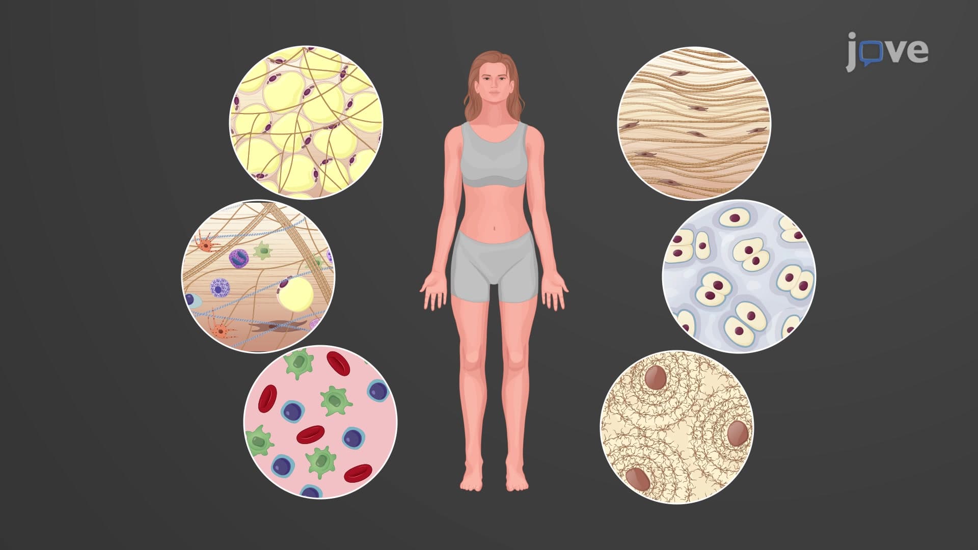
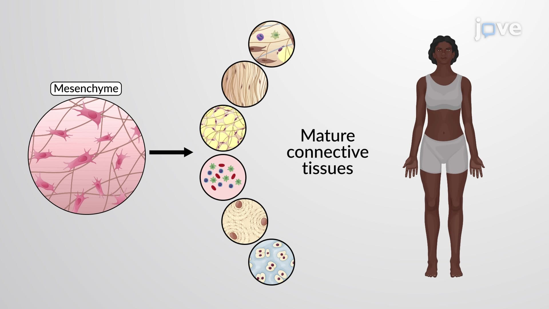
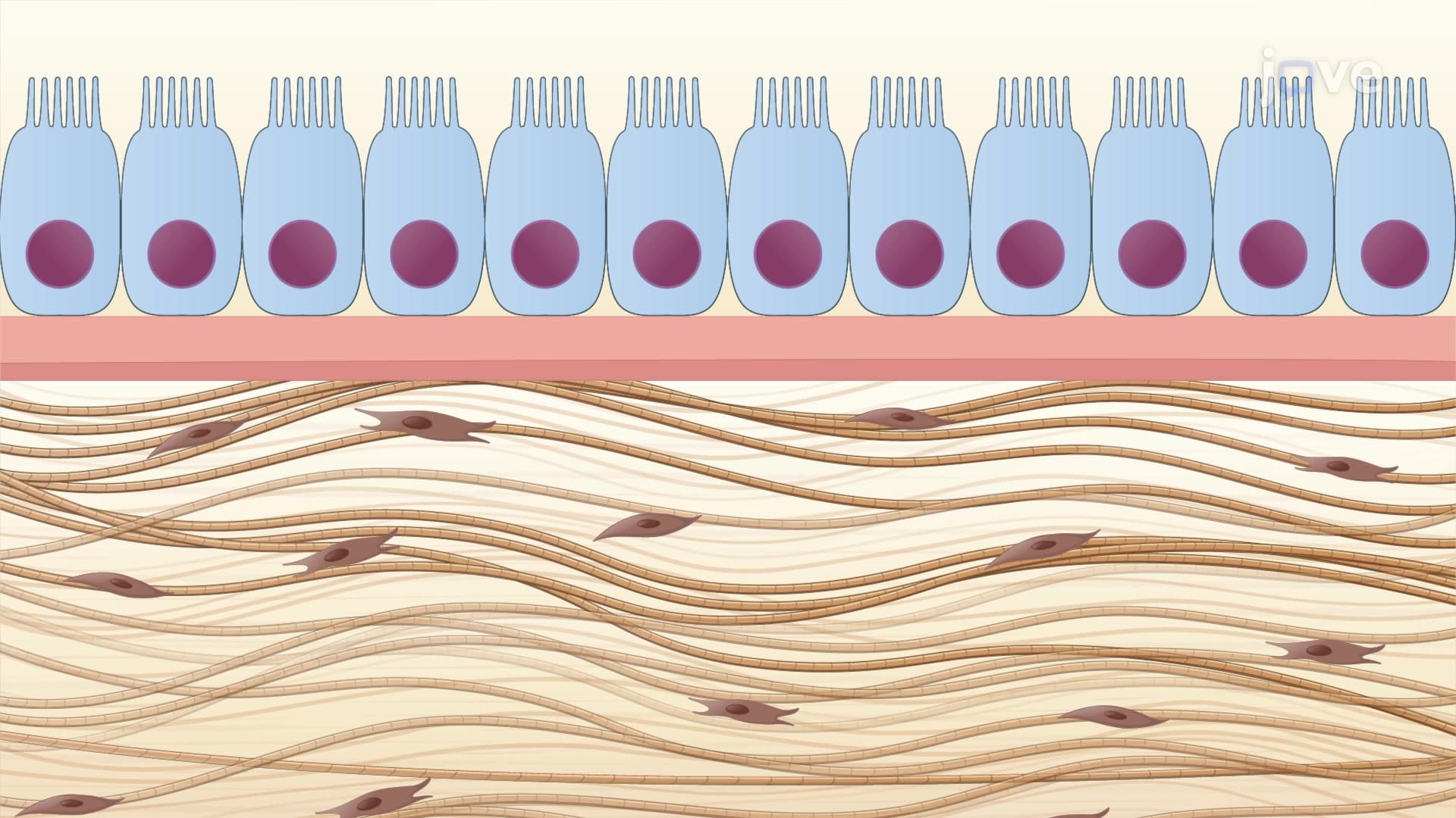
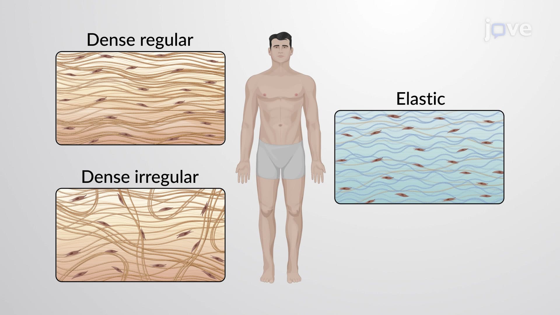
Loose connective tissue represents the most widespread connective tissue in the human body, characterized by its open, mesh-like architecture. Unlike dense connective tissue found in tendons, loose connective tissue features widely spaced fibers embedded in abundant ground substance-a gel-like matrix of water, proteins, and polysaccharides. This structural arrangement allows for flexibility while providing essential support functions.
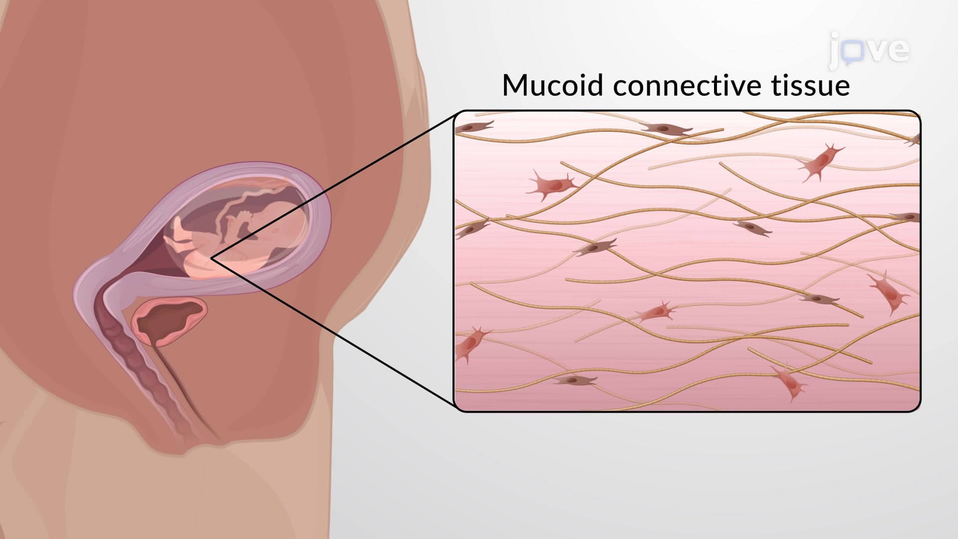
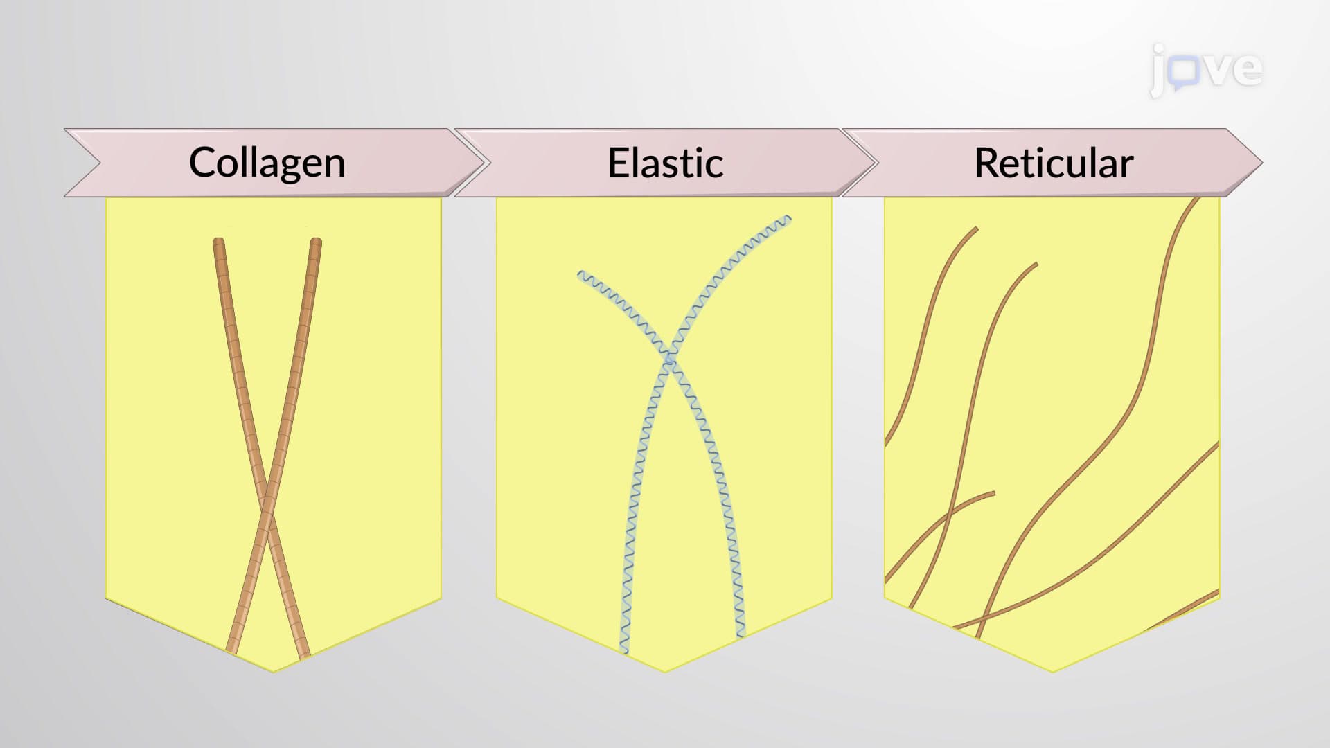
The tissue contains three primary fiber types: collagen fibers for tensile strength, elastic fibers for flexibility, and reticular fibers for delicate support networks. Scattered throughout this matrix are various cell types, with fibroblasts being the most common producers of the extracellular matrix components.
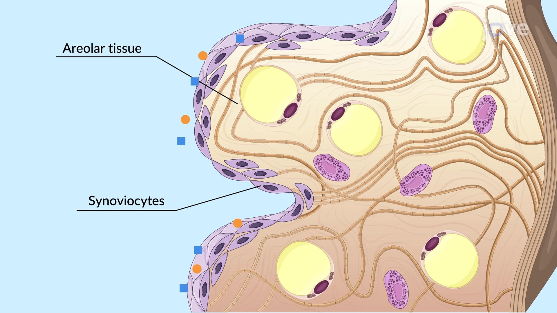
Areolar tissue serves as the body's universal packing material, filling spaces between organs and connecting different tissue types. In clinical settings at institutions like the Mayo Clinic, surgeons regularly encounter areolar tissue when separating tissue planes during procedures. This tissue creates the loose fascial planes that allow structures to move smoothly past each other.
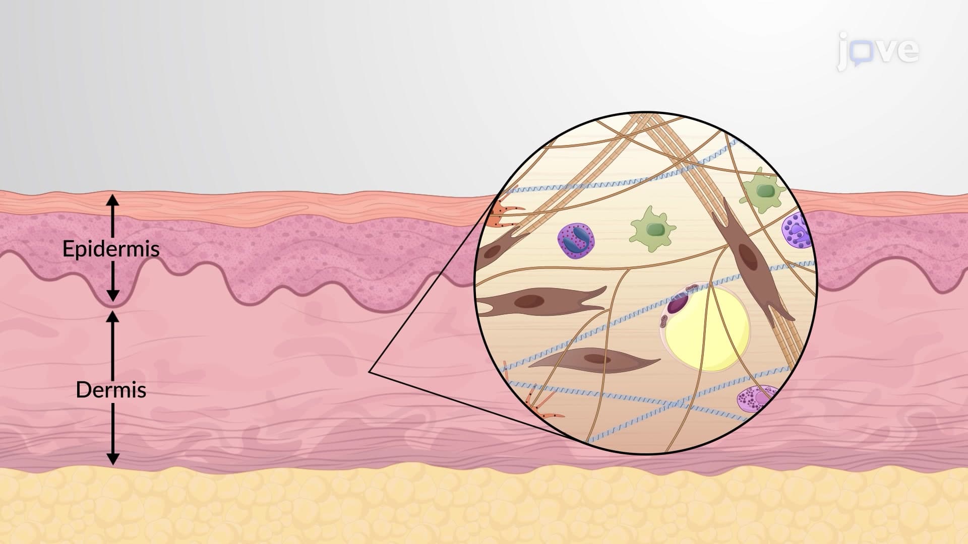
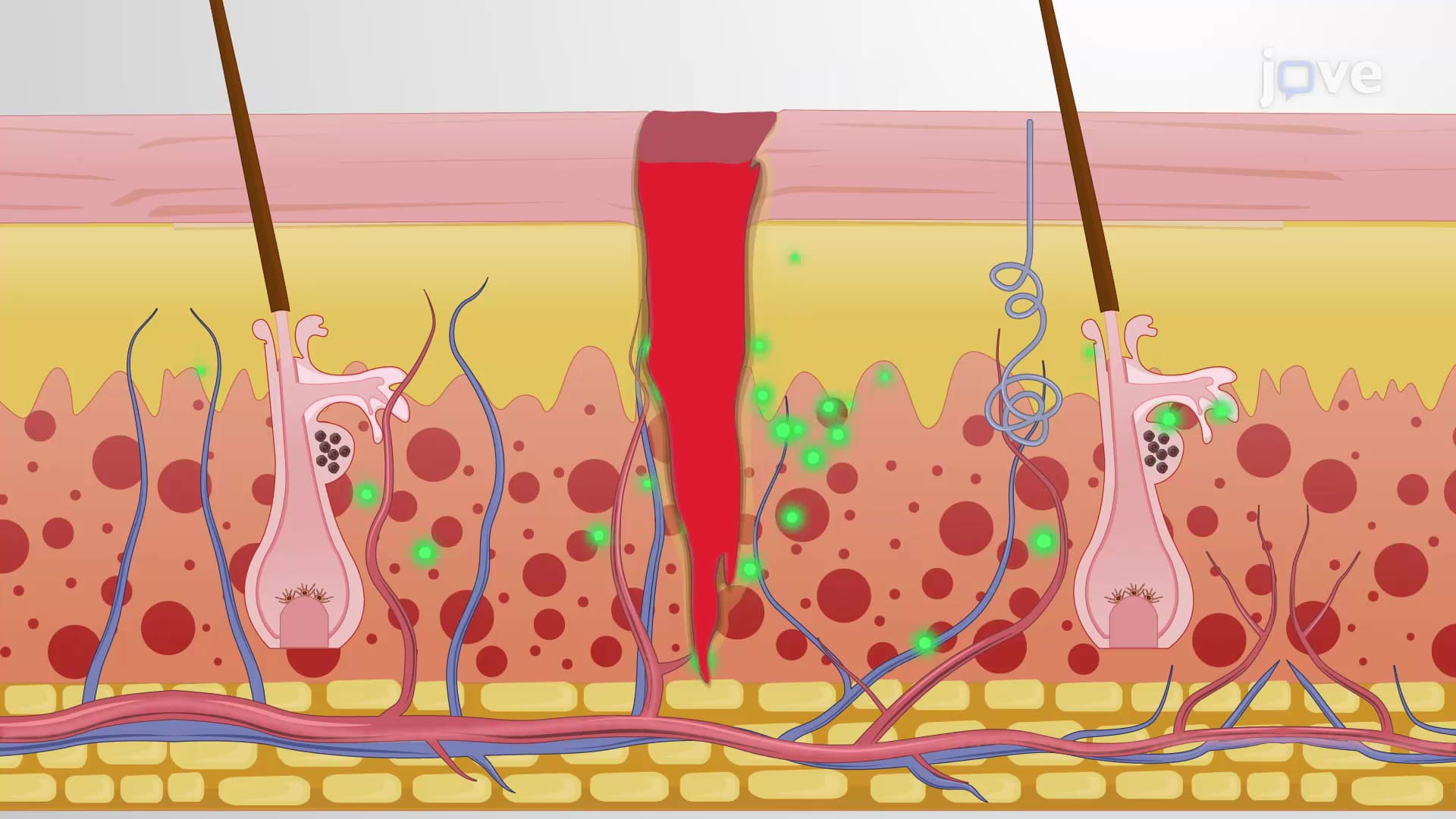
Students preparing for the MCAT will encounter areolar tissue's role in connecting epithelial tissues to underlying structures. For example, the areolar layer beneath your skin's epidermis contains blood vessels and nerve endings that wouldn't fit in the epithelial layer itself.
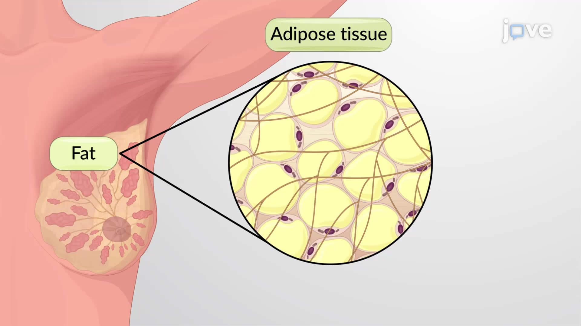
Adipose tissue consists primarily of adipocytes-specialized cells that store triglycerides in large lipid droplets. Beyond energy storage, adipose tissue provides crucial insulation and protection. The perirenal fat surrounding kidneys, commonly seen in radiological studies at US medical centers, demonstrates adipose tissue's protective function.
For AP Biology students, understanding brown versus white adipose tissue becomes important. Brown adipose tissue, found primarily in infants and hibernating mammals, generates heat through specialized mitochondria, while white adipose tissue primarily stores energy.
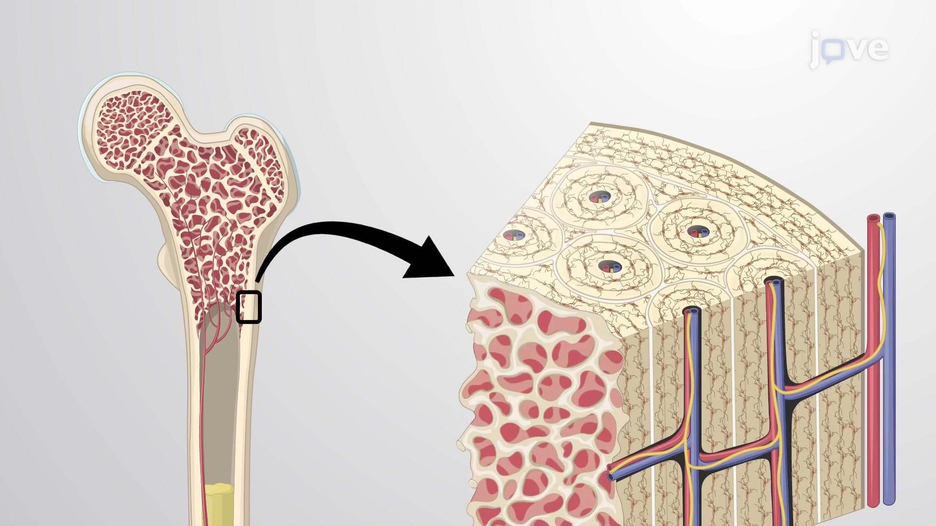
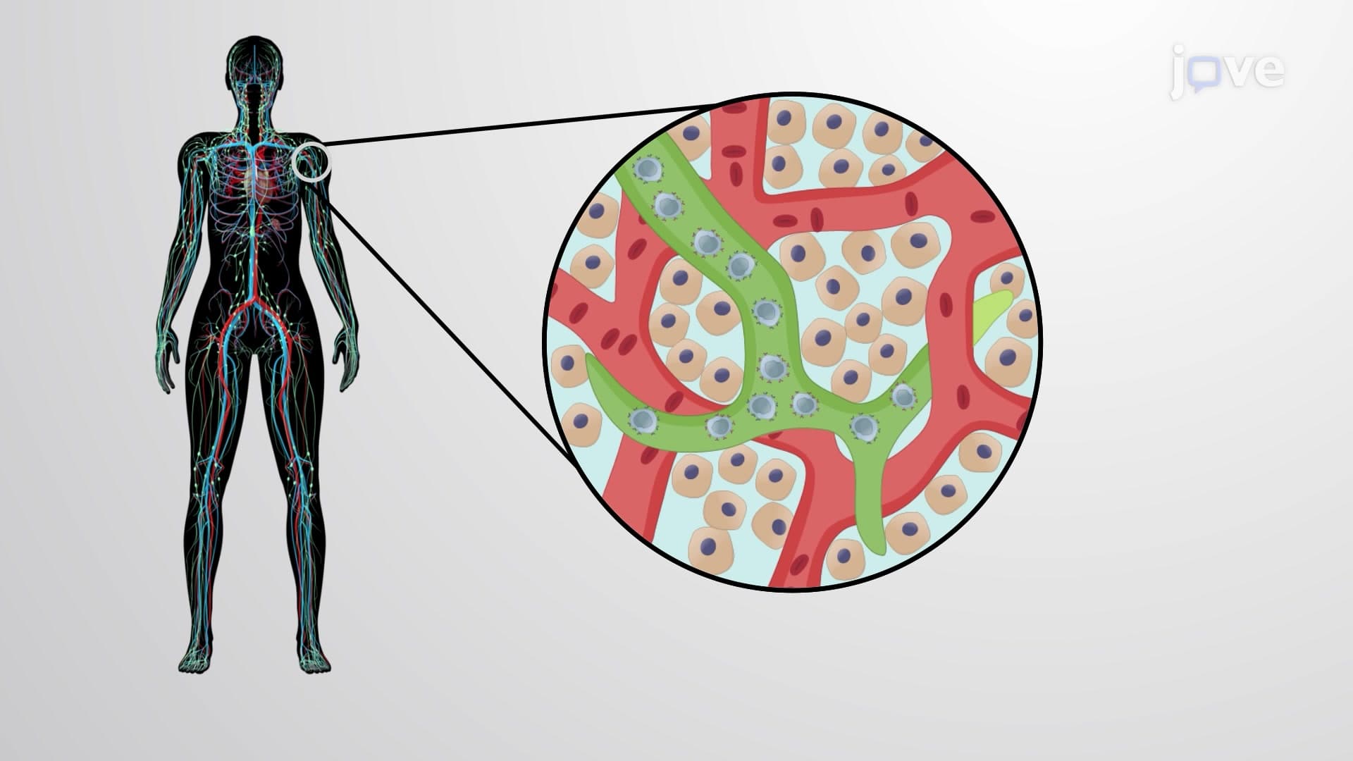
Reticular tissue forms the structural backbone of lymphoid organs including lymph nodes, spleen, and bone marrow. The fine reticular fibers create a three-dimensional network that supports immune cells and blood-forming tissues. Medical students studying for the USMLE encounter reticular tissue when learning about lymph node architecture and hematopoiesis in bone marrow.
This tissue type becomes clinically significant in diseases affecting lymphoid organs, where disruption of the reticular framework can impair organ function.
Related Micro-courses