Loading subject page…

- Anatomy and Physiology
- The Urinary System
- External Anatomy of the Kidney
Micro-course: The Urinary System
78,053 views
Learn key concepts and earn your certificate.

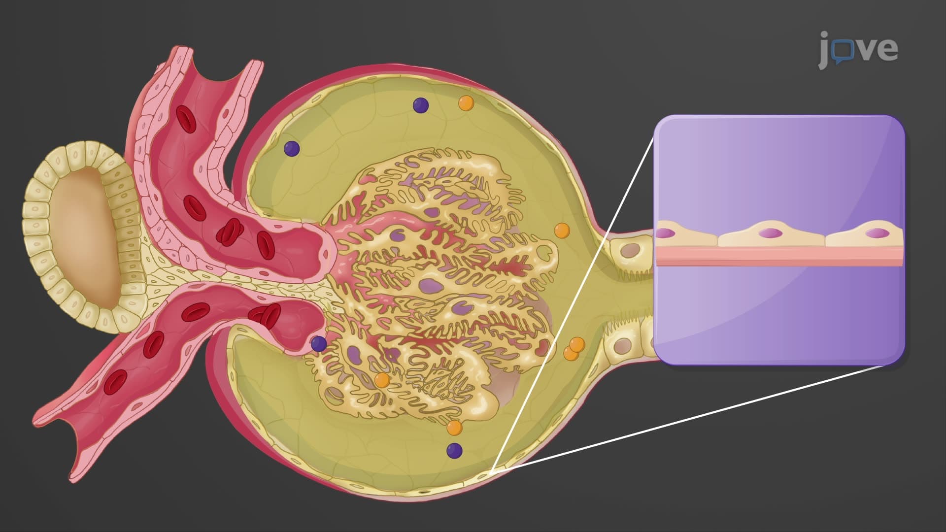

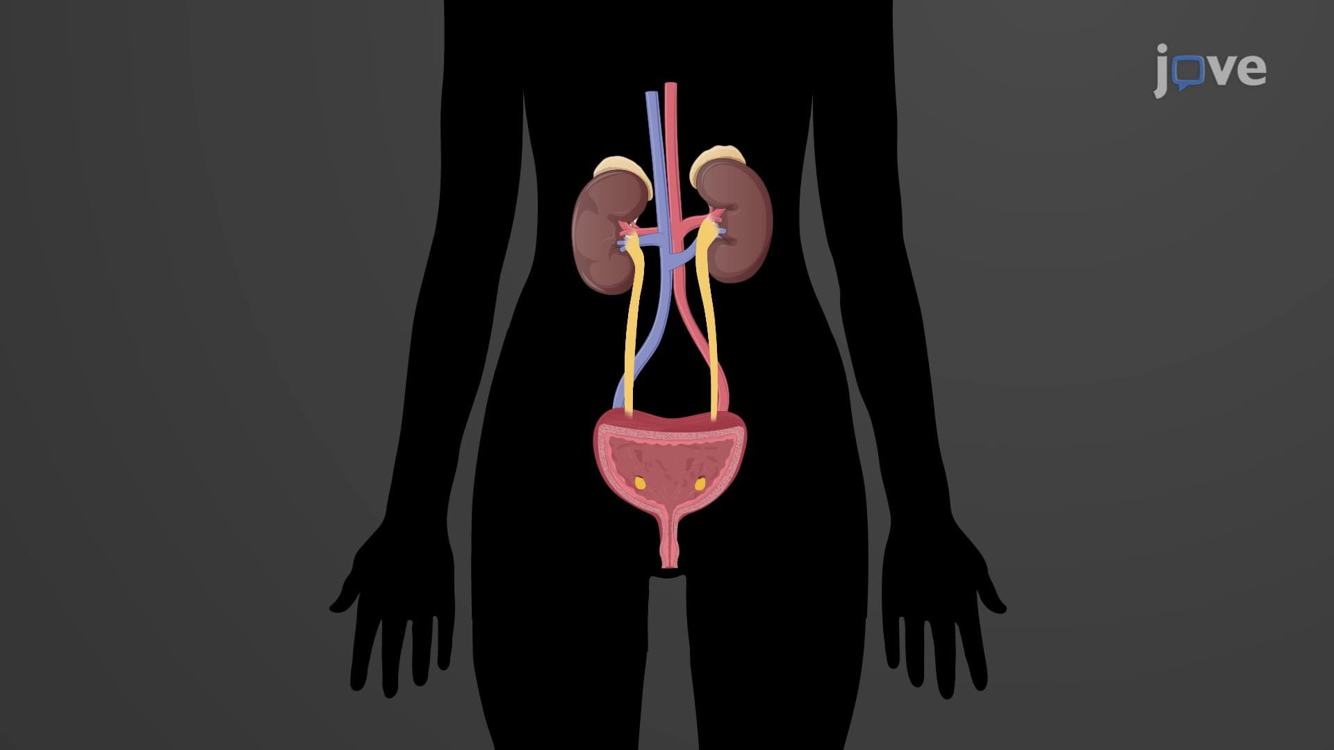
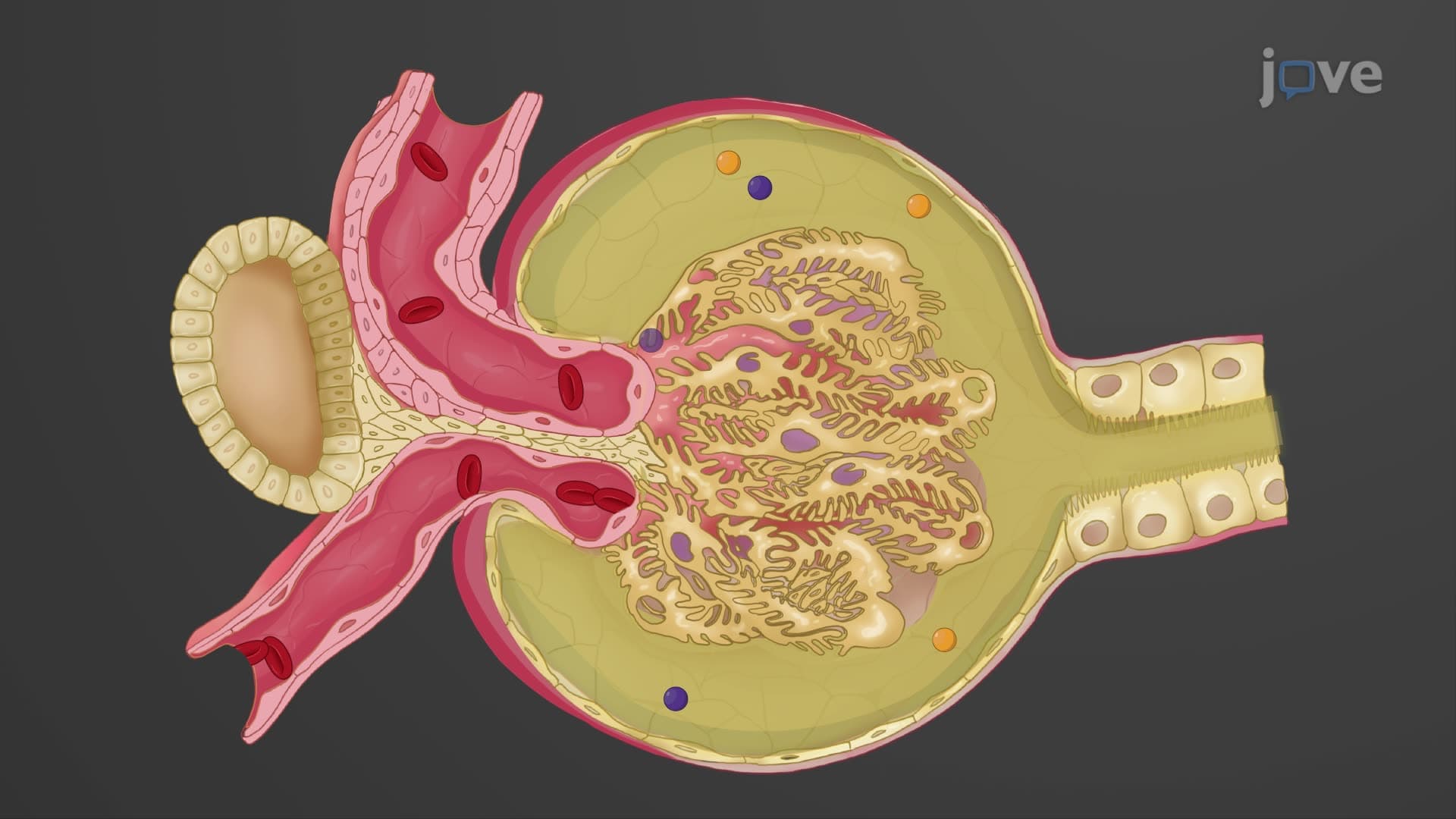
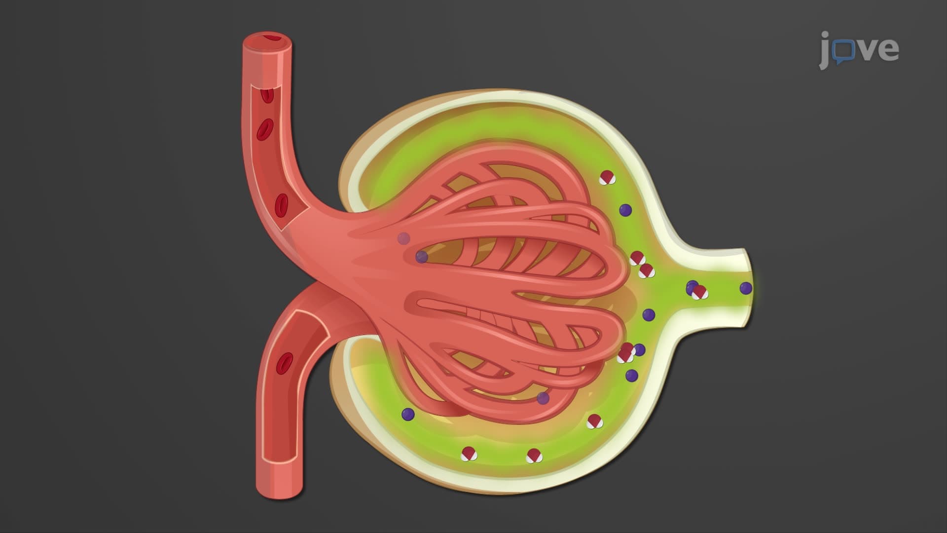


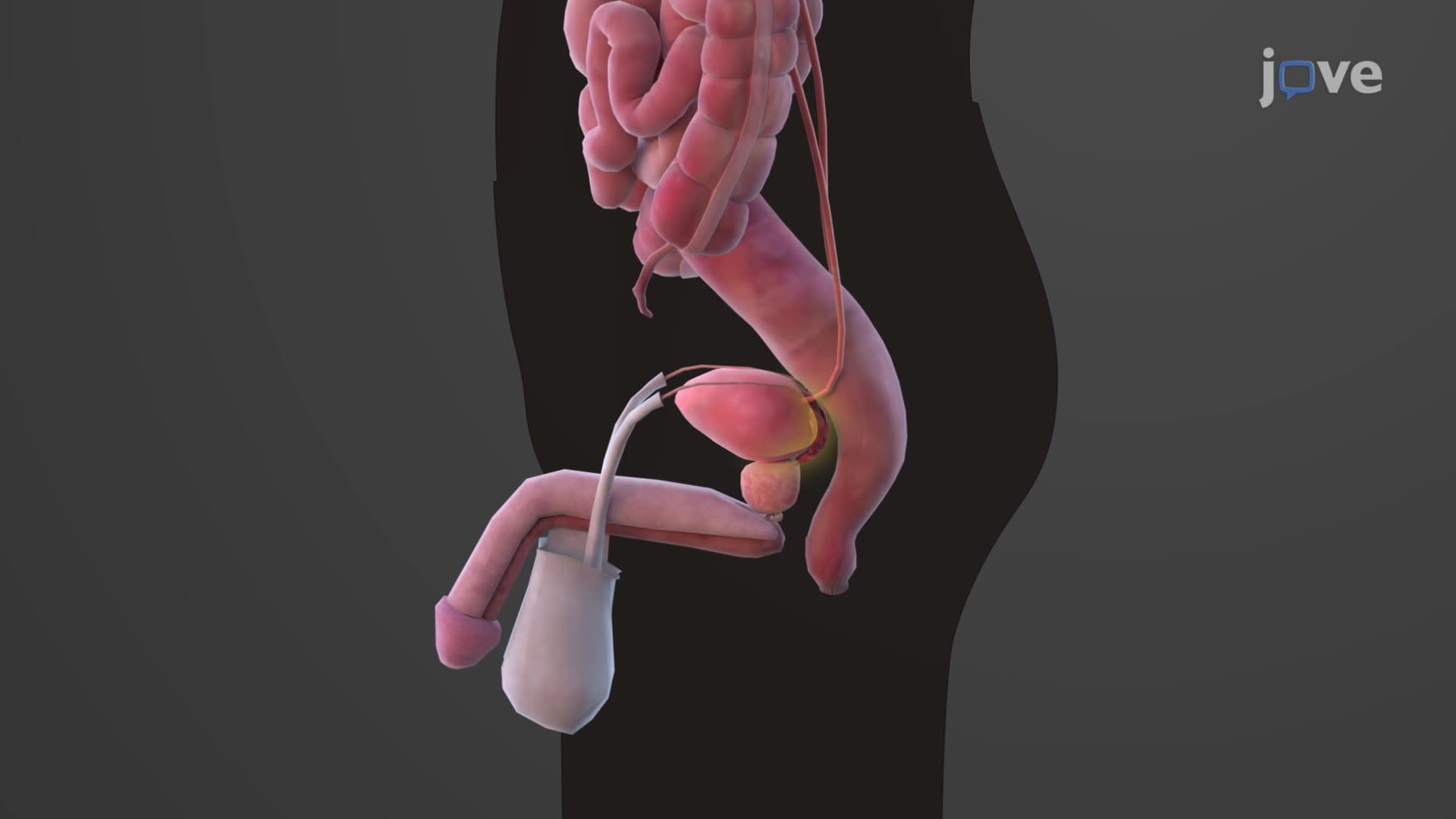
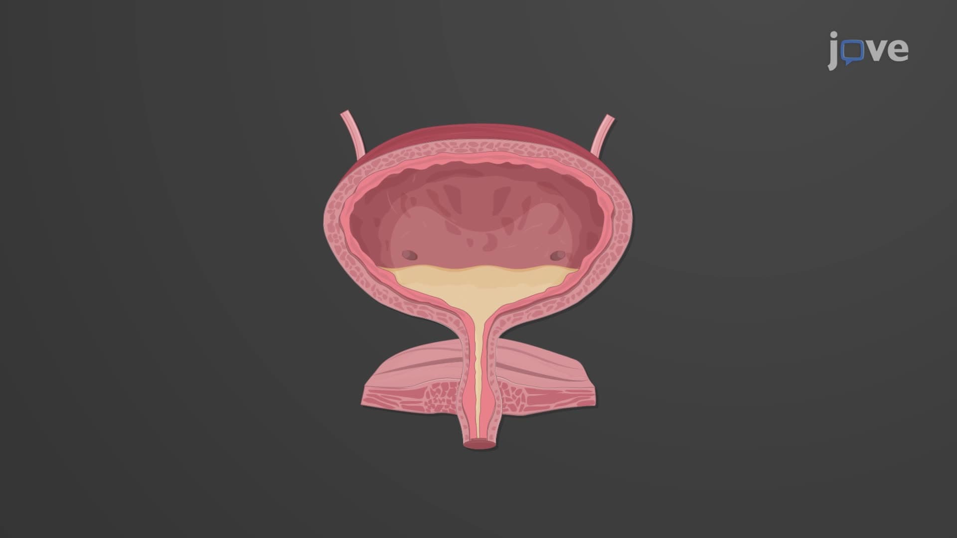

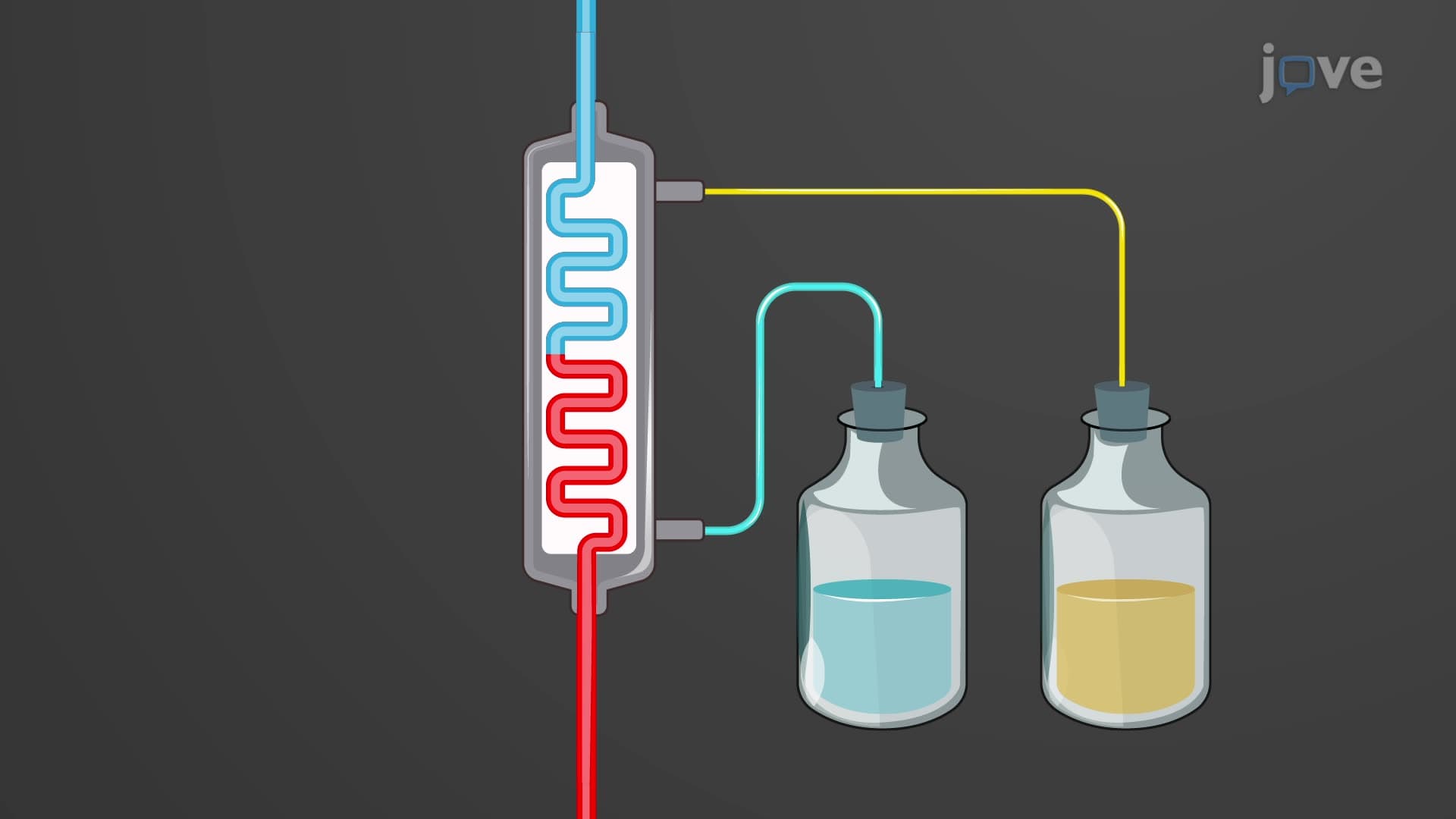
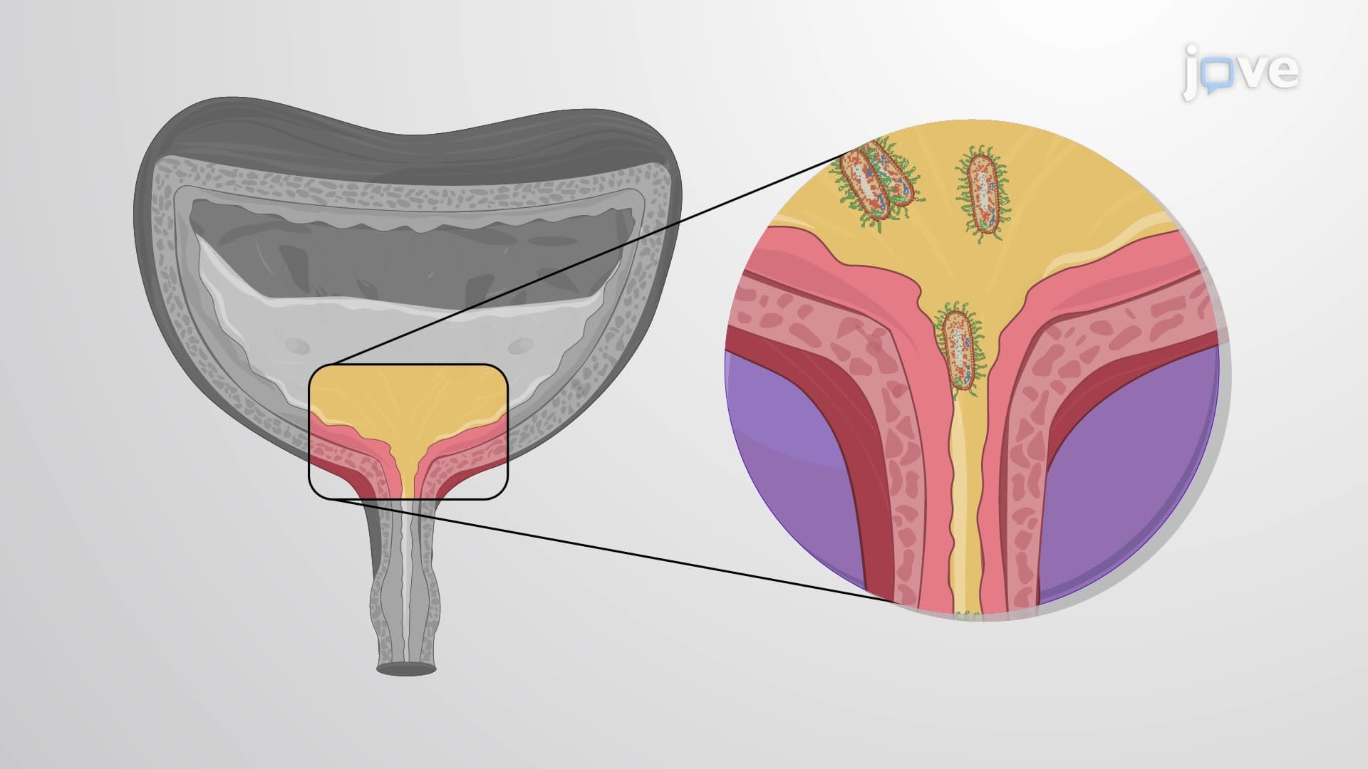
External Anatomy of the Kidney

00:00
01:21
Video Summary: What Is External Anatomy Kidney
Did you know your kidneys filter about 50 gallons of blood daily-equivalent to filling a standard bathtub? Understanding kidney external anatomy biology reveals how these bean-shaped organs, positioned behind your ribcage, accomplish this incredible feat. Medical students at Johns Hopkins University study these precise anatomical relationships to diagnose conditions like kidney stones. What is external anatomy kidney structure teaches us about protection, positioning, and function in the human body. Watch the full video on JoVE Coach to master this concept with expert-led visuals and step-by-step explanations.
- Understand what is external anatomy kidney structure, including precise location, dimensions, and protective layers
- Identify the three distinct tissue layers that surround and protect each kidney from external damage
- Analyze the significance of renal hilum positioning on the medial border for vascular and neural connections
- Compare male and female kidney weight differences and explain the biological reasoning behind these variations
- Evaluate how the kidney's external positioning between T12-L3 vertebrae provides optimal protection and function
- Apply knowledge of kidney external anatomy to interpret medical imaging and diagnostic procedures
- Recognize the relationship between external kidney structure and common pathological conditions
- Explore connections between external anatomy and internal kidney function for comprehensive understanding
Understanding What is External Anatomy Kidney Structure
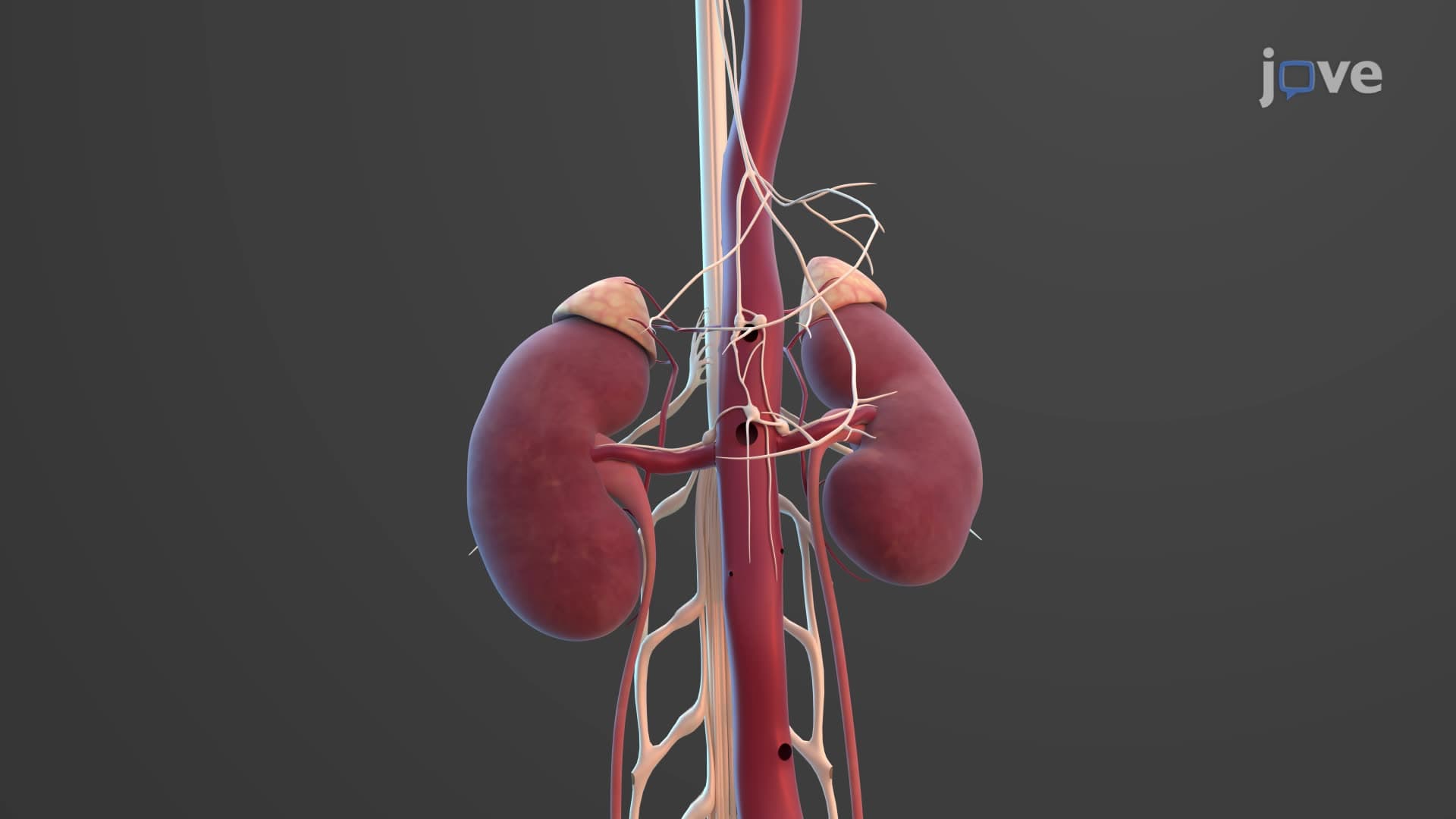
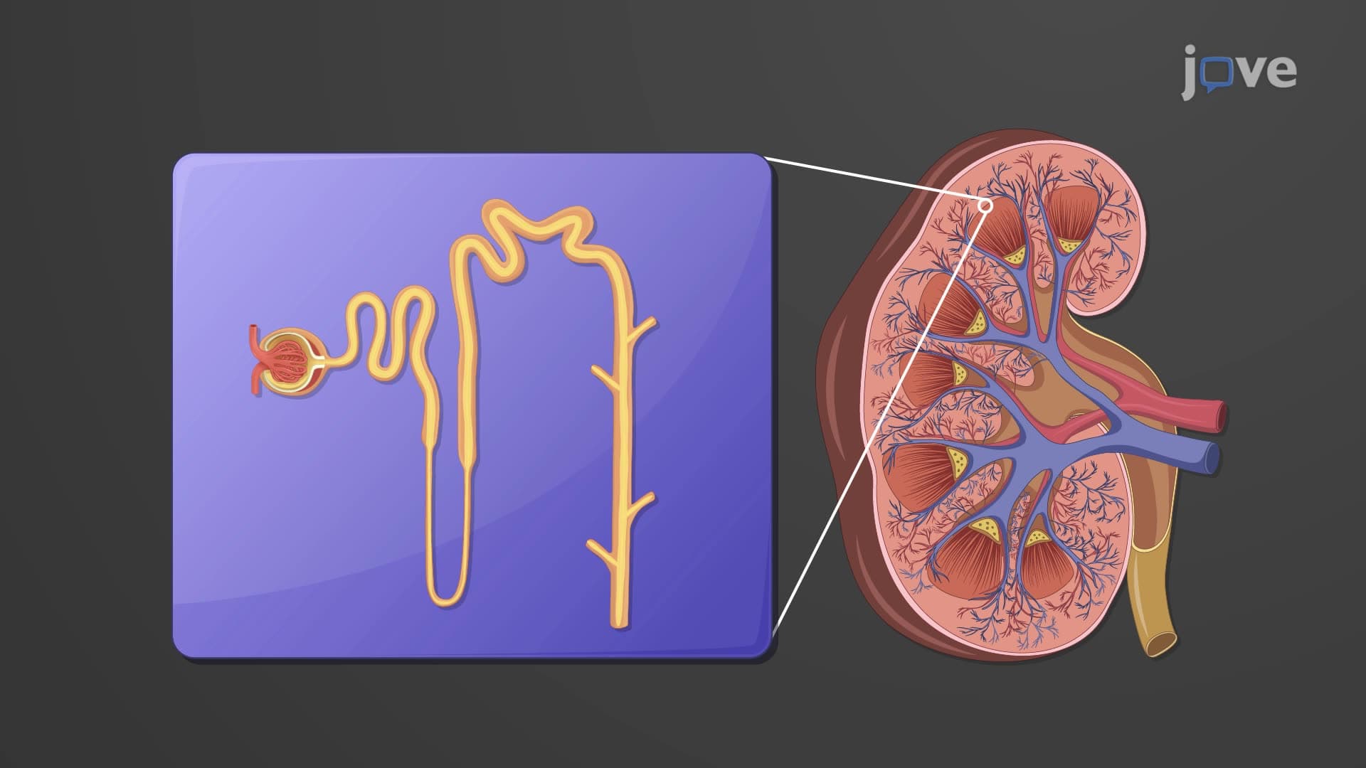
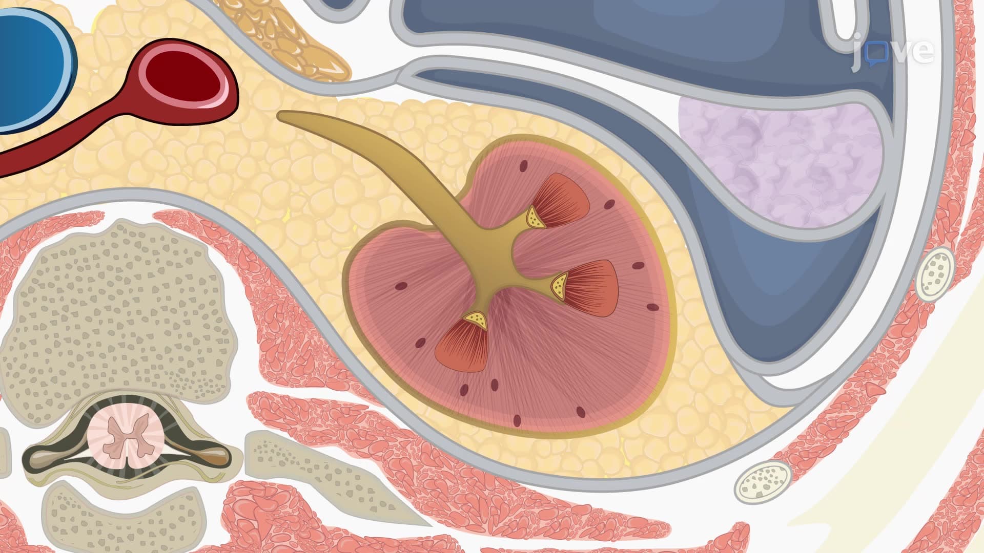
The kidney external anatomy biology forms the foundation for understanding how these vital organs maintain homeostasis in the human body. Each kidney's strategic placement in the retroperitoneal space, nestled between the T12 and L3 vertebrae, provides optimal protection while maintaining accessibility for essential functions. This positioning places them partially under the protection of ribs 11 and 12, creating a natural shield against external trauma-a design principle that emergency room physicians at Cleveland Clinic rely on when assessing blunt force injuries.
Dimensional Characteristics and Renal External Anatomy Explained
The precise measurements of kidney external anatomy reveal remarkable consistency across healthy individuals. Adult kidneys typically measure 10-12 centimeters in length (roughly the size of a computer mouse), 5-7 centimeters in width, and 3 centimeters in thickness. The weight differences between males (125-175 grams) and females (115-155 grams) reflect body size variations and hormonal influences on organ development. Pre-med students preparing for the MCAT often memorize these measurements, as they frequently appear in biological sciences passages about renal physiology.
Renal Hilum and External Connectivity
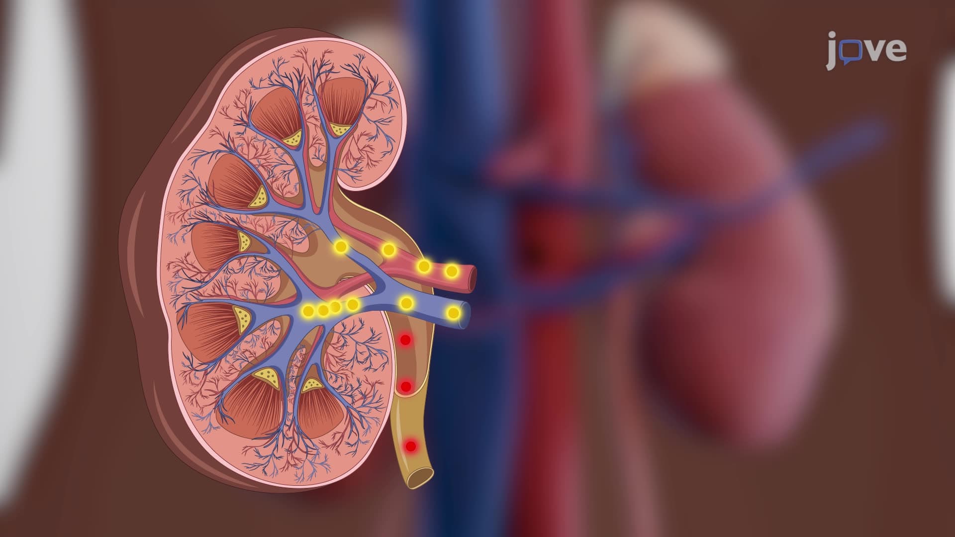
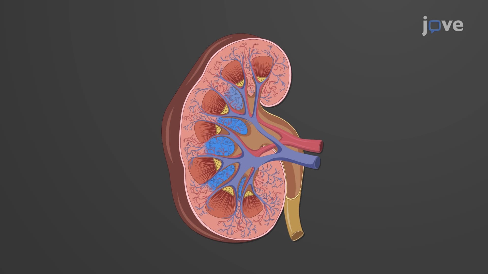
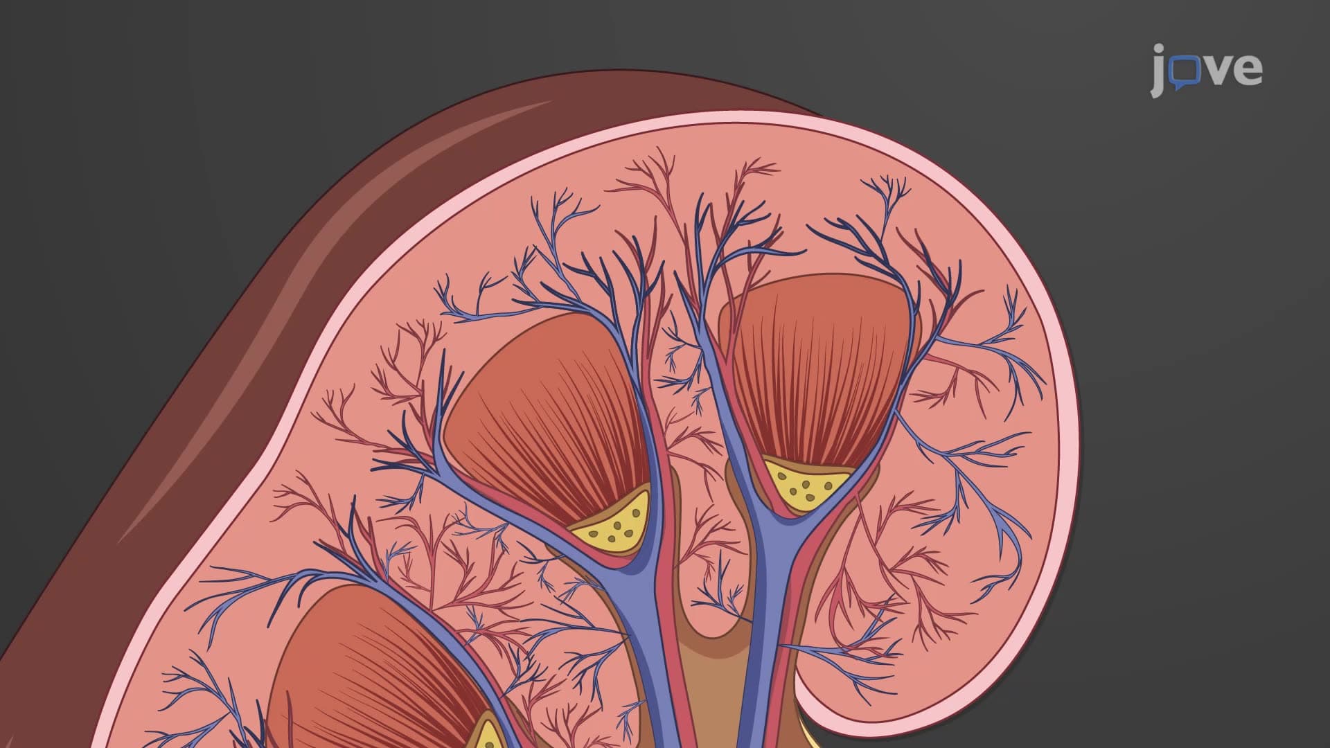
What is the external anatomy of the kidney becomes clearer when examining the renal hilum-the kidney's "gateway" located on the concave medial border. This critical structure serves as the entry and exit point for the renal artery, renal vein, ureter, lymphatic vessels, and sympathetic nerves. The hilum's positioning allows for efficient vascular access during surgical procedures, making it essential knowledge for students pursuing careers in nephrology or urology. Understanding renal hilum capsule external relationships helps explain how kidney transplants maintain proper blood flow and drainage.
Protective Tissue Layers and Kidney Gross External Anatomy
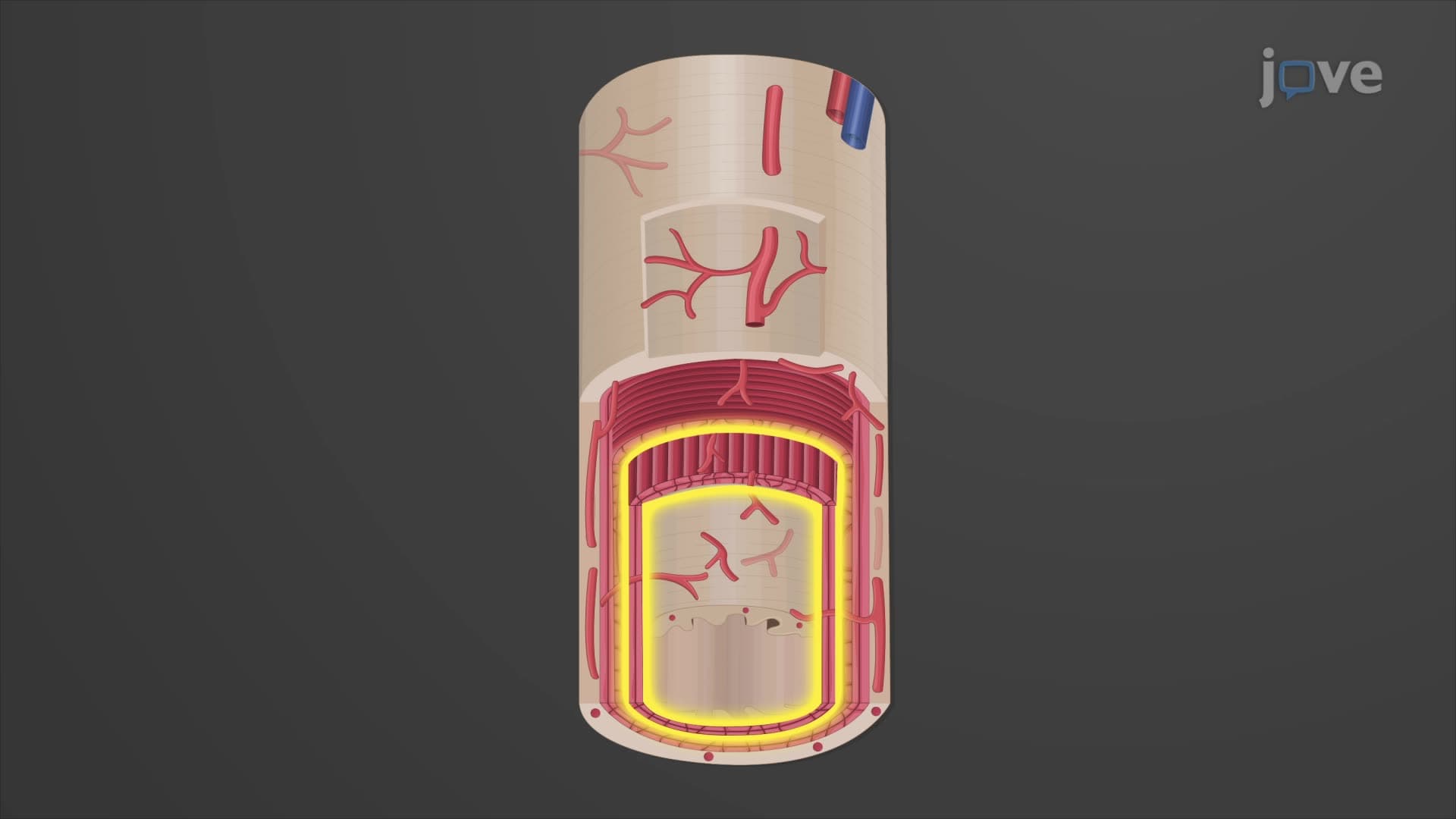
The three-layer protection system exemplifies kidney bean shape anatomy efficiency. The innermost renal capsule, composed of dense irregular connective tissue, provides immediate protection while allowing for slight expansion during filtration changes. The middle perirenal fat capsule cushions against mechanical shock and helps maintain kidney position-particularly important for athletes who experience repeated jarring motions. The outermost renal fascia anchors the entire structure to the abdominal wall, preventing dangerous kidney displacement that could compromise vascular connections. Nursing students studying for the NCLEX-RN exam must understand these layers to properly assess patients with flank pain or suspected kidney trauma.
Frequently Asked Questions
External kidney anatomy refers to the outer structural features, positioning, and protective layers of the kidneys in the human body. It's crucial for understanding kidney function, medical imaging interpretation, and diagnosing conditions like kidney stones or trauma. This knowledge forms the foundation for advanced studies in nephrology, urology, and emergency medicine.
MCAT questions often test kidney positioning relative to vertebrae, protective tissue layers, and hilum structure in passages about renal physiology or homeostasis. AP Biology exams may include kidney external anatomy in questions about organ systems, particularly focusing on structure-function relationships. Students should memorize key measurements and understand how external features relate to kidney function.
USMLE Step 1 emphasizes renal hilum contents, protective fascial layers, and anatomical relationships for clinical correlation questions. Common topics include kidney trauma assessment, surgical approach considerations, and imaging interpretation. Understanding the retroperitoneal location and rib protection helps answer questions about blunt abdominal trauma and differential diagnosis.
ER physicians at major trauma centers like Houston Methodist Hospital assess flank pain and blunt trauma by understanding kidney positioning behind ribs 11-12. They use external anatomy knowledge to interpret CT scans, determine injury severity, and decide between conservative management versus surgical intervention. This knowledge directly impacts patient outcomes in trauma situations.
Kidney external anatomy is very manageable for high school students with basic biology background. The key is connecting structure to function-understanding why kidneys need protection, specific positioning, and multiple tissue layers. Visual learning tools and anatomical models make these concepts accessible even for students new to human anatomy.
Create acronyms for the three protective layers (capsule, fat, fascia) and use size comparisons for measurements (kidney length equals computer mouse size). Practice identifying structures on anatomical diagrams and relate each feature to its function. Active recall through practice questions works better than passive reading for long-term retention.
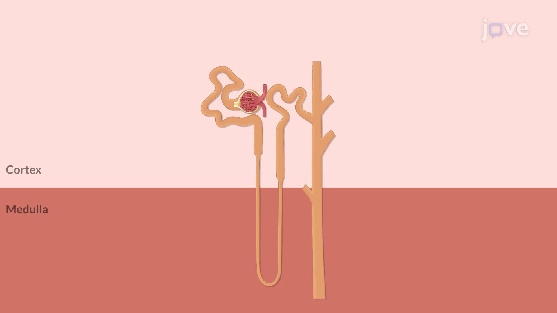
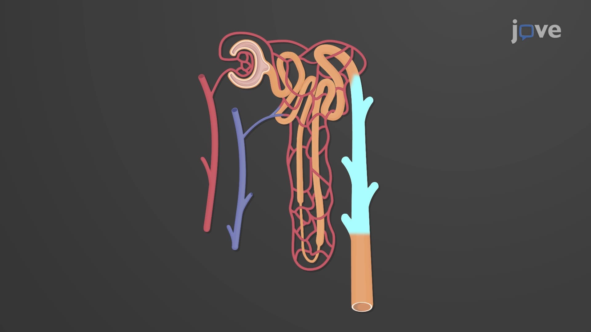
External anatomy provides the framework for understanding internal organization-the hilum leads to internal collecting systems, protective layers relate to internal pressure regulation, and positioning affects internal blood flow patterns. This foundational knowledge makes learning about nephrons, collecting ducts, and renal circulation much more logical and memorable.
Progress to internal kidney anatomy including cortex and medulla organization, then nephron structure and function. Advanced students should explore renal blood supply, filtration mechanisms, and hormonal regulation. Clinical applications include kidney disease pathophysiology, dialysis principles, and transplant considerations for pre-med track students.
Related Micro-courses

20 Concepts

37 Concepts

14 Concepts