Loading subject page…

- Anatomy and Physiology
- The Urinary System
- Internal Anatomy of the Kidney
Micro-course: The Urinary System
78,053 views
Learn key concepts and earn your certificate.

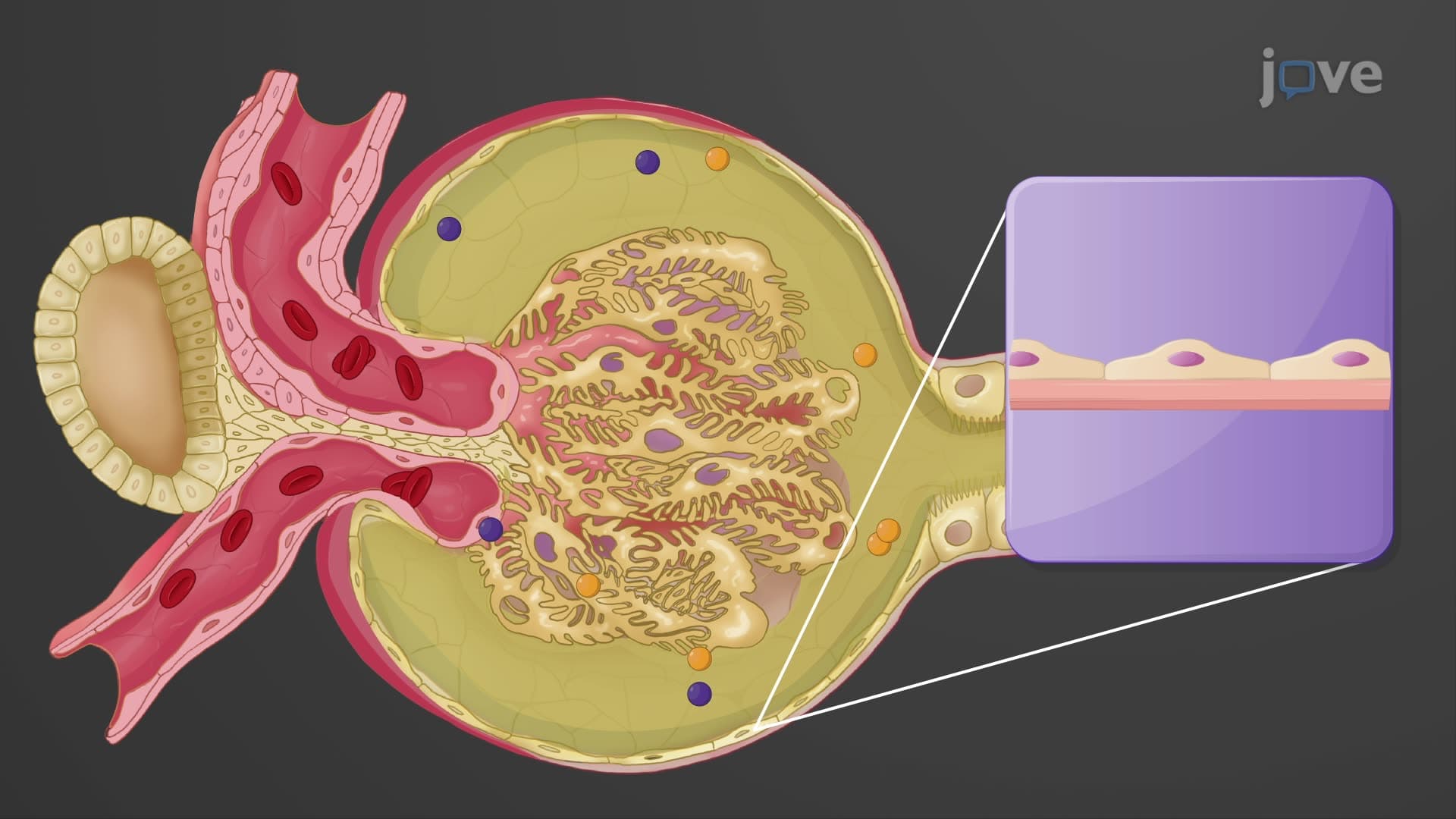

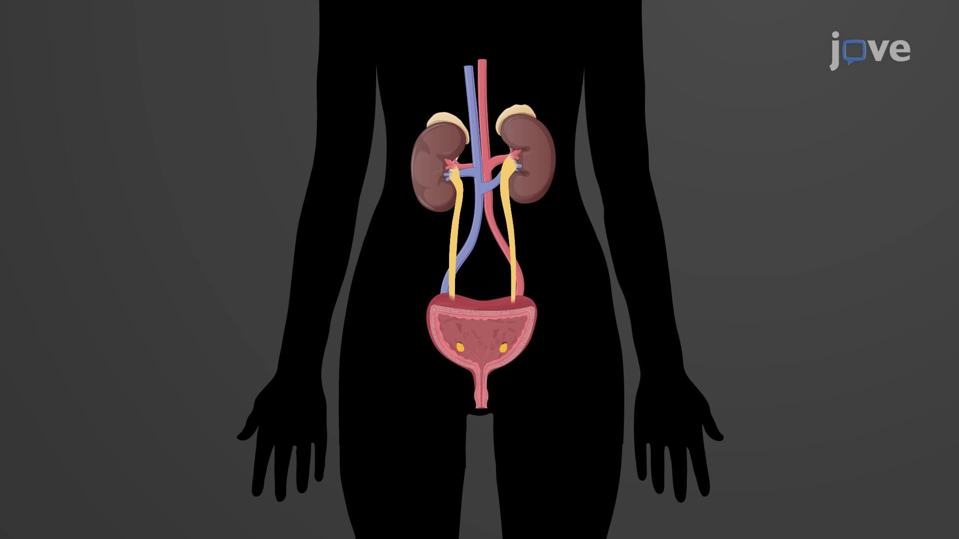
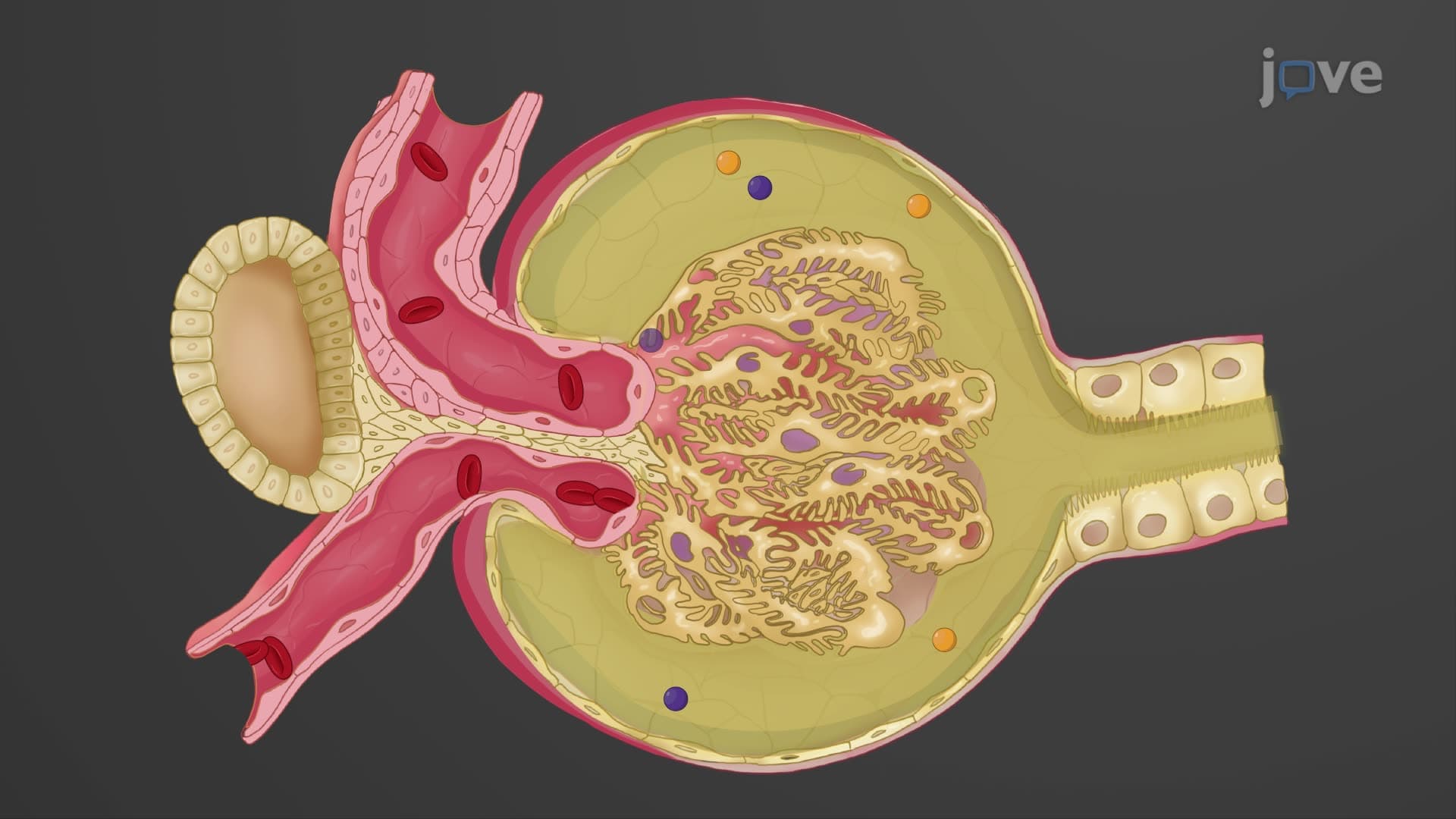
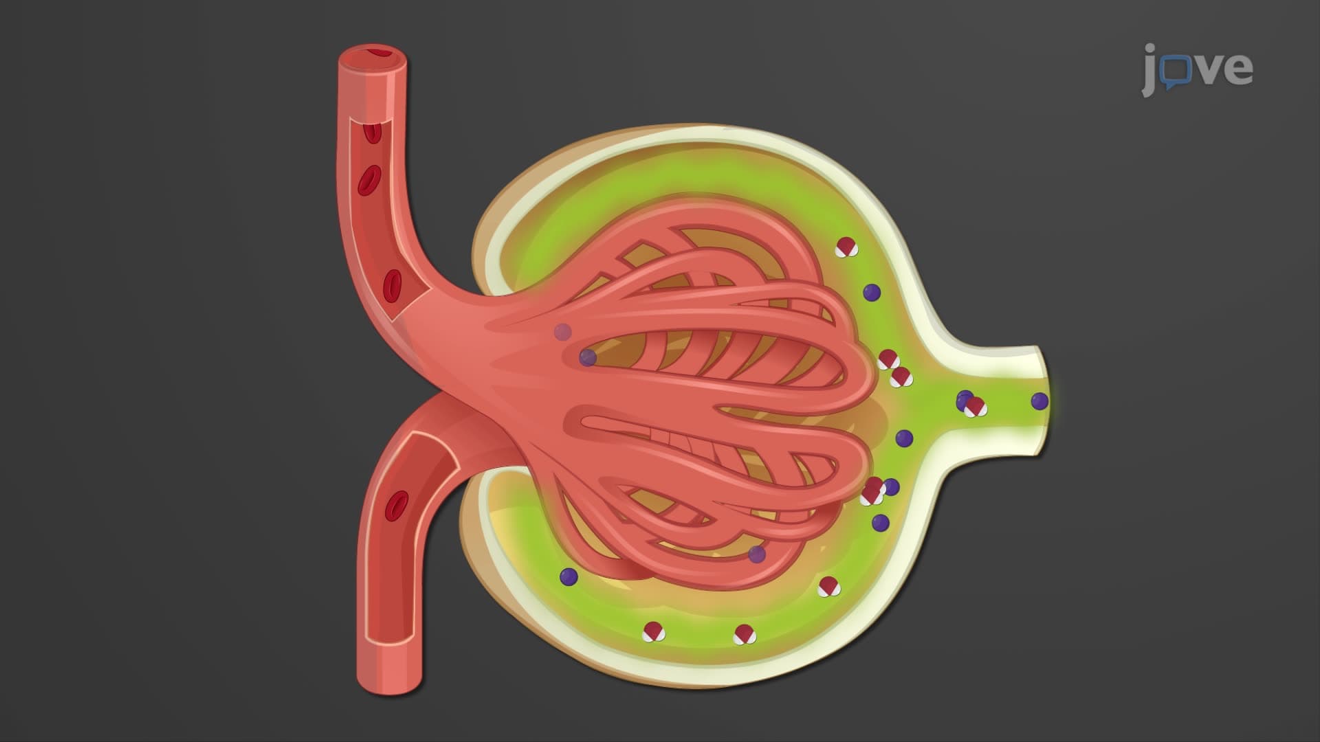


Internal Anatomy of the Kidney

00:00
01:12
Video Summary: What Is Internal Anatomy Kidney
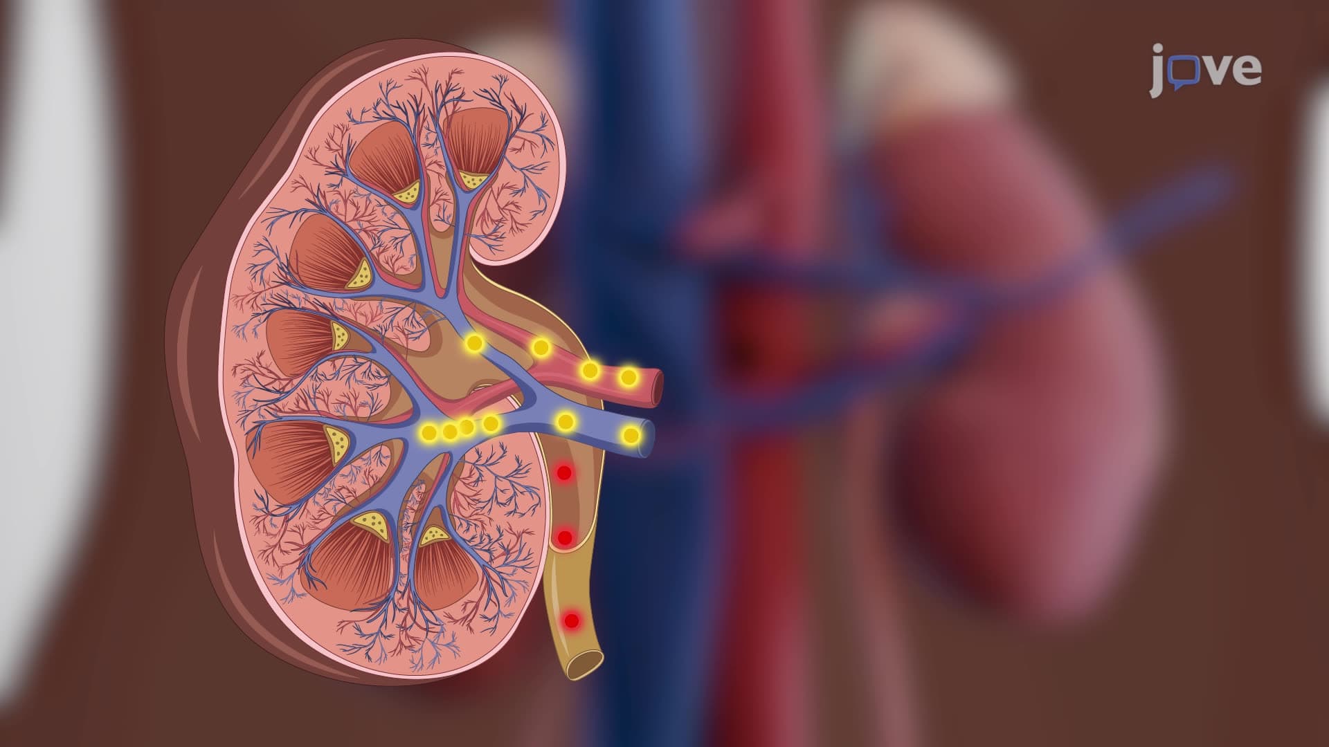
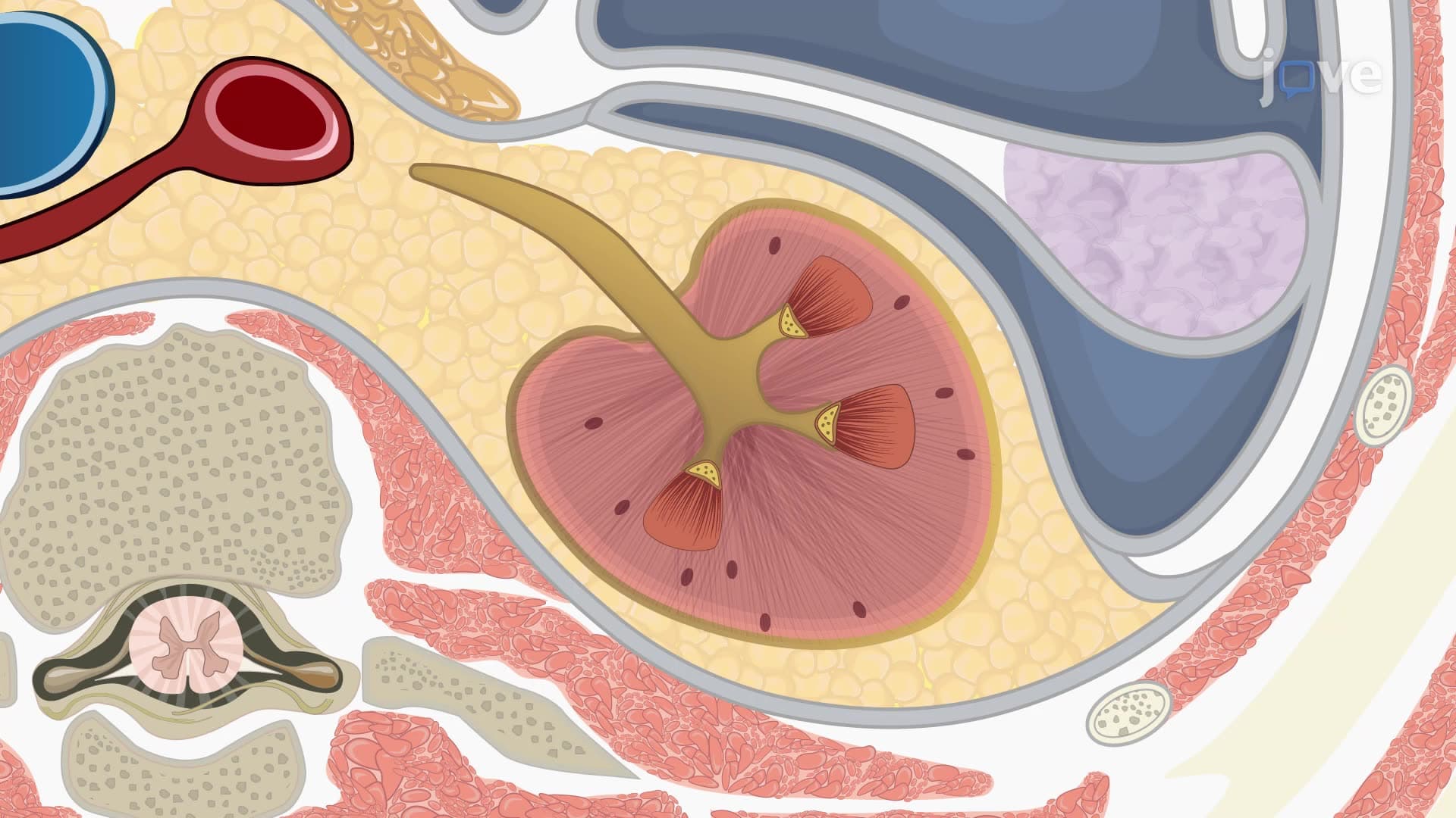
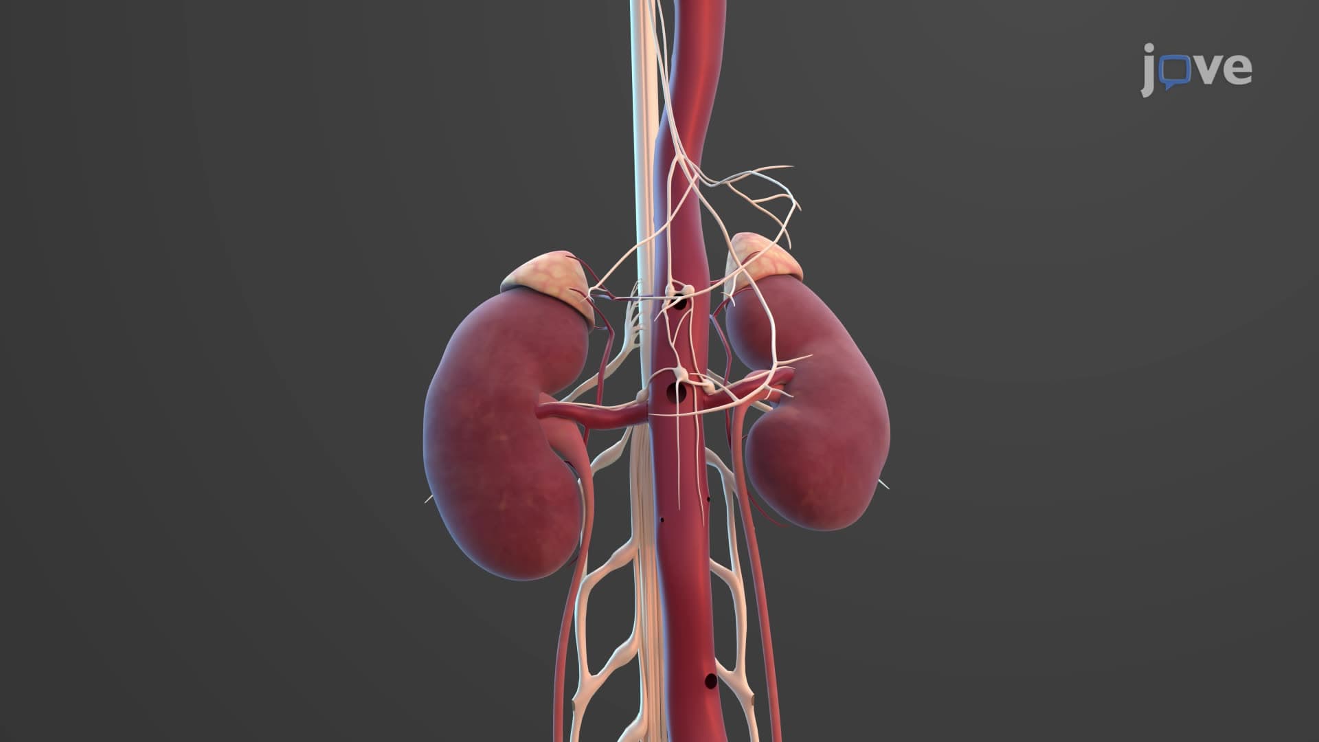
Did you know that your kidneys filter about 50 gallons of blood daily through intricate internal structures? Understanding kidney internal anatomy biology reveals how these vital organs maintain homeostasis in your body. The Mayo Clinic reports that kidney disease affects over 37 million Americans, making knowledge of what is the internal anatomy of the kidney crucial for health literacy. From the outer renal cortex to the inner medullary pyramids and collecting system, each component plays a specific role in urine formation and waste elimination. Watch the full video on JoVE Coach to master this concept with expert-led visuals and step-by-step explanations.
- Understand what is internal anatomy kidney and identify the three main regions: renal cortex, renal medulla, and renal pelvis
- Identify the structural components of medullary pyramids and their relationship to renal columns
- Analyze how renal papillae connect to the collecting system through minor and major calyces
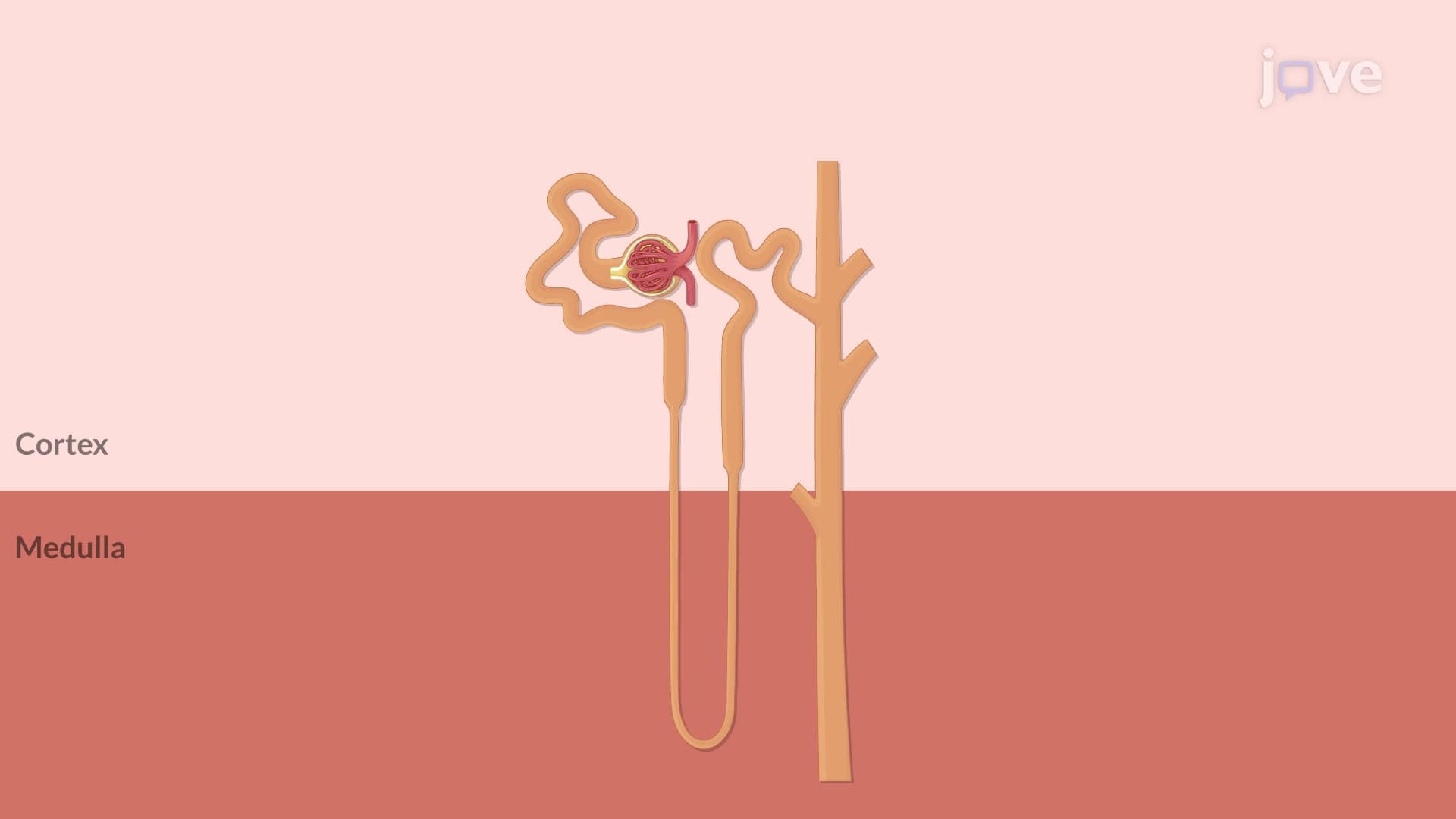
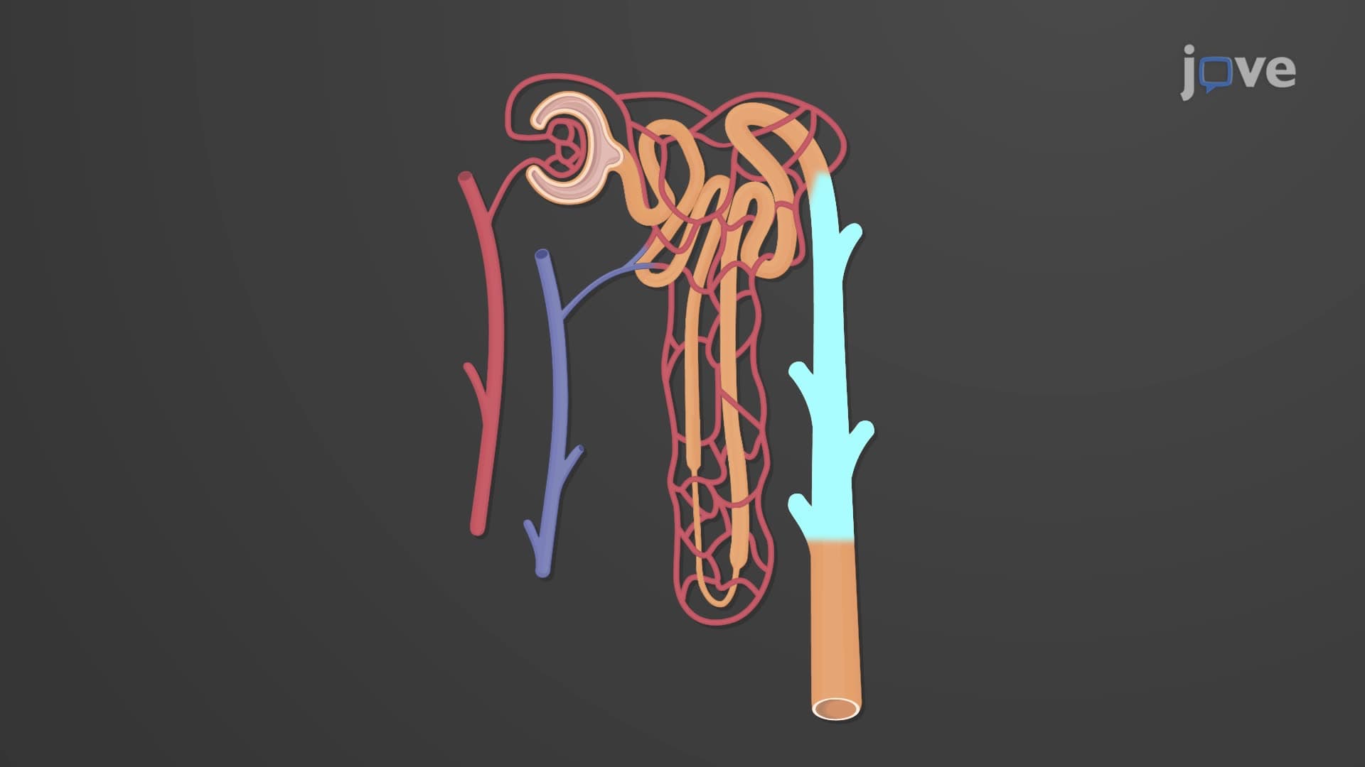
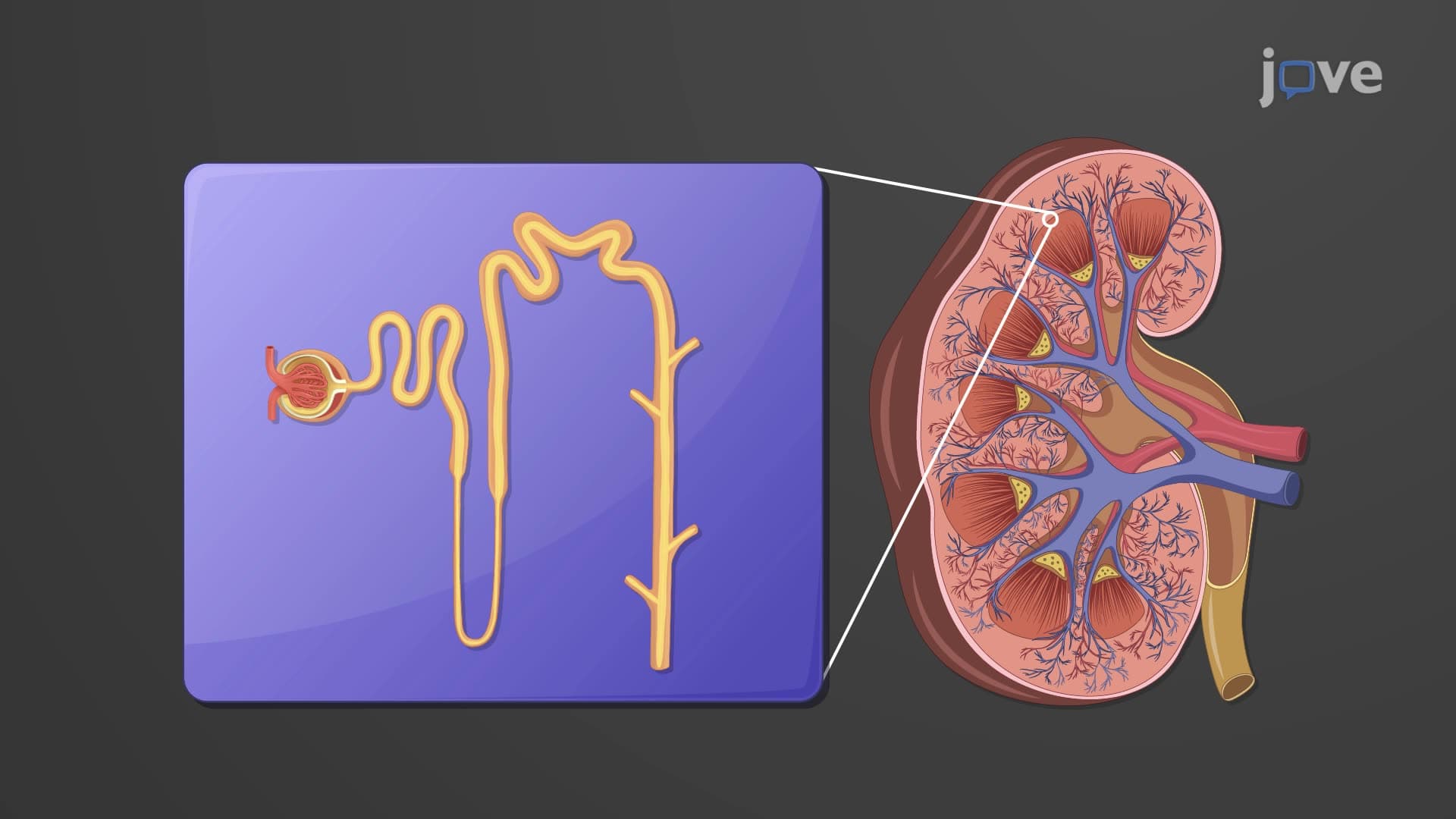
- Compare the functional differences between cortical and medullary regions in nephron organization
- Recognize the pathway of urine flow from papillae through calyces to the renal pelvis
- Evaluate the relationship between parenchyma structure and kidney function
- Apply knowledge of internal kidney anatomy to understand filtration and concentration processes
- Explore how anatomical abnormalities can lead to kidney dysfunction and disease
Understanding What is Internal Anatomy Kidney: The Three-Zone Organization
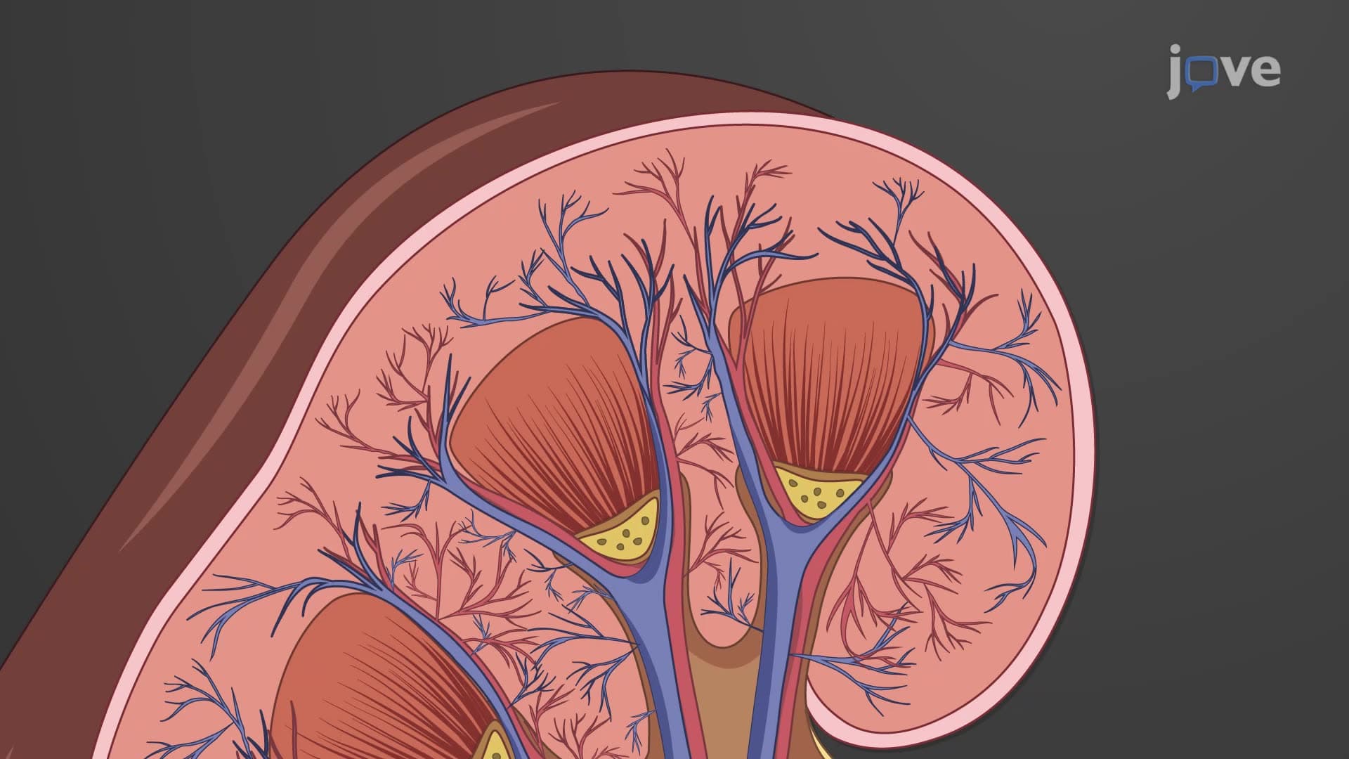
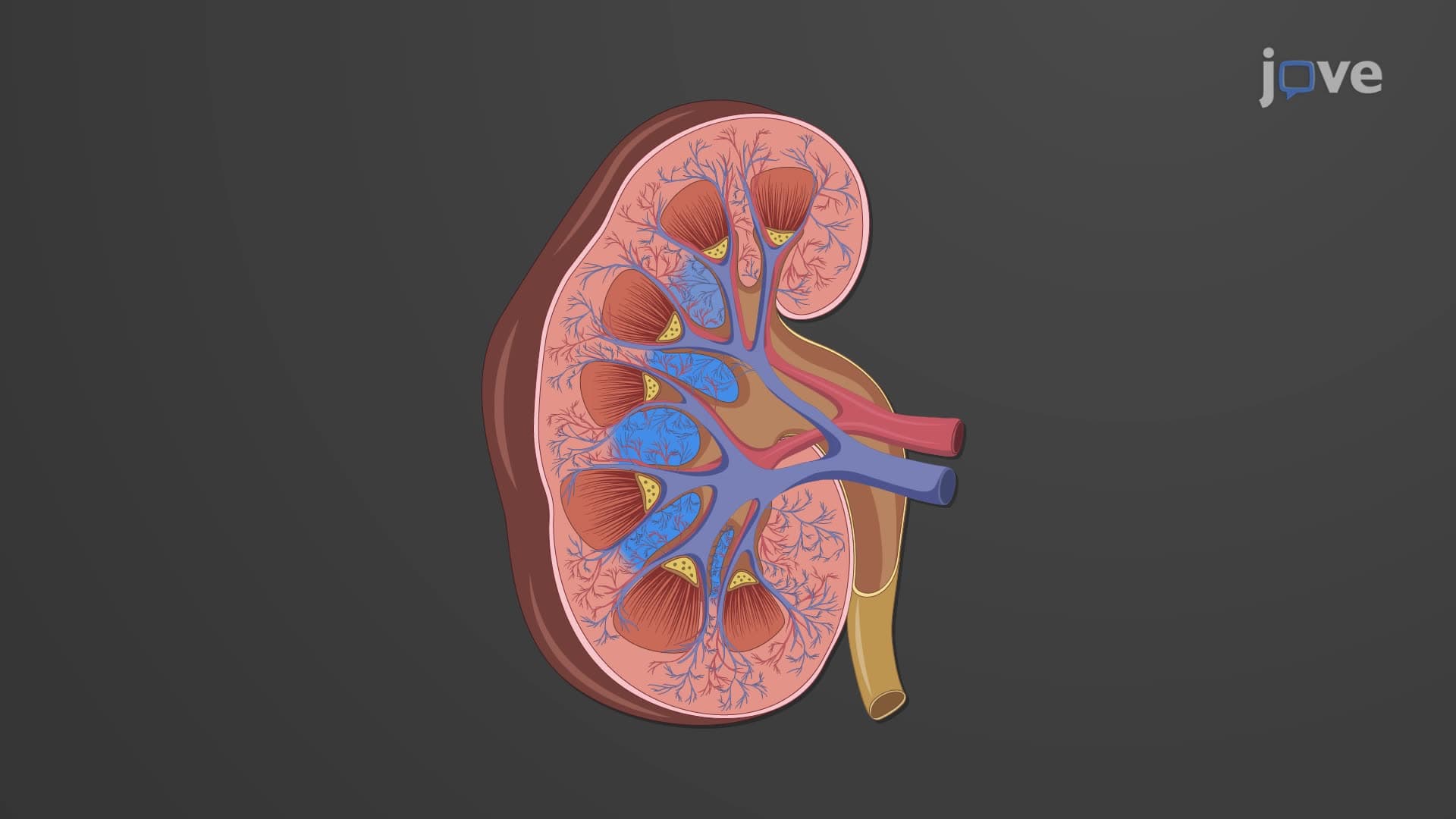
The kidney internal anatomy biology reveals a sophisticated three-zone organization essential for maintaining fluid and electrolyte balance. When you examine a frontal section of the kidney, you'll observe distinct regions that work together seamlessly. The outermost renal cortex appears light red and granular, housing the glomeruli and proximal portions of nephrons where initial filtration occurs. This region is crucial for blood pressure regulation through the renin-angiotensin system.
Renal Medulla and Pyramid Structure
The renal internal anatomy explained becomes fascinating when examining the medullary pyramids. These cone-shaped structures contain the loops of Henle and collecting ducts, creating the kidney's concentrating mechanism. American medical students studying for the MCAT often struggle with this concept, but understanding that each pyramid functions like a biological "concentrator" helps clarify the process. The Cleveland Clinic notes that damage to these pyramids can significantly impair urine concentration ability.
The Collecting System: From Papillae to Pelvis
What is the internal anatomy of the kidney becomes clearer when tracing urine flow through the collecting system. Each renal papilla serves as the drainage point for one medullary pyramid, connecting to minor calyces that merge into major calyces. This funnel-like arrangement ensures efficient urine transport to the renal pelvis. Students preparing for AP Biology exams should note that this anatomical organization prevents backflow and maintains proper drainage pressure.
Clinical Relevance and Pathophysiology
Understanding renal cortex medulla pyramid relationships proves essential in clinical settings. Kidney stones often lodge at the ureteropelvic junction, where the renal pelvis narrows into the ureter. Additionally, conditions like medullary sponge kidney affect the collecting ducts within the pyramids, demonstrating how anatomical knowledge directly applies to patient care. Pre-med students should recognize that many USMLE Step 1 questions test the correlation between anatomy and pathological processes.
Frequently Asked Questions
Internal kidney anatomy refers to the organized structure of renal cortex, medulla with pyramids, and pelvis that enables filtration and urine formation. Understanding this anatomy is crucial for comprehending how kidneys maintain fluid balance, filter waste products, and regulate blood pressure in the human body.
MCAT questions often test the relationship between nephron location and function within cortical and medullary regions. AP Biology exams typically focus on identifying structures in kidney cross-sections and explaining how anatomical organization supports filtration processes. Students should practice labeling diagrams and connecting structure to function.
USMLE Step 1 emphasizes the collecting system pathway, medullary pyramid organization, and cortical-medullary junction. Questions often involve clinical scenarios where anatomical knowledge helps diagnose conditions like kidney stones, infections, or congenital abnormalities affecting specific regions.
Medical professionals use anatomical knowledge to interpret imaging studies like CT scans and ultrasounds. For example, cortical thinning suggests chronic kidney disease, while dilated calyces indicate obstruction, helping physicians at institutions like Johns Hopkins develop targeted treatment plans for patients.
No advanced chemistry background is required to grasp basic kidney anatomy. High school biology provides sufficient foundation for understanding structural organization. However, connecting anatomy to physiological processes like filtration and concentration does benefit from understanding basic chemistry concepts taught in introductory college courses.
Create anatomical diagrams tracing urine flow from cortex through pyramids to pelvis, then to ureter. Use mnemonics like "Cortex Contains Capillaries" for the outer region, and practice identifying structures on unlabeled kidney cross-sections. Active recall through self-testing proves more effective than passive review.
Progress to nephron anatomy and physiology, focusing on glomerular filtration, tubular reabsorption, and secretion processes. Then explore kidney regulation of blood pressure, acid-base balance, and electrolyte homeostasis. Advanced students should investigate pathophysiology of common kidney diseases and their anatomical correlations.
Kidney anatomy integrates with cardiovascular system through renal blood vessels, endocrine system via hormone production, and urinary system through urine transport. Understanding these connections helps explain conditions like hypertension, diabetes complications, and fluid retention that appear frequently in medical school coursework and clinical practice.
Related Micro-courses

20 Concepts

37 Concepts

14 Concepts