Loading subject page…

- Anatomy and Physiology
- The Urinary System
- The Micturition Reflex
Micro-course: The Urinary System
78,053 views
Learn key concepts and earn your certificate.
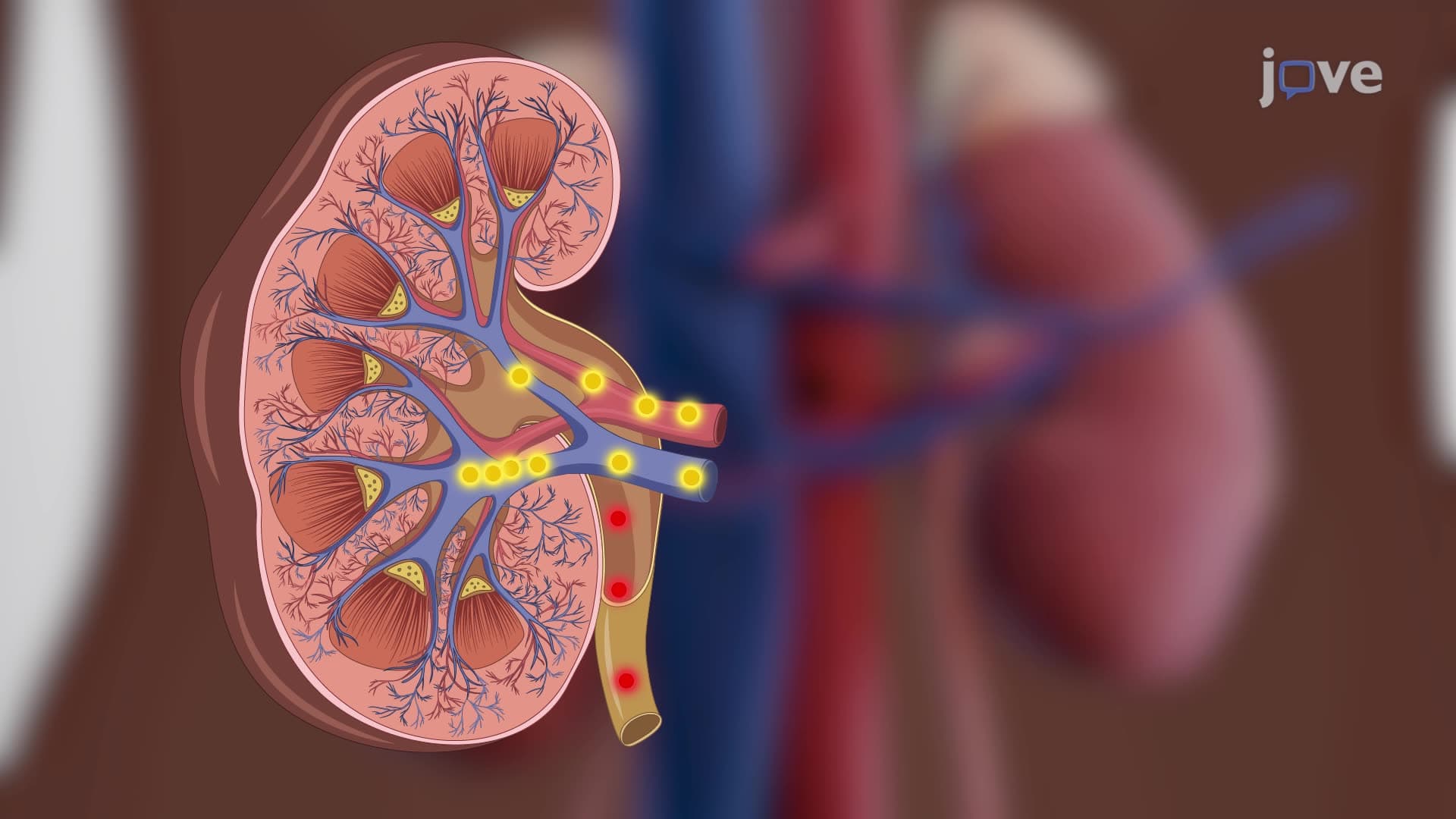

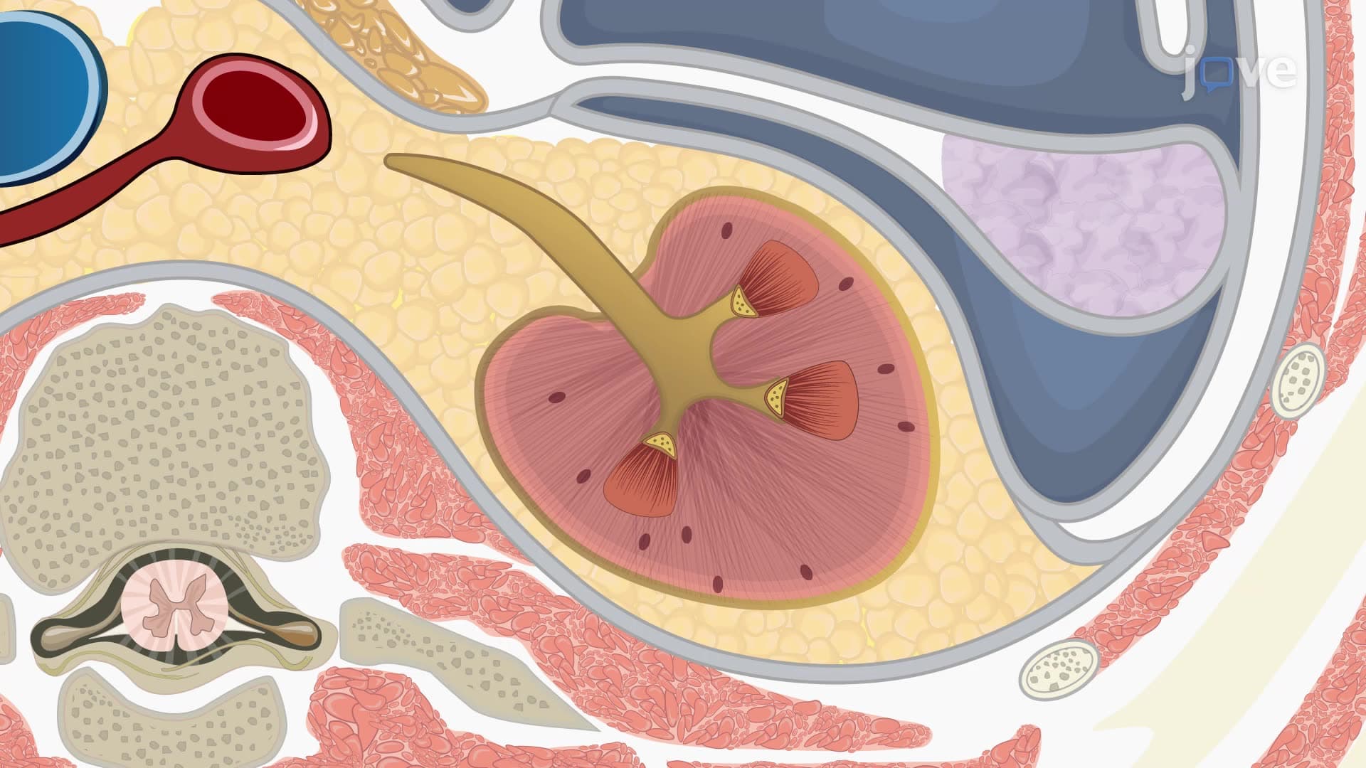
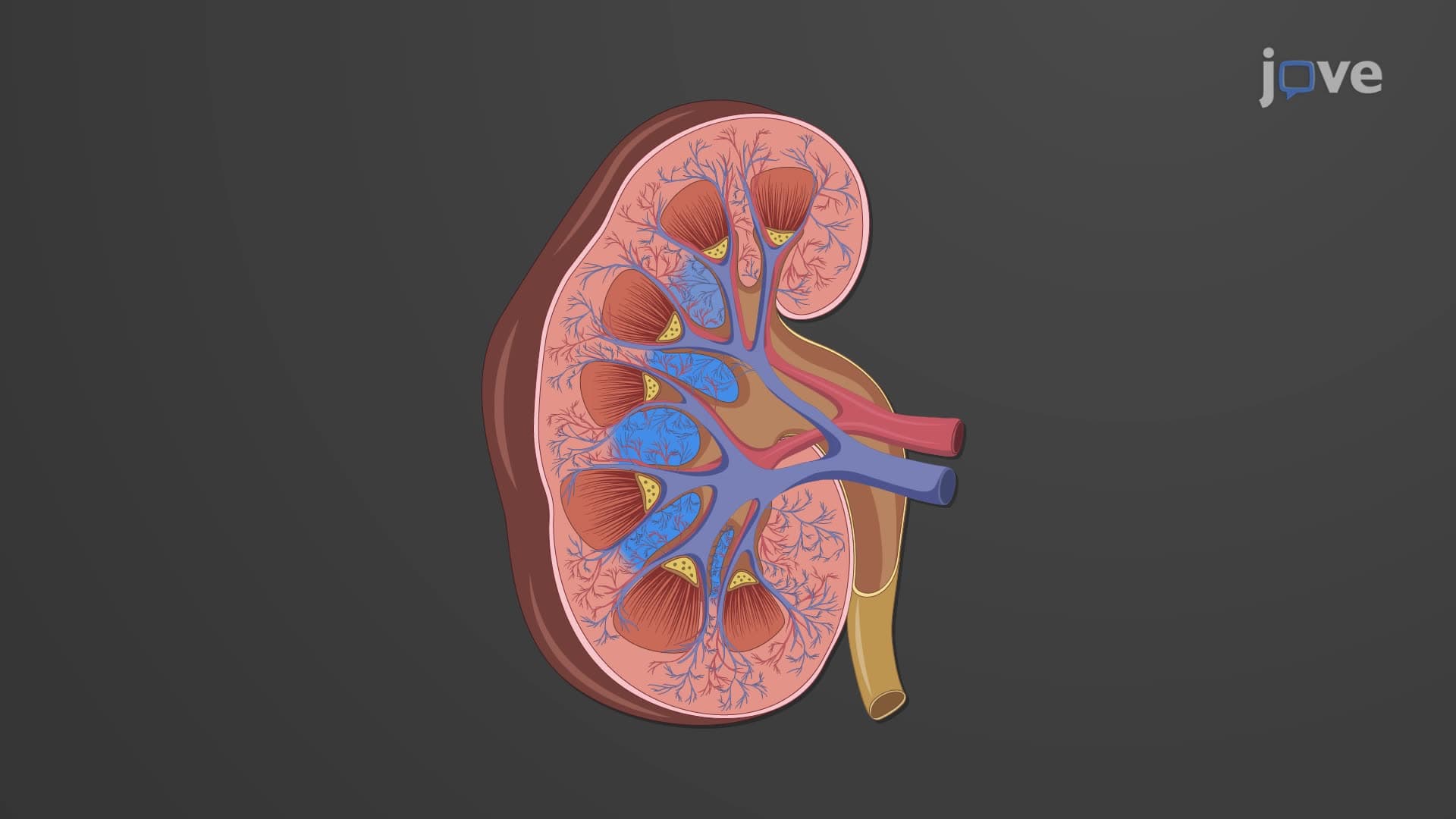
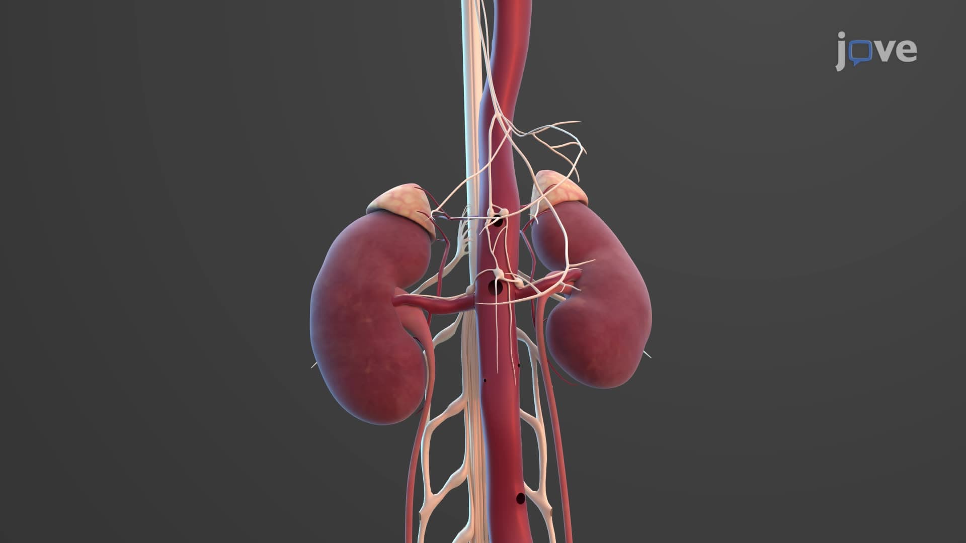


The Micturition Reflex

00:00
01:26
Video Summary: What Is the Micturition Reflex
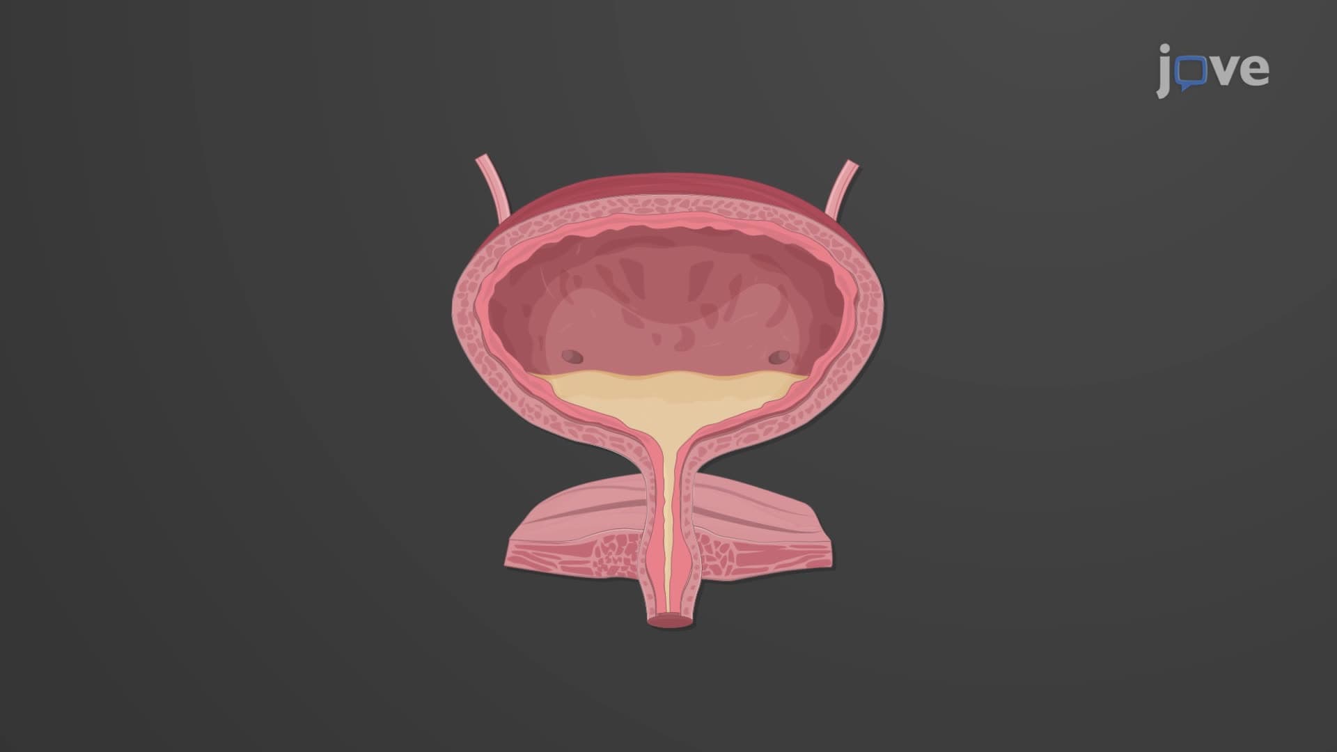
Ever wonder how your body "knows" when to hold it during a three-hour AP Biology exam versus when it's safe to use the restroom? The micturition reflex biology involves a sophisticated neural control system that coordinates bladder filling and emptying through two distinct spinal reflexes. In clinical settings like Johns Hopkins Hospital, understanding this reflex helps diagnose conditions from pediatric bedwetting to adult incontinence. The micturition reflex seamlessly switches between storage and voiding modes based on bladder volume and conscious control. Watch the full video on JoVE Coach to master this concept with expert-led visuals and step-by-step explanations.
- Understand the micturition reflex as a dual-phase neurological process controlling bladder function through storage and voiding reflexes
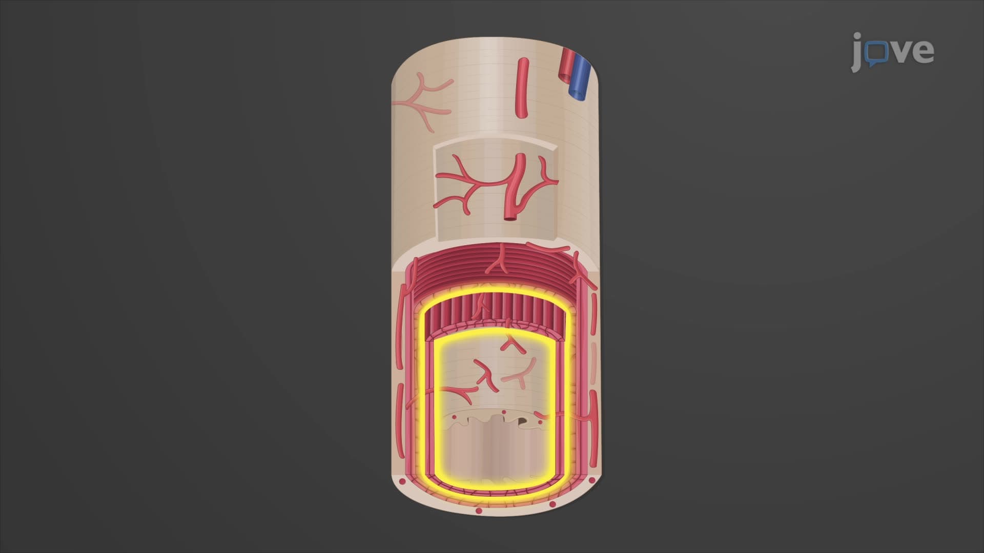
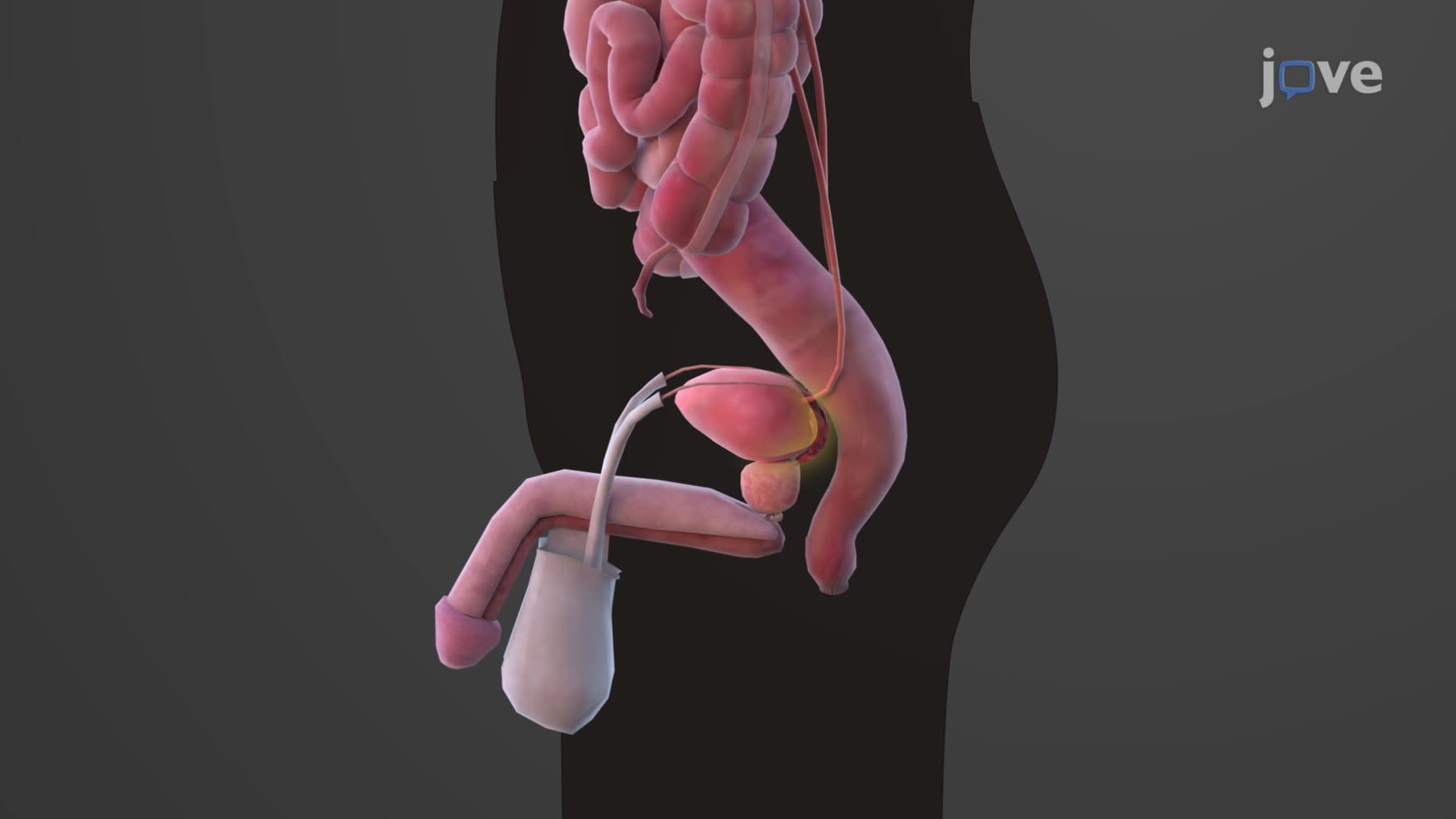
- Identify the key anatomical structures involved: detrusor muscle, internal urethral sphincter, external urethral sphincter, and pontine control centers
- Analyze how sympathetic and parasympathetic nervous systems create opposing effects during different phases of bladder control
- Compare involuntary spinal reflexes with voluntary cortical control mechanisms in micturition regulation
- Evaluate the developmental timeline of bladder control from infancy through early childhood
- Apply knowledge of stretch receptors and neural pathways to understand clinical conditions like neurogenic bladder
- Recognize the critical volume threshold (200 mL) that triggers the transition from storage to voiding reflex
- Explore how pontine micturition center coordinates with spinal segments to orchestrate complex bladder functions
Understanding the Micturition Reflex: A Two-Phase Control System
The micturition reflex biology represents one of the most elegant examples of autonomic nervous system coordination in human physiology. This complex process involves precise timing between neural circuits, smooth muscle contractions, and voluntary control mechanisms that most students encounter in AP Biology, college anatomy courses, and pre-medical studies.
Phase 1: The Storage Reflex and Sympathetic Dominance
During bladder filling, the urination reflex explained physiology begins with the storage phase. As urine enters from the kidneys, stretch receptors in the bladder wall detect increasing volume. When bladder capacity remains below 200 mL, the sacral spinal cord (specifically segments S2-S4) activates the pontine storage center in the brainstem. This triggers sympathetic nervous system responses that keep urine contained.
The sympathetic pathways release norepinephrine, which has two critical effects: inhibiting the detrusor muscle (the bladder's main smooth muscle) and stimulating the internal urethral sphincter to contract. Think of this like a dual-lock system-the bladder stays relaxed while the internal sphincter stays tightly closed. Students preparing for the MCAT often encounter questions about this reciprocal relationship between sympathetic and parasympathetic control.
Phase 2: The Voiding Reflex and Parasympathetic Activation
What is the micturition reflex in physiology becomes clearer when examining the voiding phase. Once bladder volume exceeds approximately 200 mL, stretch receptors send stronger signals to the pontine micturition center. This neurological switch activates parasympathetic pathways that release acetylcholine, causing detrusor muscle contraction and internal sphincter relaxation.
However, urination still requires voluntary control of the external urethral sphincter, which consists of skeletal muscle innervated by the pudendal nerve. College students studying for USMLE Step 1 frequently see questions about this voluntary override mechanism, as it represents the key difference between reflexive and conscious bladder control.
Clinical Applications and Developmental Considerations
The micturition reflex pathway has significant clinical implications, particularly in pediatric medicine and neurology. Children under age two lack the corticospinal connections necessary for voluntary external sphincter control, explaining why toilet training typically begins around this developmental milestone. Conditions like spina bifida can disrupt these neural pathways, leading to neurogenic bladder dysfunction that requires medical management.
Understanding these mechanisms proves essential for students pursuing healthcare careers, as bladder dysfunction affects millions of Americans and frequently appears on nursing exams like NCLEX-RN and HESI A2.
Frequently Asked Questions
The micturition reflex is a neurologically controlled process that manages bladder filling and emptying through two phases: storage and voiding. During storage, sympathetic nerves keep the detrusor muscle relaxed while contracting the internal sphincter. When bladder volume exceeds 200 mL, parasympathetic activation reverses these effects, enabling urination with voluntary external sphincter control.
MCAT questions often focus on sympathetic versus parasympathetic effects, neurotransmitter roles (norepinephrine vs. acetylcholine), and voluntary control mechanisms. AP Biology exams typically emphasize the reflex arc components and homeostatic regulation. Students should memorize the 200 mL threshold and understand how stretch receptors initiate neural responses.
USMLE Step 1 frequently tests pontine micturition center functions, spinal cord segments (S2-S4), and clinical correlations like neurogenic bladder. Step 2 emphasizes pediatric development of bladder control and pathological conditions. Focus on understanding both normal physiology and common dysfunctions affecting different age groups.
NCLEX-RN questions often involve urinary retention, incontinence management, and catheter care decisions. Understanding normal micturition physiology helps nurses recognize when bladder dysfunction indicates neurological problems, medication side effects, or post-surgical complications. This knowledge directly impacts patient assessment and intervention strategies.
Yes, urodynamic studies at medical centers like Mayo Clinic directly measure detrusor muscle contractions and sphincter responses during bladder filling. These tests help diagnose conditions ranging from overactive bladder to spinal cord injuries. Medical students often observe these procedures during urology rotations to understand normal versus abnormal micturition patterns.
Not at all-high school biology and basic anatomy provide sufficient background for understanding this concept. Focus on learning the two main neurotransmitters (norepinephrine and acetylcholine), their opposing effects, and the anatomical structures involved. Most students find this topic more straightforward than complex metabolic pathways or genetic regulation mechanisms.
Create a simple flowchart showing bladder filling → stretch receptors → spinal cord → pontine centers → sympathetic/parasympathetic responses. Use mnemonics like "Sympathetic = Storage" and "Parasympathetic = Peeing" to remember neurotransmitter effects. Practice drawing the pathway from memory and connecting it to clinical scenarios for deeper retention.
Explore related topics like kidney function and urine formation, other autonomic reflexes (such as defecation), and neurological control of smooth muscle throughout the body. These concepts build naturally from micturition physiology and frequently appear together on comprehensive exams covering urogenital and nervous system integration.
Related Micro-courses

20 Concepts

37 Concepts

14 Concepts