Loading video…


 16 Concepts
 1.6M views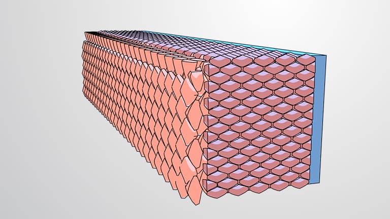

Video Summary: What are Tissues
Did you know that your heart beats over 100,000 times daily thanks to specialized cardiac muscle tissue? Body tissues biology forms the foundation of how multicellular organisms like humans organize millions of cells into functional units. From the protective epithelial tissue in your skin to the connective tissue supporting your bones, understanding what are tissues reveals how the Mayo Clinic's pathologists can diagnose diseases by examining tissue samples under microscopes. Watch the full video on JoVE Coach to master this concept with expert-led visuals and step-by-step explanations.
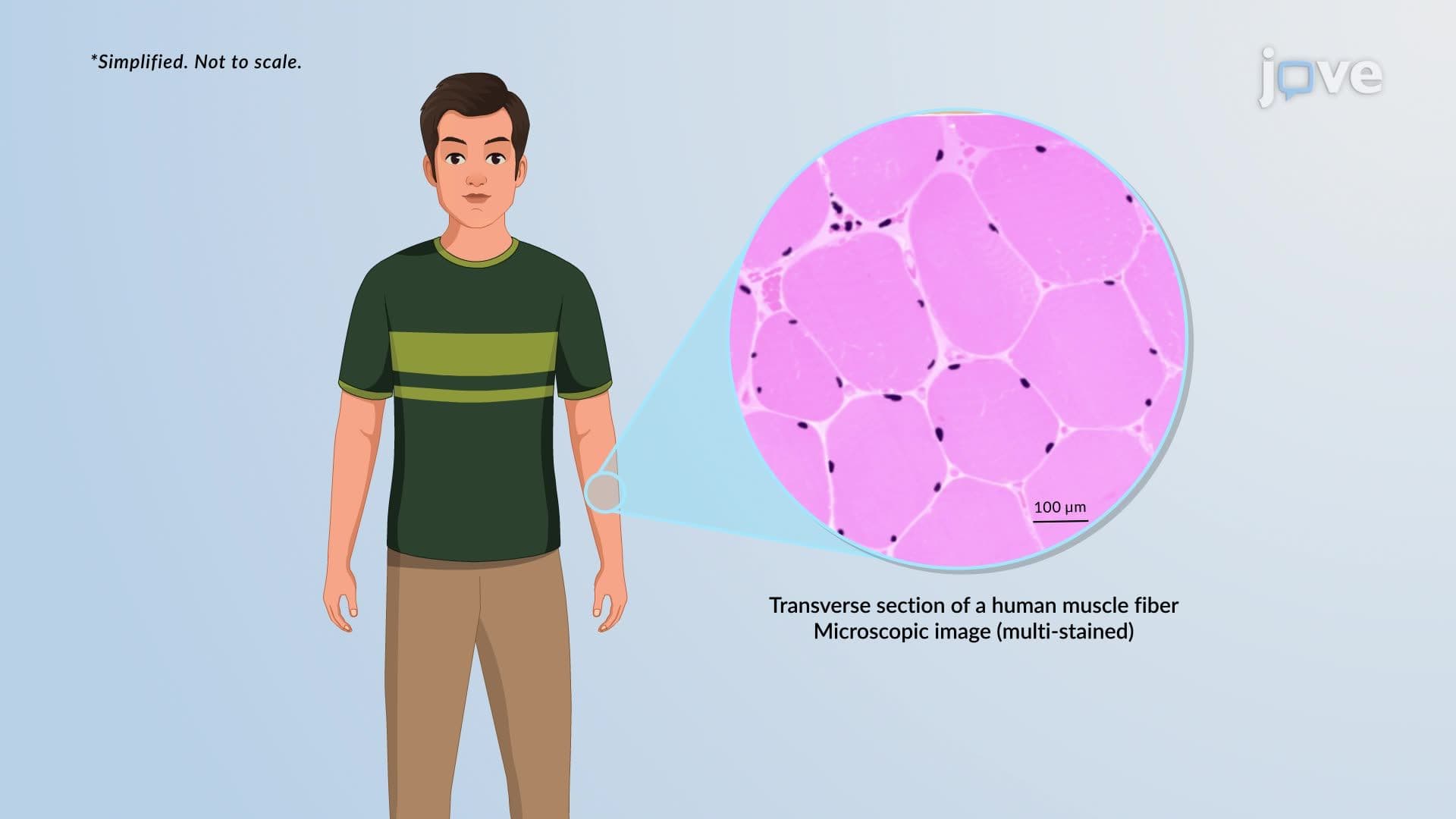
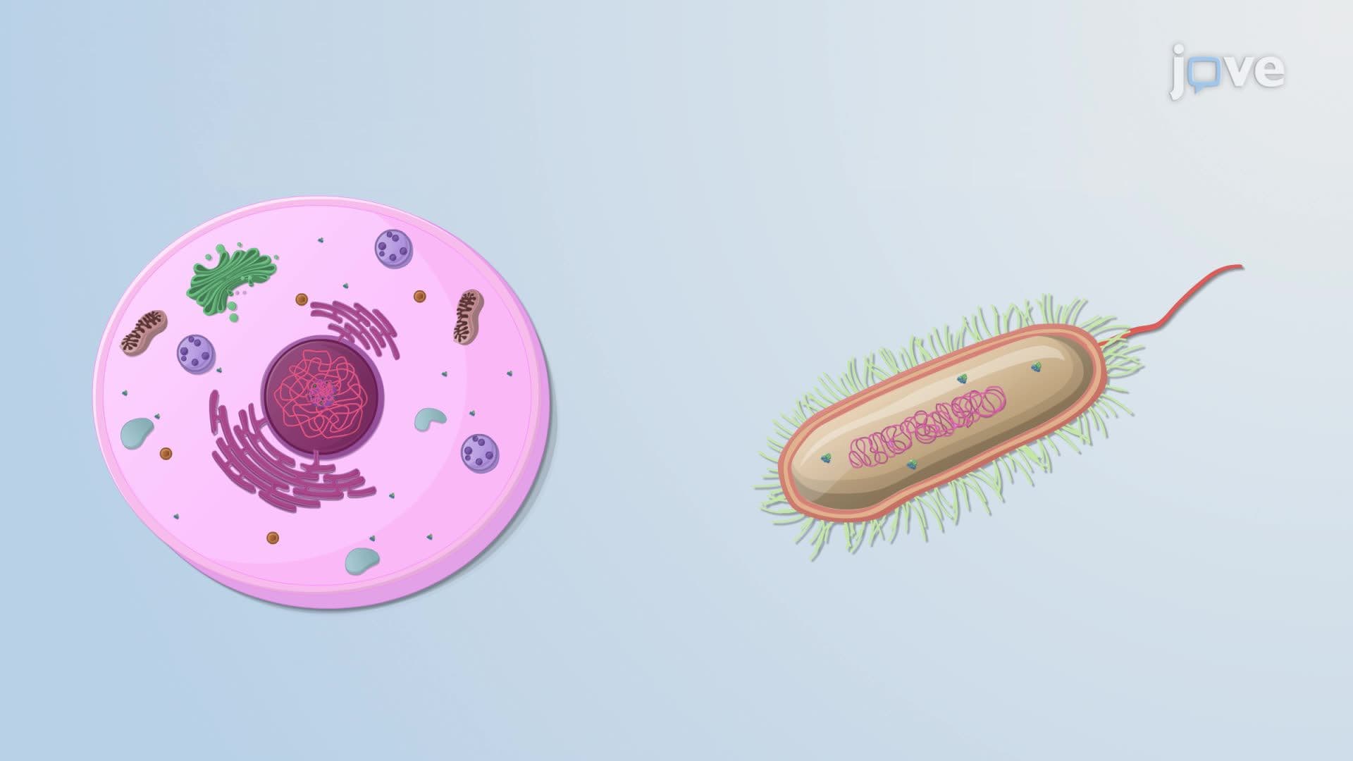
What are tissues? They represent a crucial organizational level in multicellular organisms, bridging the gap between individual cells and complex organs. In the human body, tissues function as specialized communities where cells with similar structures work together to perform specific functions. This tissue organization hierarchy allows for the incredible complexity we observe in organisms like humans, where trillions of cells coordinate seamlessly.
The concept of tissues becomes particularly important when studying for exams like the AP Biology test or college anatomy courses, where understanding biological organization levels is fundamental to success.
Body tissues biology recognizes four major tissue categories, each serving distinct roles in human physiology. Nervous tissue controls information processing and transmission, forming the basis of our brain, spinal cord, and peripheral nerves. For example, when you touch a hot stove, nervous tissue rapidly transmits pain signals to your brain within milliseconds.
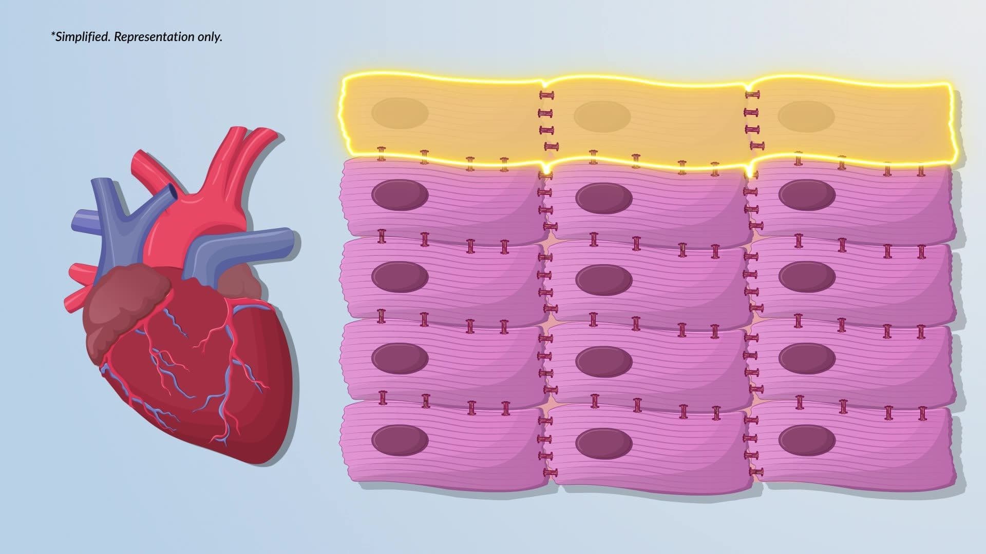
Muscle tissue enables movement through three specialized types: skeletal muscle (attached to bones for voluntary movement), cardiac muscle (found exclusively in the heart), and smooth muscle (lining organs like the digestive tract). Consider how Olympic athletes at the U.S. Training Centers rely on precisely coordinated skeletal muscle tissue to achieve peak performance.
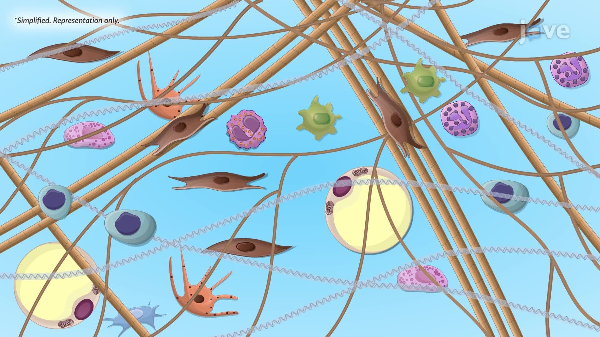
Epithelial tissue creates protective boundaries throughout the body, covering surfaces and lining cavities. Your skin's outer layer exemplifies stratified epithelial tissue, while your lung air sacs feature simple epithelial arrangements for efficient gas exchange. Finally, connective tissue provides structural support, including bone, cartilage, blood, and the supportive framework beneath epithelial layers.
Histology tissue biology reveals fascinating organizational patterns within epithelial tissues. These tissues display distinct polarity, with an apical surface facing the external environment and a basal surface attached to underlying connective tissue. This arrangement proves critical in organs like the kidneys, where epithelial cells in nephrons must selectively transport substances between blood and urine.
Cell shapes vary dramatically based on function. Squamous cells appear flattened and thin, perfect for efficient diffusion in lung alveoli. Cuboidal cells, roughly cube-shaped, excel in secretion and absorption in kidney tubules. Columnar cells, tall and narrow, maximize surface area for absorption in the small intestine. Pre-med students preparing for the MCAT often focus on these shape-function relationships.
Understanding tissue function body relationships becomes essential when examining how organs operate. Most organs contain all four tissue types working in coordination. In the heart, cardiac muscle tissue provides contractile force, nervous tissue regulates rhythm, epithelial tissue lines chambers, and connective tissue forms supportive structures and valves.
This integration concept frequently appears on college anatomy exams and USMLE Step 1 questions, where students must predict tissue dysfunction consequences. For instance, understanding how connective tissue disorders like Ehlers-Danlos syndrome affect multiple organ systems requires grasping fundamental tissue interactions and support mechanisms.
Related Micro-courses