Loading video…


 12 Concepts
 824.1K views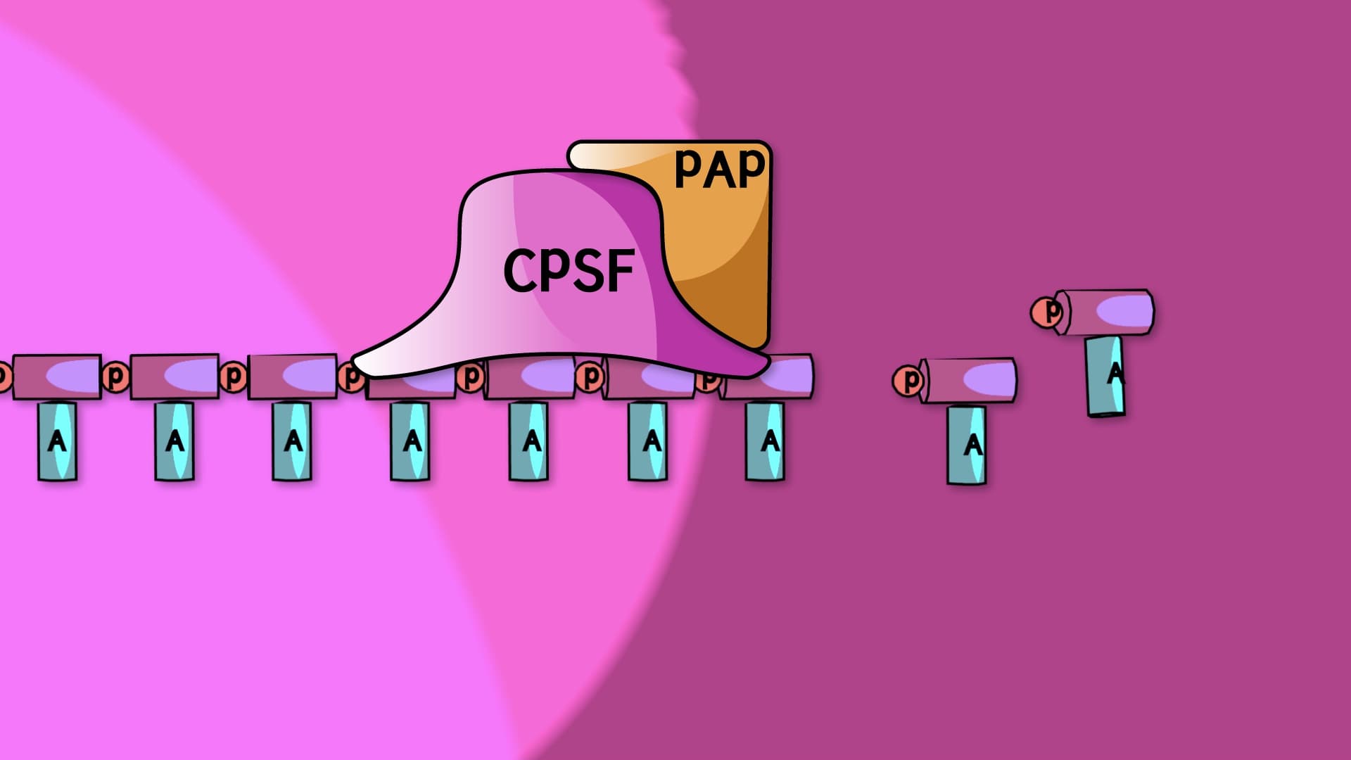

Video Summary: What Is Pre Mrna Processing
Ever wonder how your cells prevent genetic chaos when making proteins? Pre mRNA processing acts as a sophisticated quality control system, transforming raw genetic transcripts into functional messengers through three critical modifications. Just like how the FDA requires specific labeling and safety checks before medications reach patients, eukaryotic cells must process pre-mRNA through capping, polyadenylation, and splicing before translation can begin. Watch the full video on JoVE Coach to master this concept with expert-led visuals and step-by-step explanations.
Pre mRNA processing represents one of the most sophisticated quality control mechanisms in molecular biology. When RNA polymerase II transcribes a gene in eukaryotic cells, the initial product-called precursor mRNA or pre-mRNA-is essentially a rough draft that requires extensive editing before becoming a functional protein-coding message. This processing occurs in the nucleus and involves three coordinated modifications that transform the raw transcript into mature mRNA ready for translation.
The first modification involves adding a 7-methylguanosine cap to the 5' end of the pre-mRNA transcript. This cap forms through an unusual 5'-5' triphosphate linkage, creating a distinctive structure that serves multiple critical functions. The cap protects the mRNA from 5' exonuclease degradation, which would otherwise rapidly destroy the transcript. Additionally, the cap serves as a recognition signal for cap-binding proteins and eukaryotic initiation factors that guide the mRNA to ribosomes during translation initiation. Students preparing for the MCAT often encounter questions about cap structure and function, particularly regarding how cap-binding complex formation facilitates ribosome recruitment.
The 3' end modification involves recognizing a polyadenylation signal sequence (typically AAUAAA in mammals) located 10-30 nucleotides upstream of the cleavage site. This sequence recruits cleavage and polyadenylation specificity factor (CPSF), which directs endonuclease cleavage of the pre-mRNA. Following cleavage, poly(A) polymerase adds approximately 200-250 adenine residues to form the poly-A tail. This tail enhances mRNA stability by protecting against 3' exonuclease activity and facilitates nuclear export through interactions with poly(A)-binding proteins. In clinical contexts, mutations affecting polyadenylation signals can cause diseases like thalassemia, where defective processing leads to reduced globin protein production.
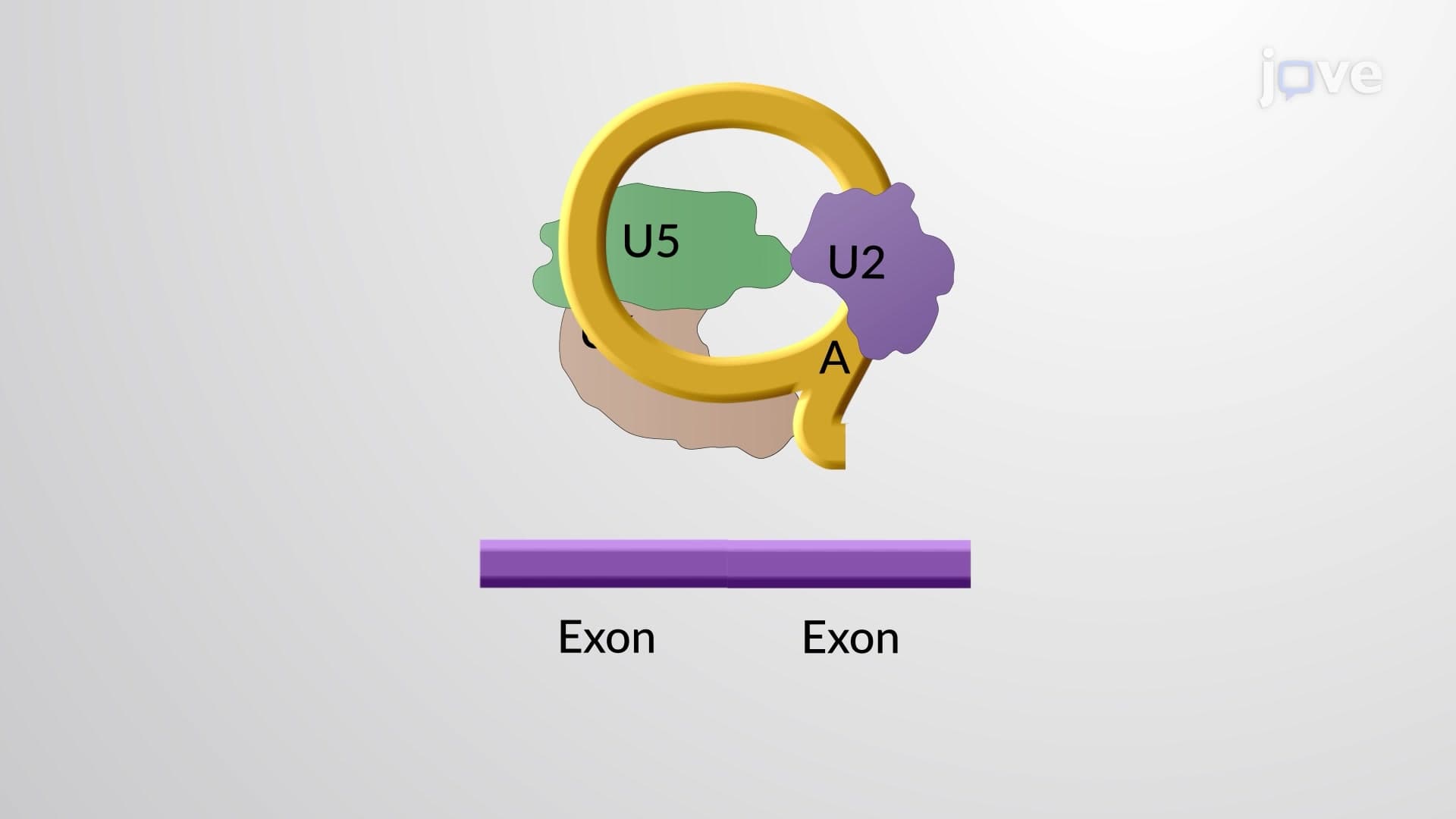
The most complex processing step involves spliceosome-mediated intron removal and exon joining. The spliceosome, a dynamic ribonucleoprotein complex composed of U1, U2, U4, U5, and U6 small nuclear RNPs (snRNPs), recognizes conserved splice sites at intron boundaries. The process occurs through two transesterification reactions that precisely remove introns while joining exons in the correct order. Alternative splicing allows a single gene to produce multiple protein isoforms, dramatically expanding the proteome. For example, the DSCAM gene in humans can theoretically produce over 38,000 different protein variants through alternative splicing. AP Biology students frequently encounter questions about splicing mechanisms and how alternative splicing contributes to protein diversity in complex organisms.
Related Micro-courses