Loading video…


 12 Concepts
 767K views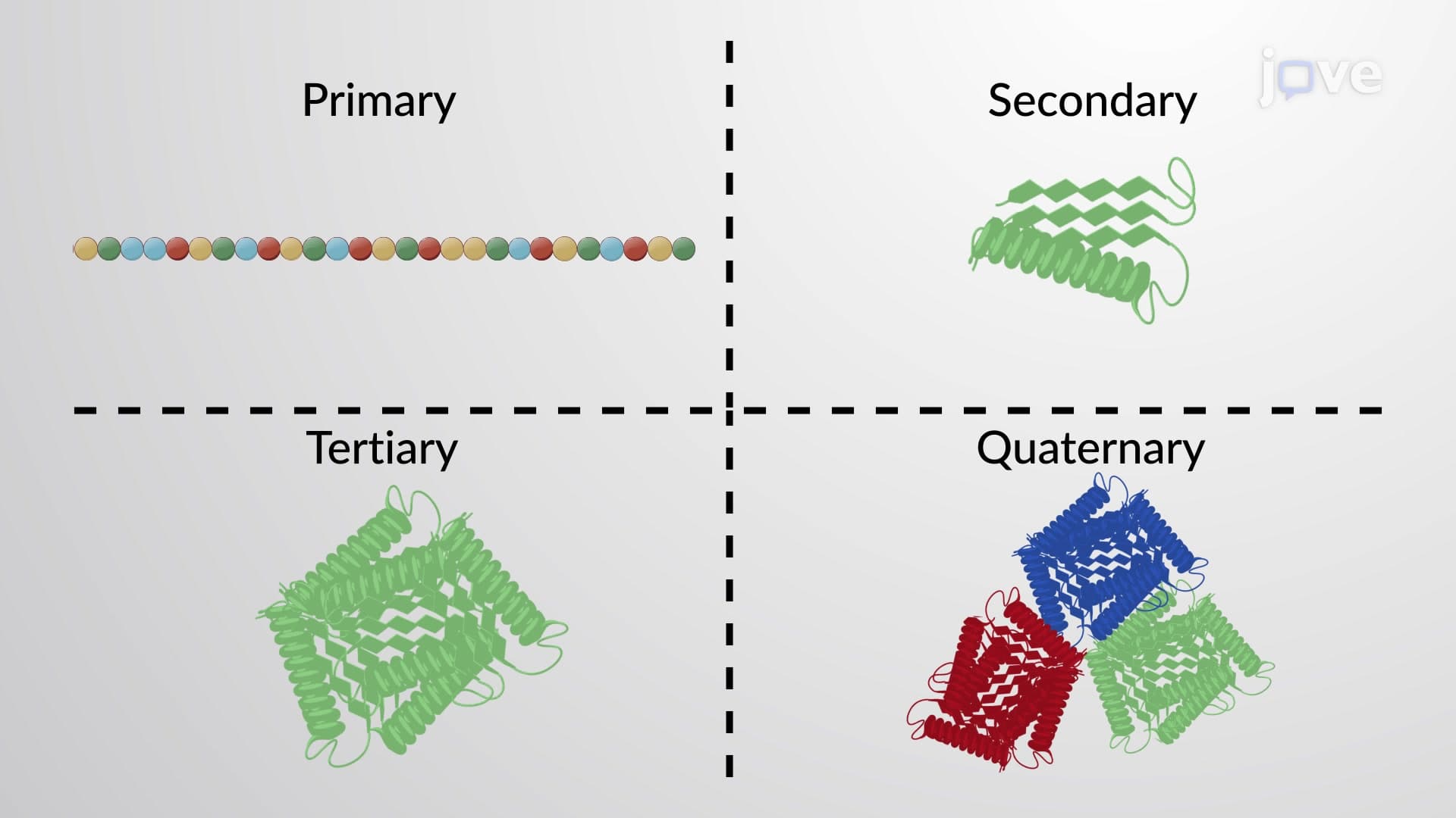

Video Summary: What Is Protein and Protein Structure
Did you know that hemoglobin, the protein carrying oxygen in your blood, is made of over 570 amino acids folded into a precise three-dimensional shape? Understanding protein and protein structure reveals how these molecular machines perform life-sustaining functions in every cell. From insulin regulating blood sugar in diabetes patients to antibodies fighting COVID-19, proteins demonstrate the critical relationship between structure and biological function. What is Protein And Protein Structure becomes clear when examining how amino acid sequences determine everything from enzyme activity to membrane transport. Watch the full video on JoVE Coach to master this concept with expert-led visuals and step-by-step explanations.
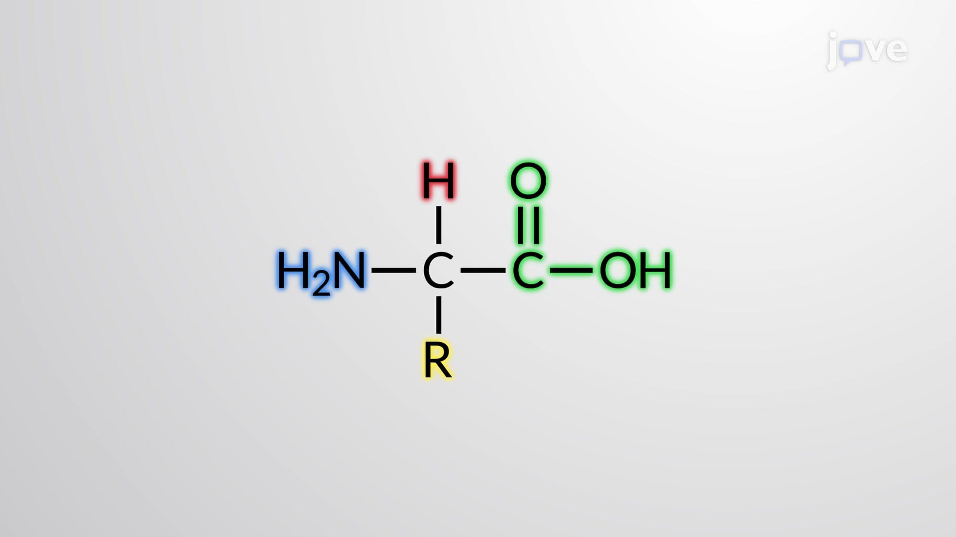
Proteins represent nature's most versatile macromolecules, serving as enzymes, structural components, transport molecules, and signaling mediators. The protein and protein structure concept centers on how twenty different amino acids combine to create thousands of distinct proteins, each with unique functions. This molecular diversity stems from both the sequence of amino acids and their three-dimensional arrangements, making protein structure analysis essential for understanding biological processes.
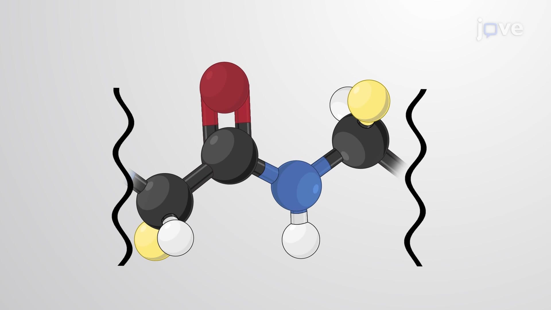
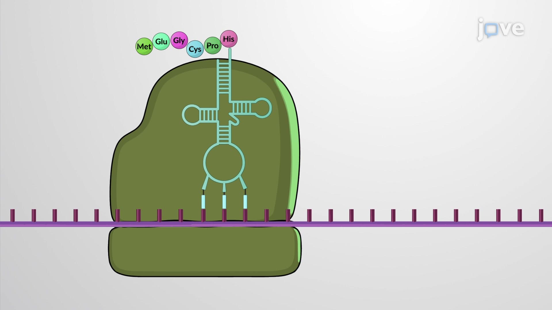
Primary structure establishes the fundamental blueprint-the linear sequence of amino acids connected by peptide bonds. Reading from the N-terminus (amino end) to the C-terminus (carboxyl end), this sequence determines all higher-order structures. Consider insulin, discovered at the University of Toronto, whose 51-amino acid sequence must be precisely maintained for blood glucose regulation.
Secondary structures emerge through hydrogen bonding between backbone atoms, creating regular patterns like alpha-helices and beta-pleated sheets. Alpha-helices spiral like DNA's double helix, while beta-sheets form extended structures resembling accordion pleats. These patterns appear in proteins throughout your body-keratin in hair contains alpha-helices, while silk fibroin demonstrates beta-sheet strength.
Tertiary structure represents the complete three-dimensional folding pattern resulting from R-group interactions. Hydrophobic amino acids cluster together excluding water, while polar residues interact with aqueous environments. Disulfide bridges between cysteine residues provide additional stability, as seen in antibodies fighting infections.
Quaternary structure involves multiple polypeptide chains assembling into functional units. Hemoglobin exemplifies this complexity with four subunits (two alpha, two beta) working cooperatively to transport oxygen from lungs to tissues. This structural hierarchy appears frequently on AP Biology exams and MCAT questions, where students must connect molecular details to physiological functions.
Protein location correlates directly with surface properties. Cytoplasmic proteins like glycolytic enzymes display hydrophilic surfaces interacting with cellular water, while maintaining hydrophobic cores for stability. Conversely, membrane proteins possess hydrophobic external regions inserting into lipid bilayers, with hydrophilic domains extending into aqueous compartments. Understanding these relationships proves crucial for predicting protein behavior and designing therapeutic interventions targeting specific cellular compartments.
Related Micro-courses