Loading video…


 13 Concepts
 1.3M views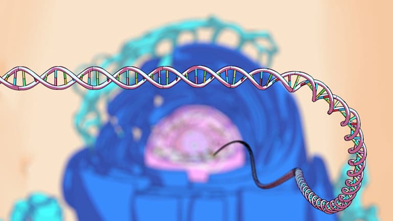

Video Summary: What Is DNA Packaging
Every human cell contains 6 feet of DNA squeezed into a nucleus just 0.00001 inches wide-imagine fitting 30 miles of thread into a tennis ball! DNA packaging is the remarkable process that makes this possible through hierarchical folding mechanisms. The Mayo Clinic's genetic research labs study these packaging errors linked to cancer development. What is DNA packaging becomes clear through understanding nucleosomes, chromatin fibers, and chromosome formation. Watch the full video on JoVE Coach to master this concept with expert-led visuals and step-by-step explanations.
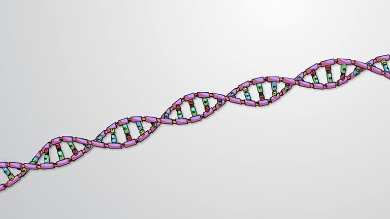
What is DNA packaging represents one of biology's most elegant engineering solutions. Human diploid cells contain approximately 3.2 billion base pairs of DNA, creating a linear molecule stretching over 6 feet when fully extended. Yet this massive information repository must fit within a nucleus measuring just 10 micrometers in diameter-roughly 1/2500th the width of a human hair.
The National Human Genome Research Institute estimates that without proper packaging, cellular DNA would be 200,000 times longer than the cell itself. This spatial constraint drives the evolution of sophisticated packaging mechanisms that maintain both DNA integrity and accessibility for essential cellular processes.
The first packaging level involves histone proteins-positively charged molecules that attract negatively charged DNA through electrostatic interactions. The core histone octamer contains two copies each of H2A, H2B, H3, and H4 proteins, forming a protein disk around which DNA wraps 1.65 times.
Each nucleosome packages approximately 147 base pairs of DNA, creating the characteristic "beads on a string" appearance visible under electron microscopy. Stanford University's chromatin research demonstrates that nucleosome positioning significantly influences gene accessibility, with tightly wrapped regions showing reduced transcriptional activity.
Nucleosomes connected by linker DNA segments further condense into 30-nanometer chromatin fibers through interactions with histone H1 and other architectural proteins. This secondary packaging reduces DNA length by an additional 6-fold, creating the chromatin structure visible in non-dividing cell nuclei.
Research at Harvard Medical School reveals that chromatin modifications-including histone methylation and acetylation-regulate fiber compaction and gene expression patterns. These epigenetic modifications create "open" euchromatin regions for active genes and "closed" heterochromatin regions for silenced sequences.
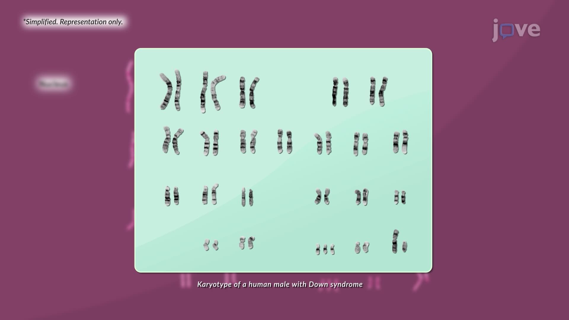
During mitosis and meiosis, chromatin fibers undergo extreme condensation facilitated by condensin proteins and topoisomerase enzymes. This final packaging level creates the familiar X-shaped chromosomes visible under light microscopy, achieving a 10,000-fold compaction compared to naked DNA.
The University of California system's cell biology programs emphasize understanding chromosome packaging for AP Biology and MCAT preparation, as packaging defects contribute to genetic disorders like cancer and developmental abnormalities. Clinical applications include analyzing chromosome structure in genetic counseling and developing targeted cancer therapies that exploit packaging vulnerabilities in tumor cells.
Related Micro-courses