Loading concepts…

 11 Concepts
 7K Views
 Popular topics
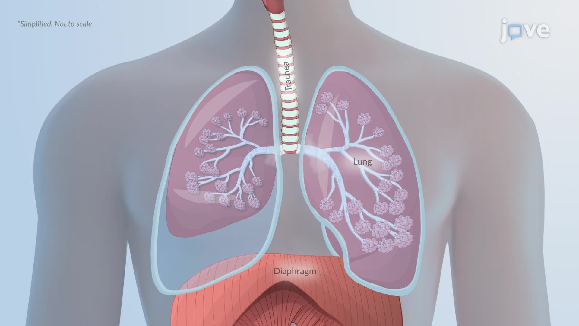


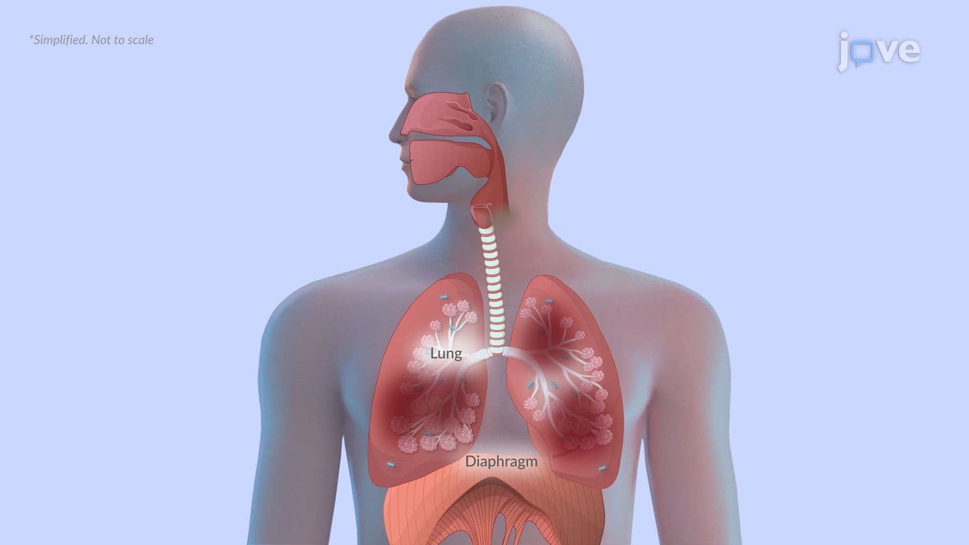
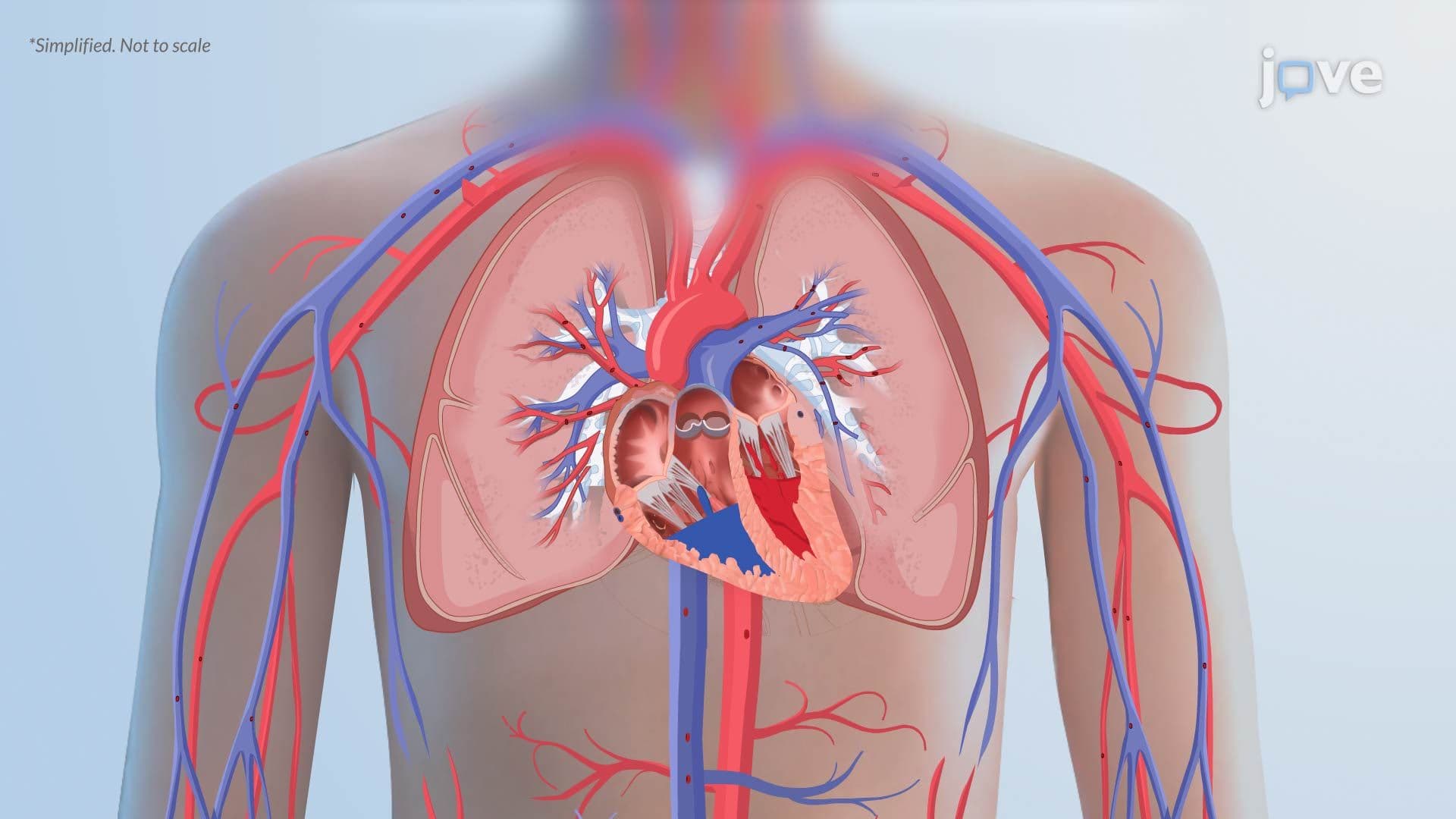
Respiratory System Disorders represent some of the most clinically significant conditions covered in pre-health and nursing education. This micro-course, supported by JoVE Coach, examines the pathophysiology and clinical features of common respiratory diseases, including asthma, COPD, pneumonia, pneumothorax, atelectasis, pulmonary edema, and pulmonary embolism, equipping students with the mechanistic understanding required for US licensing and board exams.
1. Pneumothorax: Pressure Mechanics and Tension Pneumothorax The pleural space normally sustains a slight negative pressure that keeps the lungs passively expanded. When air breaches this space, through a ruptured alveolus or penetrating chest trauma, negative pressure is lost, and the elastic lung recoils inward, collapsing partially or fully. Reduced alveolar surface area lowers oxygen exchange, producing dyspnea and chest pain. In a tension pneumothorax, air accumulates without escape, compressing the lung, shifting the mediastinum, and compressing the great veins. The resulting drop in venous return causes obstructive shock, a rapid, life-threatening emergency requiring immediate needle decompression, as taught in US emergency medicine curricula.
2. Atelectasis: Alveolar Collapse and V/Q Mismatch Atelectasis describes the collapse of alveoli, reducing or eliminating ventilation to affected lung tissue. Because the lung's elastic fibers cause the empty air sacs to retract, airflow ceases while blood perfusion often continues, creating a ventilation-perfusion (V/Q) mismatch. Oxygen transfer into the bloodstream falls, causing hypoxemia. Initially, carbon dioxide levels stay normal because CO₂ diffuses more readily, and unaffected alveoli compensate through hyperventilation. However, extensive or prolonged atelectasis overwhelms compensation, leading to hypercapnia. Clinically, patients present with rapid breathing, cough, cyanosis, and chest pain, findings commonly tested on the NCLEX and USMLE.
3. Asthma: Pathophysiology, Triggers, and Inflammation Asthma is a chronic inflammatory airway disorder marked by hyperresponsiveness and airflow obstruction that is at least partially reversible. Underlying inflammation causes airway wall swelling and excess mucus secretion, narrowing the airway lumen and making exhalation difficult. Both genetic predisposition and environmental exposures contribute, individuals with family histories of allergies or asthma carry greater risk. Triggers broadly divide into allergic (pollen, dust mites, pet dander, mold, cockroach allergens) and non-allergic categories (tobacco smoke, cold air, exercise, chemical fumes). Viral respiratory infections, particularly influenza and RSV, are a leading trigger in children, a pattern frequently highlighted in US pediatric nursing content.
4. Asthma: Clinical Manifestations and Acute Exacerbation Hallmark asthma symptoms include a persistent dry cough that worsens at night, expiratory wheezing caused by turbulent airflow through narrowed bronchi, exertional or resting shortness of breath, and chest tightness. During an acute exacerbation (asthma attack), these symptoms intensify rapidly, producing severe dyspnea, continuous wheezing, tachycardia, tachypnea, and visible use of accessory respiratory muscles. A short-acting beta-agonist rescue inhaler is the first-line response. Failure to respond requires emergent hospitalization. Understanding the graduated severity of asthma attacks is essential for NCLEX priority-setting questions and USMLE clinical vignettes involving respiratory emergencies in the US.
5. COPD: Introduction, Risk Factors, and Alpha-1 Antitrypsin Deficiency Chronic obstructive pulmonary disease (COPD) is defined by persistent respiratory symptoms and irreversible or partially reversible airflow limitation, most commonly caused by long-term inhalation of harmful particles or gases. Tobacco smoke remains the dominant risk factor in the US, but occupational dust, chemical fumes, and indoor biomass fuel combustion also contribute significantly. Repeated childhood respiratory infections and tuberculosis elevate risk in certain populations. A critical genetic factor is alpha-1 antitrypsin (AAT) deficiency, which impairs the lung's ability to neutralize destructive proteolytic enzymes, predisposing affected individuals to early-onset COPD even without heavy smoking history, a testable concept on both USMLE and NCLEX respiratory pharmacology sections.
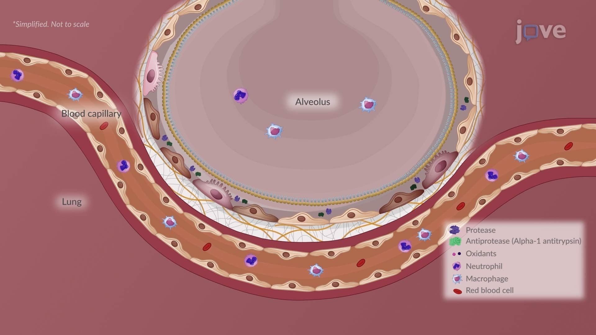
6. COPD, Emphysema: Alveolar Destruction and Air Trapping Emphysema involves permanent destruction of alveolar walls, producing abnormally enlarged distal airspaces. Toxic gases, especially cigarette smoke, generate oxidants that inactivate alpha-1 antitrypsin, allowing proteases such as neutrophil elastase to degrade the elastin scaffolding of alveolar septa. As walls break down, smaller airspaces merge into large, non-functional cavities called bullae within the parenchyma and blebs near the pleural surface. Loss of elastic recoil impairs exhalation, trapping air and causing hyperinflation. The overinflated lungs flatten the diaphragm, reducing its mechanical efficiency. Clinically, emphysema patients present with progressive dyspnea, a barrel-shaped chest, pursed-lip breathing, and minimal sputum production.
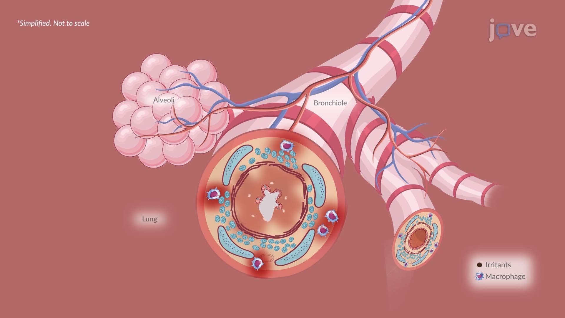
7. COPD, Chronic Bronchitis: Airway Remodeling and Mucus Hypersecretion Chronic bronchitis develops from prolonged exposure to airborne irritants that trigger persistent bronchial inflammation involving neutrophils, macrophages, and lymphocytes. Injured ciliated epithelium undergoes squamous metaplasia, severely impairing mucociliary clearance, the airway's natural defense mechanism. Simultaneously, goblet cells and submucosal glands undergo hyperplasia, generating excessive thick mucus that obstructs airways and creates a breeding ground for bacterial infection. Progressive airway remodeling, including smooth muscle hypertrophy and fibrosis, permanently narrows the bronchi, especially during expiration. V/Q mismatch and chronic hypoxemia can lead to cyanosis and secondary polycythemia (elevated red blood cell count), explaining the classic "blue bloater" clinical description in US medical education.
8. COPD: Combined Clinical Manifestations COPD encompasses both chronic bronchitis and emphysema, each contributing distinct clinical features. Chronic bronchitis produces a chronic productive cough, wheezing, exertional dyspnea progressing to resting dyspnea, and cyanosis from chronic hypoxemia. Emphysema primarily causes progressive breathlessness with minimal sputum, barrel-chest deformity, diaphragmatic flattening, tachypnea, and use of accessory muscles. Pursed-lip breathing, a common adaptive strategy in emphysema, maintains positive airway pressure during exhalation to reduce air trapping. Together, these findings help differentiate the "pink puffer" (emphysema-dominant) from the "blue bloater" (bronchitis-dominant), a classic distinction emphasized in US pathophysiology and respiratory therapy courses.
9. Pulmonary Edema: Cardiogenic vs. Noncardiogenic Mechanisms Pulmonary edema occurs when fluid accumulates in the alveoli, compromising gas exchange. In cardiogenic pulmonary edema, commonly associated with left heart failure, a leading cause of hospitalization in the US, left ventricular dysfunction raises pulmonary capillary hydrostatic pressure, forcing protein-poor fluid into the interstitium and alveoli. In noncardiogenic pulmonary edema, such as that caused by acute respiratory distress syndrome (ARDS) or toxic inhalation, increased capillary permeability allows protein-rich fluid to leak out, drawing additional water osmotically. In both types, alveolar flooding disrupts surfactant, raises surface tension, and reduces lung compliance. Untreated progression leads to respiratory failure marked by severe dyspnea, hypoxemia, and hypercapnia.
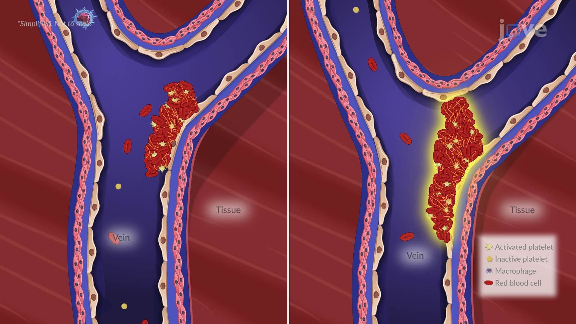
10. Pulmonary Embolism: Virchow's Triad and Deep Vein Thrombosis Pulmonary embolism (PE) occurs when a blood clot, most often originating from deep vein thrombosis (DVT) in the lower extremities, travels through the venous circulation and lodges in a pulmonary artery. The foundational framework for understanding clot formation is Virchow's triad: venous stasis, hypercoagulability, and endothelial injury. Venous stasis is seen with prolonged immobility, post-orthopedic surgery or extended travel, common clinical scenarios in US hospital settings. Hypercoagulability arises from conditions such as antithrombin III deficiency, pregnancy, or oral contraceptive use. Endothelial injury results from trauma or central venous catheter placement. PE is a high-yield topic on USMLE and NCLEX due to its acute severity and preventable risk factors.
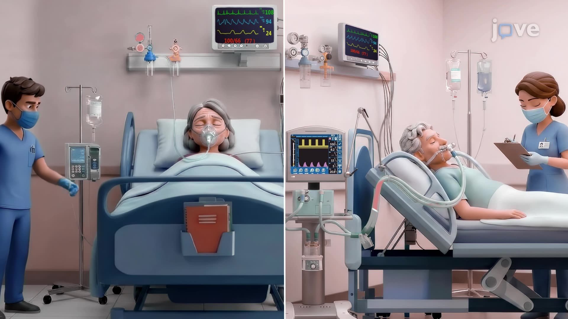
11. Pneumonia: Classification, Causative Agents, and Risk Factors Pneumonia is a lower respiratory tract infection that causes inflammation of the lung parenchyma and alveolar exudate accumulation, impairing gas exchange. Classification by acquisition site guides clinical management: community-acquired pneumonia (CAP) occurs outside healthcare settings; hospital-acquired pneumonia (HAP) develops at least 48 hours after admission; ventilator-associated pneumonia (VAP) affects mechanically ventilated patients. *Streptococcus pneumoniae* is the leading bacterial cause of CAP in the US. Viruses like influenza and RSV predominate in older adults, immunocompromised patients, and children. Multidrug-resistant organisms, including MRSA and *Pseudomonas aeruginosa*, are more common in HAP and VAP. Risk factors include advanced age, smoking, alcohol use, malnutrition, and dysphagia.