Loading video…

 11 Concepts
 7K views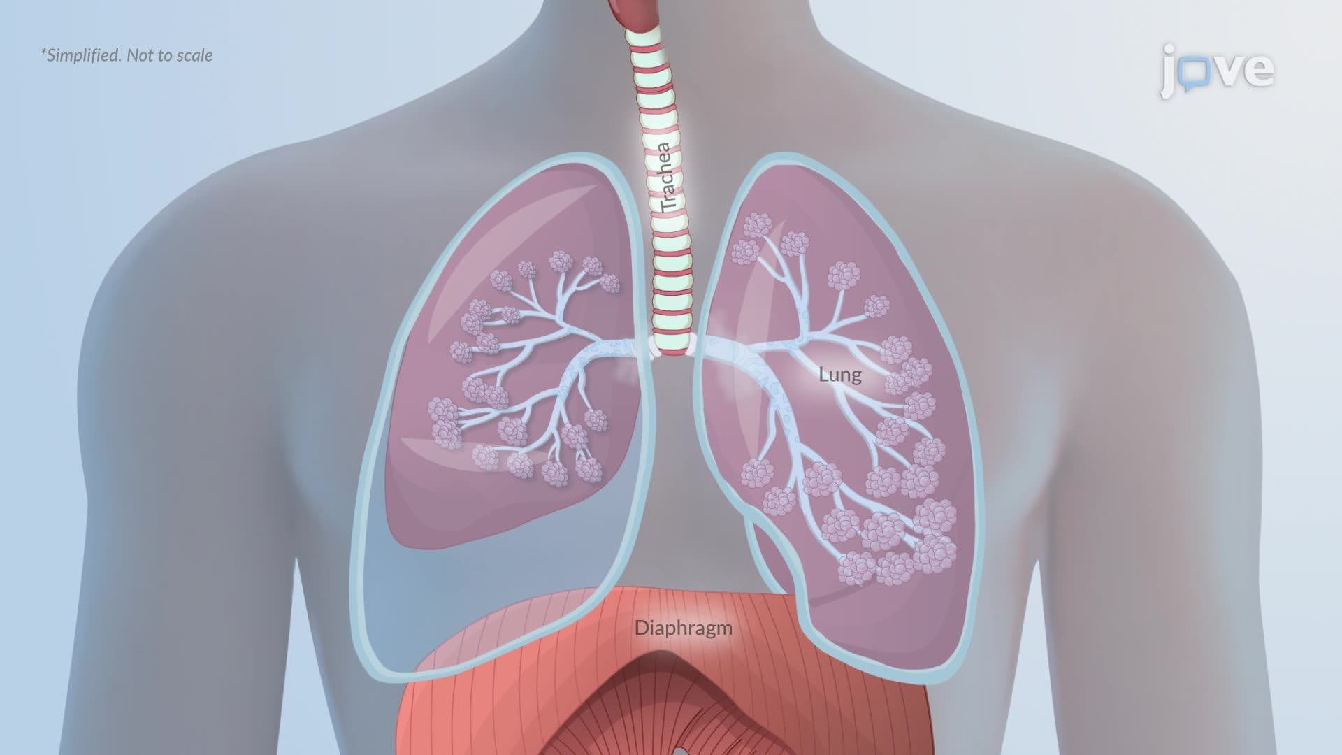

Video Summary: Pathophysiology in Atelectasis Ii
Did you know a collapsed lung can trick your body into thinking everything is fine, until oxygen levels drop dangerously low? Pathophysiology in Atelectasis II unpacks exactly how alveolar collapse disrupts the ventilation-perfusion balance, triggering hypoxemia and, in severe cases, hypercapnia. Think of a post-surgical ICU patient at Mayo Clinic struggling to breathe after anesthesia, that's atelectasis in action. Watch the full video on JoVE Coach to master this concept with expert-led visuals and step-by-step explanations.
Atelectasis is more than a "collapsed lung", it is a cascade of physiological failures that begins at the microscopic level and can escalate into life-threatening respiratory compromise. To truly understand pathophysiology in atelectasis II, you need to trace the problem from the alveolus outward: from structural collapse, to gas exchange failure, to whole-body consequences.
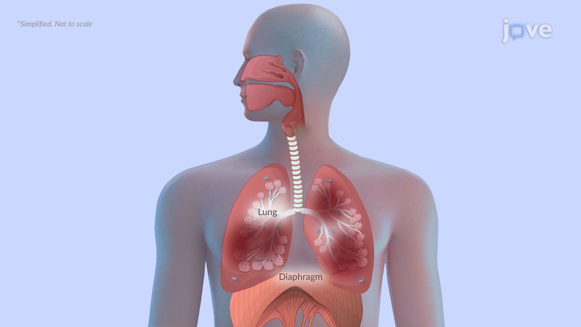
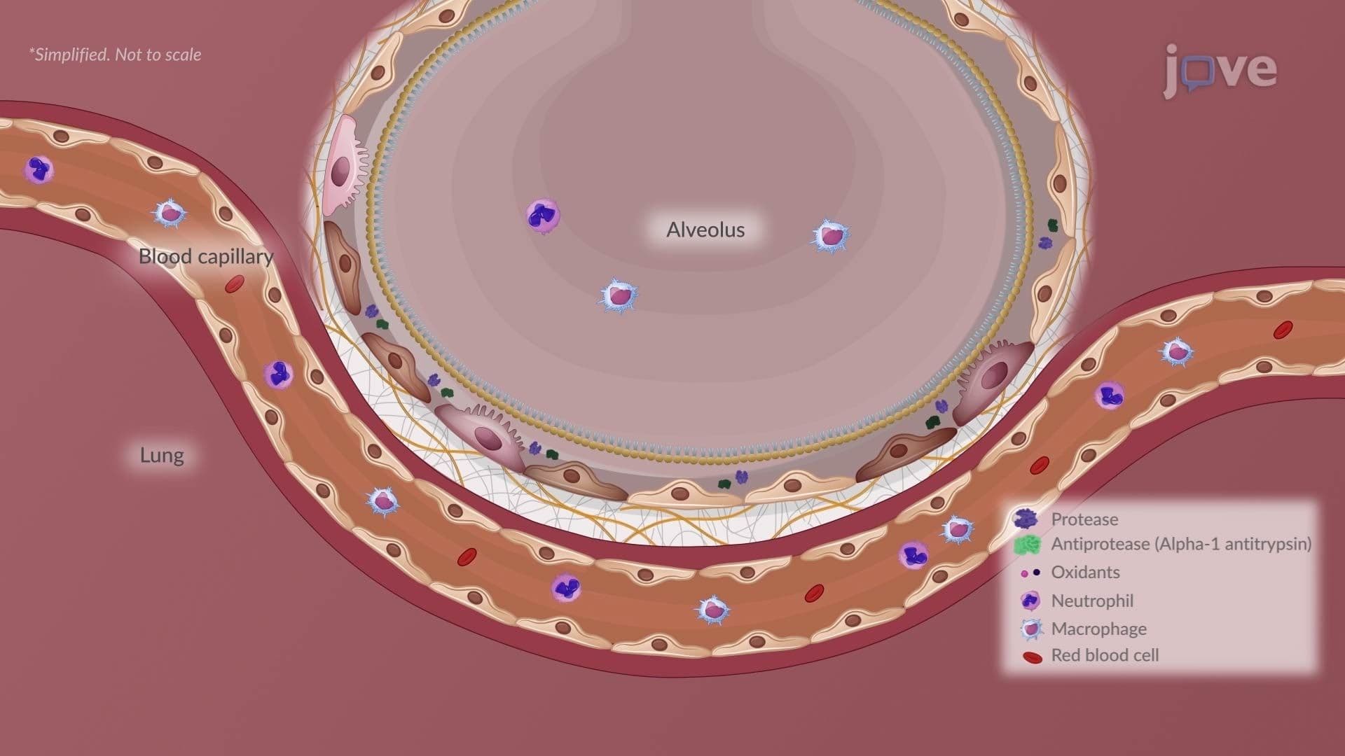
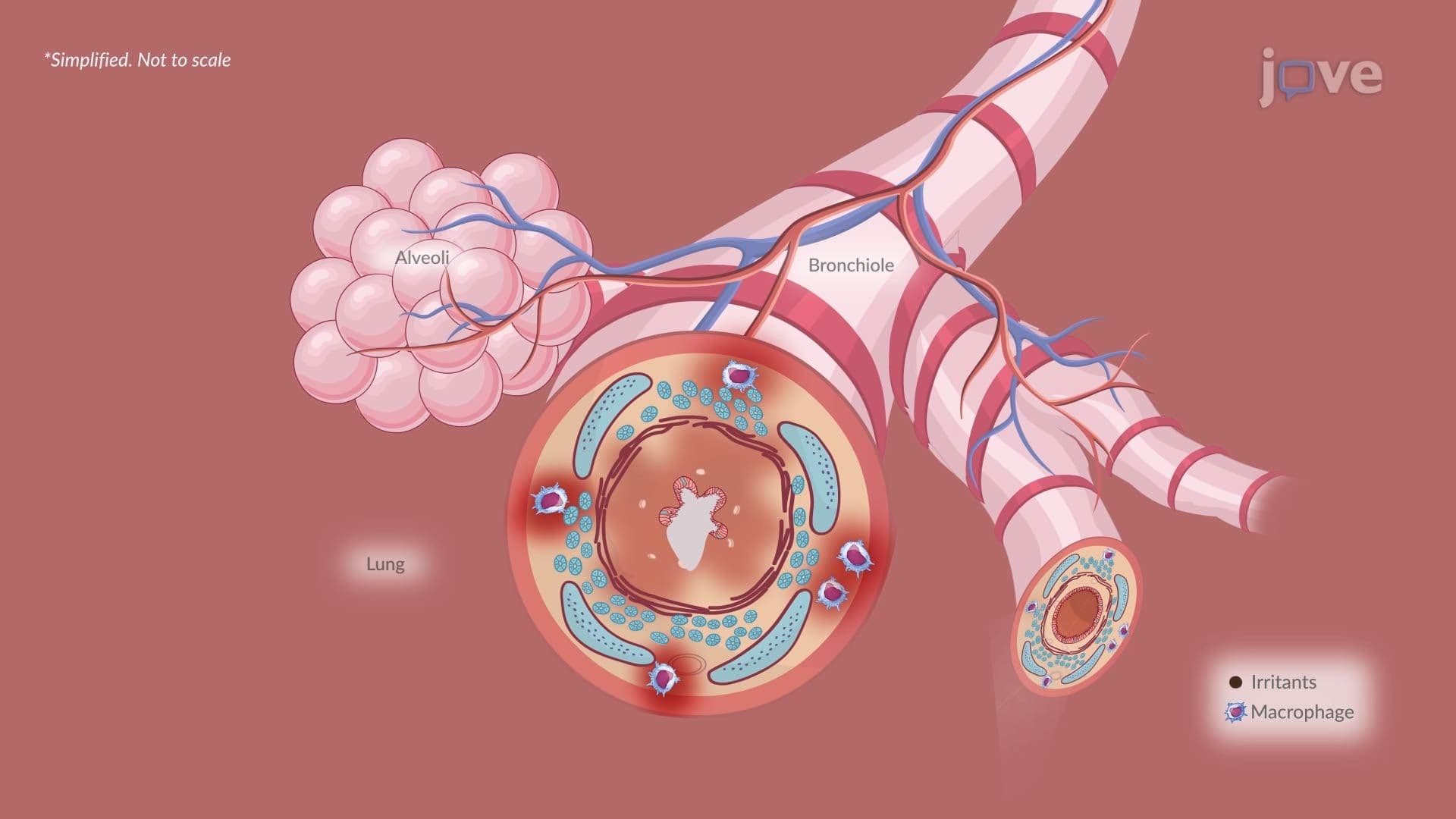
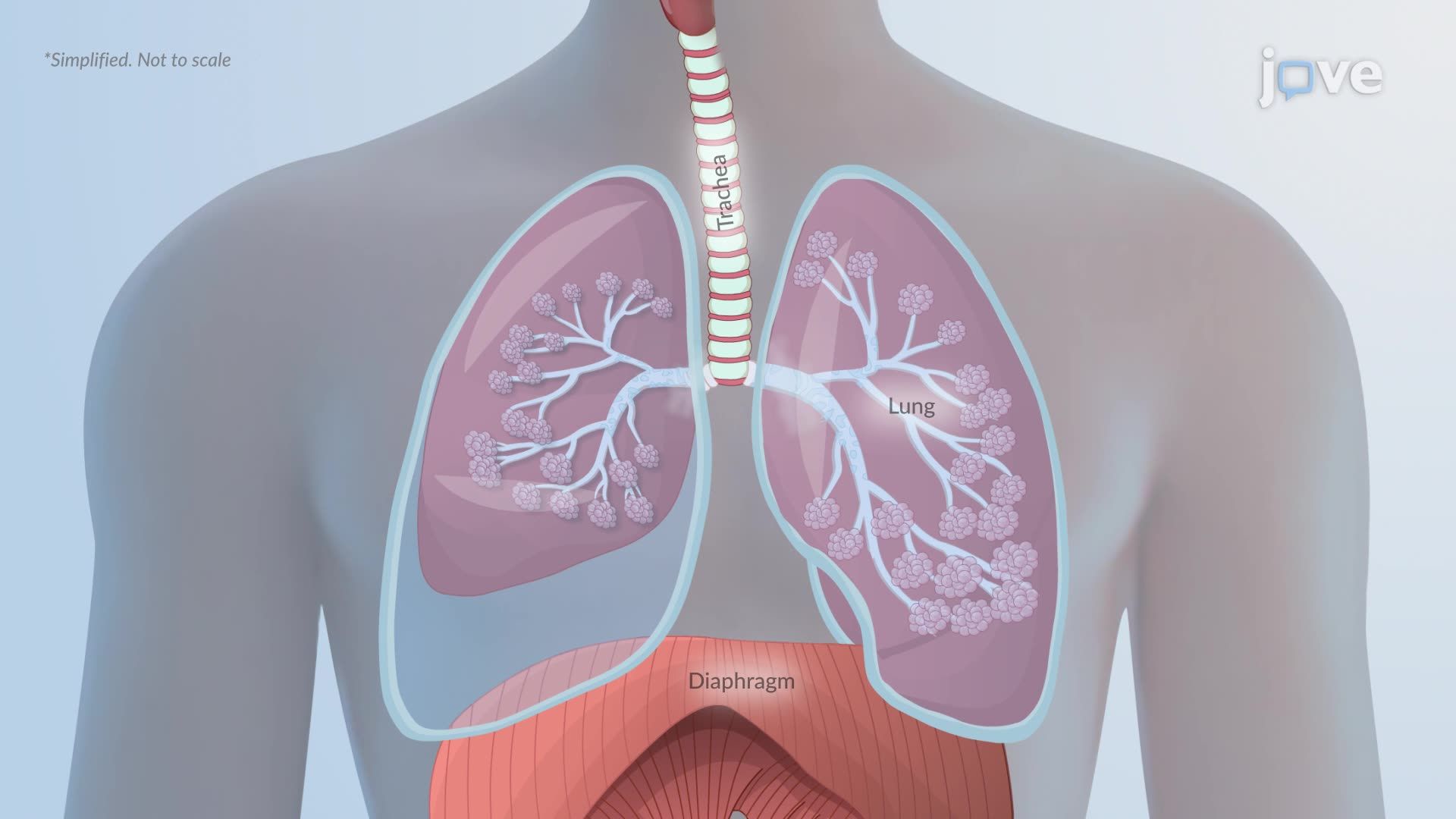
Lung tissue is inherently elastic. When alveoli, the tiny air sacs responsible for gas exchange, lose their air content, surface tension and elastic recoil cause them to shrink inward. This is the structural hallmark of atelectasis. The collapse can be caused by obstruction (a mucus plug blocking airflow, as seen in post-operative patients or those with asthma), compression (a tumor or pleural effusion pressing on lung tissue), or loss of surfactant (common in neonatal respiratory distress syndrome, or NRDS).
Once the alveoli collapse, ventilation, the movement of air in and out, drops dramatically or ceases entirely in that region. This is the first critical failure point, and it sets the stage for everything that follows.
Here is where pathophysiology in atelectasis becomes especially important for exams like the MCAT and USMLE Step 1. Even after ventilation stops in the collapsed region, blood flow (perfusion) often continues. The pulmonary capillaries surrounding those collapsed alveoli keep receiving deoxygenated blood, but there is no fresh air to exchange with. This creates what clinicians call a ventilation-perfusion (V/Q) mismatch, specifically a shunt, perfusion without ventilation.
Blood passes through the collapsed area without picking up oxygen, then mixes with oxygenated blood from healthy lung regions. The result is hypoxemia: abnormally low oxygen levels in the blood. This is why a patient with atelectasis may show a dropping pulse oximetry reading even before they report feeling short of breath. In US hospitals, post-surgical patients, particularly those recovering from abdominal or thoracic procedures, are routinely monitored for exactly this pattern.
Carbon dioxide (CO₂) is a much more diffusible gas than oxygen. In the early stages of atelectasis, healthy alveoli can compensate by ramping up ventilation, a process called compensatory hyperventilation, to offload the extra CO₂ load. This is why CO₂ levels (measured as PaCO₂) may remain surprisingly normal in initial blood gas readings, even when oxygen is already falling.
However, if atelectasis becomes extensive or prolonged, this compensation collapses. The remaining healthy alveoli cannot work hard enough to clear CO₂ for the entire lung, and hypercapnia (elevated CO₂) develops. Hypercapnia shifts blood pH downward, leading to respiratory acidosis, a serious complication that requires prompt clinical intervention. This progression is highly relevant to understanding conditions like COPD, where chronic hypercapnia is a hallmark finding, and helps answer the common exam question: *What causes respiratory system disorders to progress from manageable to critical?*
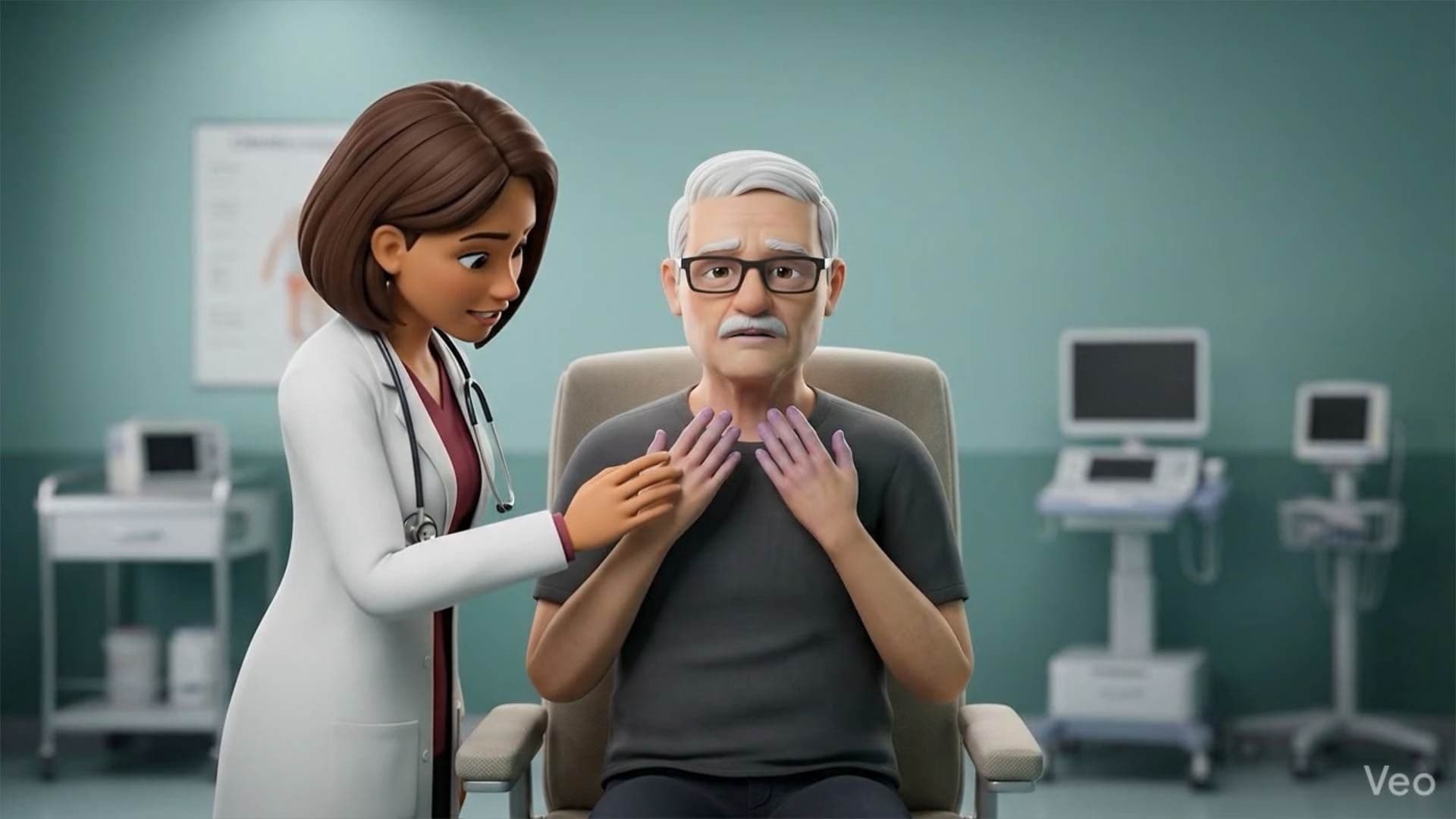
The downstream effects of hypoxemia and hypercapnia explain the classic clinical signs: shortness of breath, tachypnea (rapid breathing), coughing, chest pain, and cyanosis (a bluish discoloration of the skin or lips due to low oxygen saturation). These signs overlap significantly with pneumonia, pulmonary embolism, and restrictive lung diseases, which is precisely why understanding the specific mechanism of atelectasis helps clinicians and students differentiate between conditions.
For AP Biology and college-level anatomy and physiology courses, atelectasis serves as an ideal case study connecting cellular respiration, gas laws (Dalton's Law, Boyle's Law), and systemic homeostasis. Understanding how one structural failure, a collapsed alveolus, can cascade into whole-body oxygen deprivation gives students a model for analyzing *any* respiratory pathology, from asthma management to the risk factors for COPD to the gas exchange disruptions seen in lung cancer.
Related Micro-courses