Loading video…

 11 Concepts
 7K views

Video Summary: Pathophysiology in Pneumothorax Ii
Did you know a single rupture in your lung lining can collapse an entire lung within minutes? Pathophysiology in Pneumothorax II breaks down exactly how this happens, from lost negative pleural pressure to full cardiovascular collapse in tension pneumothorax. Emergency physicians at trauma centers like Johns Hopkins see this life-threatening condition regularly. Watch the full video on JoVE Coach to master this concept with expert-led visuals and step-by-step explanations.
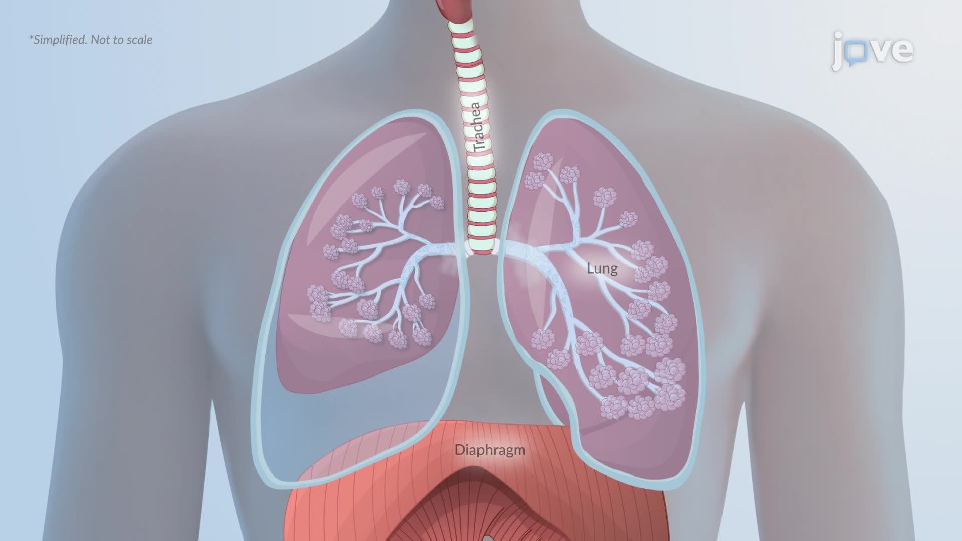
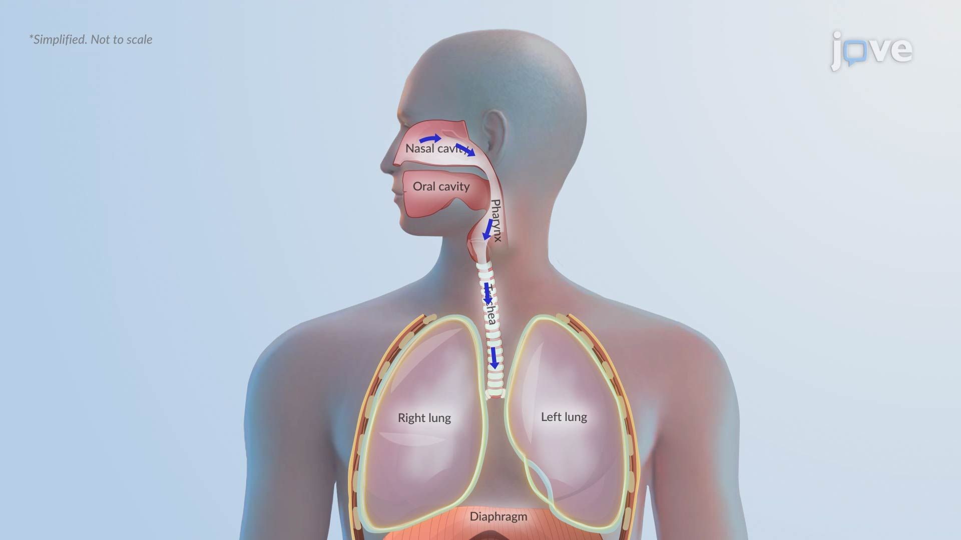
The human body is remarkably precise. The pleural space, the thin cavity between the lung and chest wall, normally maintains a slightly negative pressure, roughly −3 to −5 cmH₂O at rest. This gentle suction keeps the lungs inflated against the chest wall, making breathing possible. In Pathophysiology in Pneumothorax II, the central focus is what happens when that pressure balance collapses, and why the consequences can escalate rapidly from a breathing problem to a cardiovascular emergency.
The lungs naturally want to recoil inward due to their elastic tissue. The chest wall, by contrast, tends to spring outward. The negative pressure in the pleural space acts like a handshake between these two opposing forces, keeping the lungs open. When this seal is broken, whether by a ruptured bleb (a small air sac on the lung surface, common in tall, thin young adults), a penetrating chest wound, or a complication from COPD or asthma, outside air rushes into the pleural space. The lung recoils inward and collapses partially or completely.
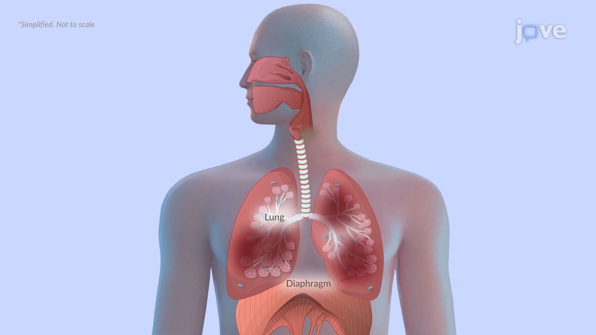
When alveoli collapse, they can no longer participate in gas exchange. Oxygen cannot enter the bloodstream effectively, and carbon dioxide cannot be expelled. This ventilation-perfusion (V/Q) mismatch is the root cause of the shortness of breath and chest pain patients experience. In clinical settings across the US, this presentation often prompts emergency chest X-rays and pulse oximetry monitoring. Students preparing for the MCAT or AP Biology exam should note that V/Q mismatch is a tested mechanism underlying hypoxemia in multiple respiratory disorders, including pneumonia and pulmonary embolism.
A simple pneumothorax is serious, but tension pneumothorax is a true emergency. If air enters the pleural space through a one-way "check valve" mechanism and cannot escape, pressure builds with every breath. The affected lung compresses further, and the accumulated air begins pushing the mediastinum, the central compartment containing the heart and great vessels, toward the opposite side. This is called a mediastinal shift. In US emergency medicine, this shift is a hallmark sign visible on imaging and a red flag for immediate needle decompression.
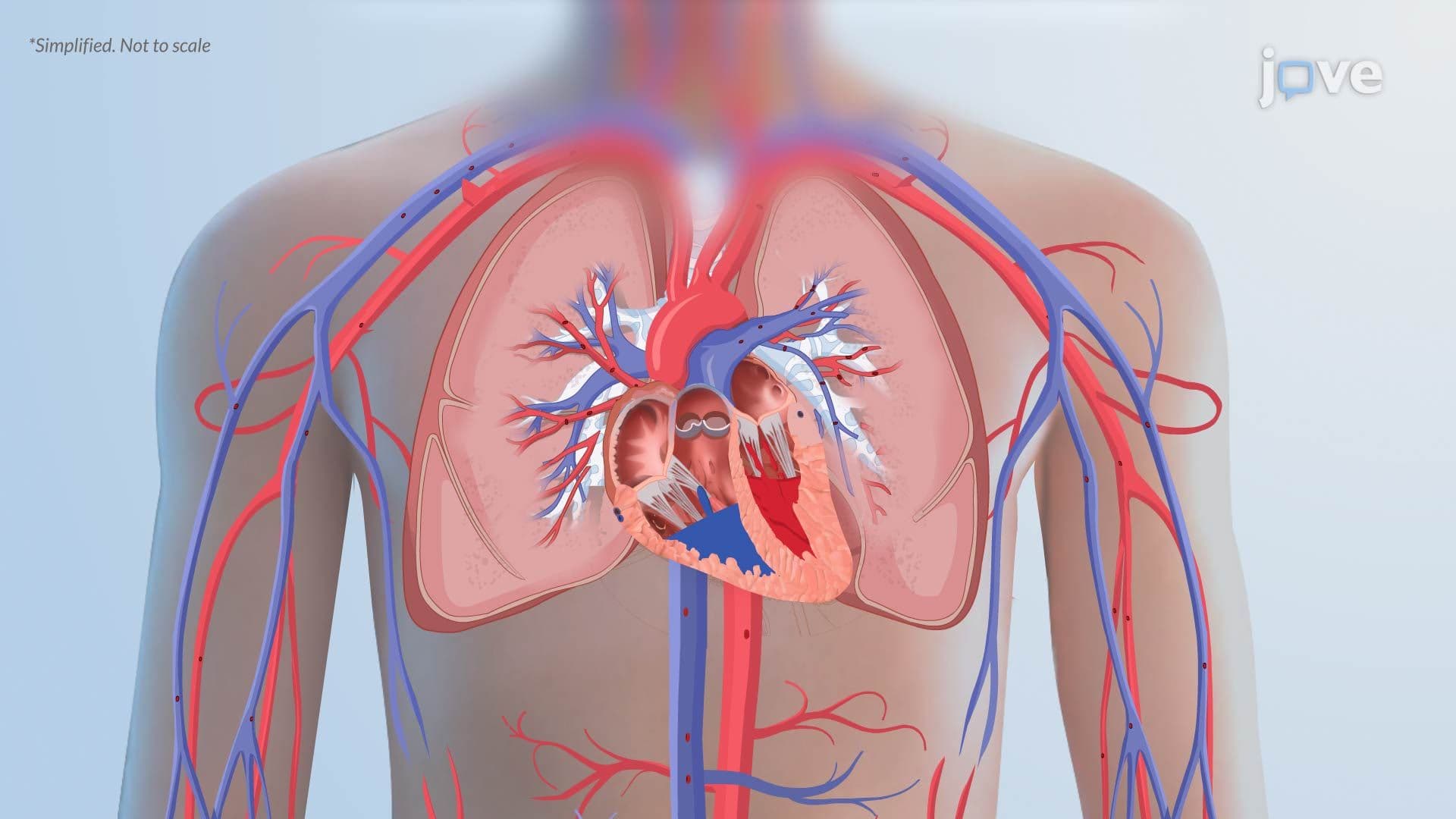
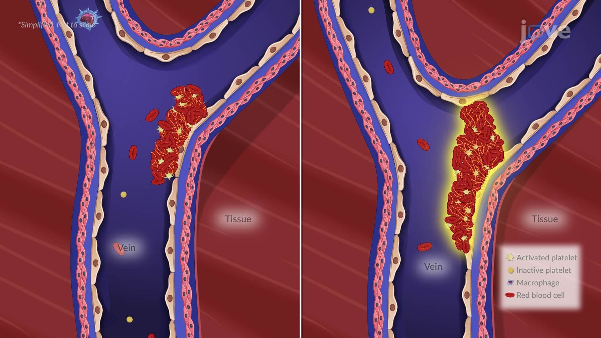
As intrathoracic pressure rises, it compresses the superior and inferior vena cava, the major veins returning blood to the right side of the heart. Venous return drops sharply, cardiac output falls, and the patient develops hypotension and signs of obstructive shock. This chain reaction, from air leak to cardiovascular collapse, is exactly why tension pneumothorax carries a high mortality rate if untreated.
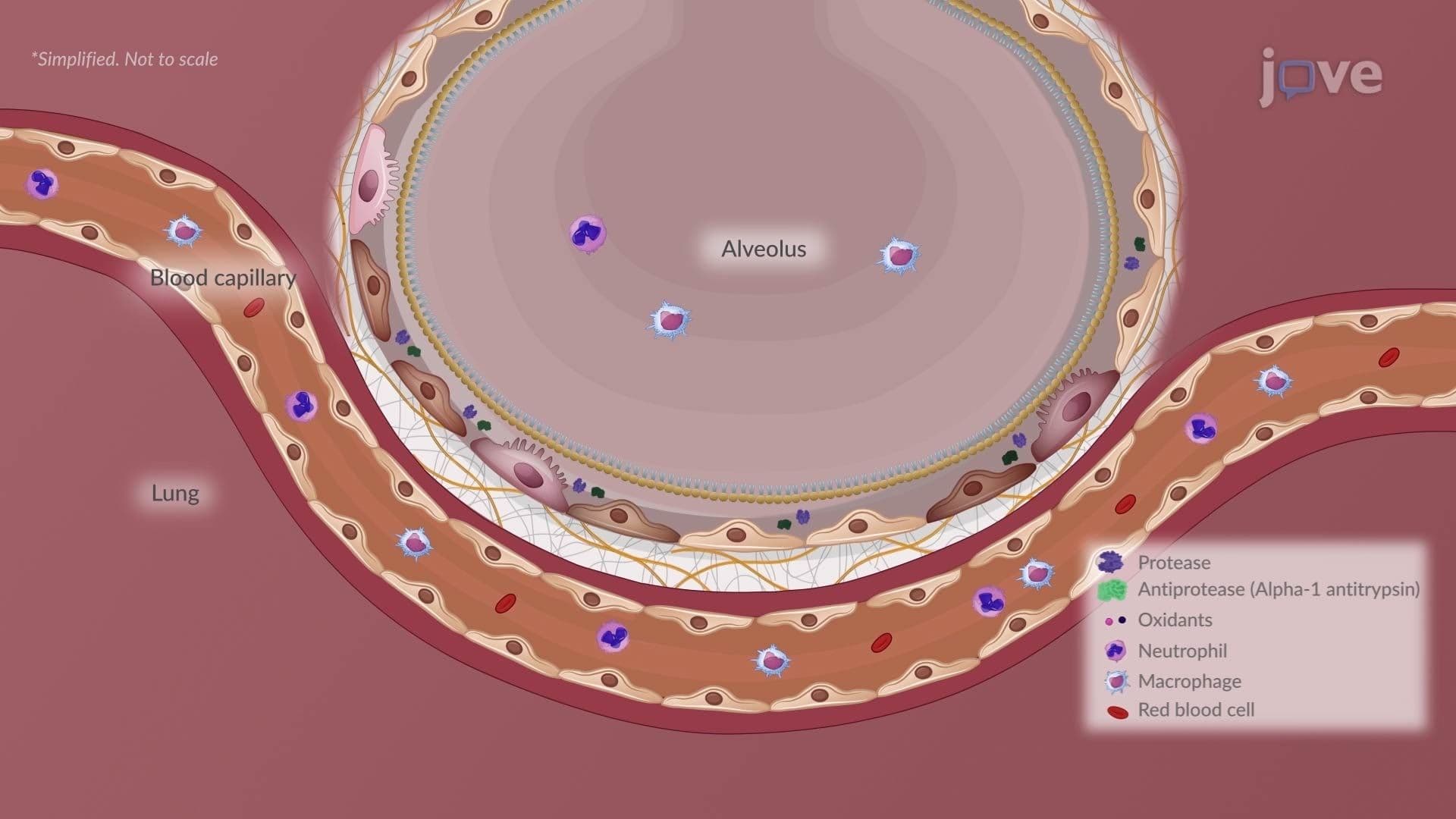
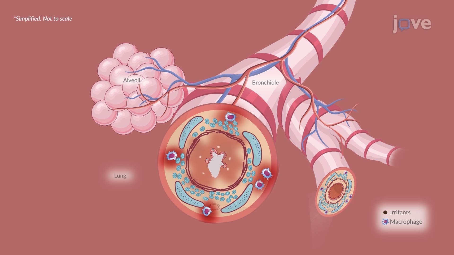
Understanding pneumothorax fits within a larger picture of respiratory pathology. Patients with COPD develop emphysematous blebs that can rupture spontaneously. Those with restrictive lung diseases have reduced lung compliance, altering pressure dynamics. Lung cancer tumors can erode airways or pleura, triggering secondary pneumothorax. For college-level anatomy and physiology courses, as well as NCLEX and USMLE Step 1 prep, linking pneumothorax to these underlying conditions strengthens diagnostic reasoning and earns points on clinical vignette questions.
Related Micro-courses