Loading concepts…

 18 Concepts
 4.3K Views
 Popular topics



Pathophysiology basics form the foundation of every health science curriculum in the United States, explaining how diseases disrupt normal body functions and produce recognizable clinical signs. This micro-course, supported by JoVE Coach, covers disease language, cellular adaptation, injury and death, inflammation, and tissue healing. Students gain a mechanistic understanding of conditions ranging from myocardial infarction to chronic inflammation, building essential knowledge for nursing, pre-med, and allied health programs.
1. The Language of Pathophysiology Understanding disease begins with its vocabulary. *Etiology* identifies the cause, for example, bacterial infection as the cause of pneumonia. *Iatrogenic* conditions arise from medical treatment itself, such as a catheter-associated urinary tract infection. Diseases present on a spectrum: *acute* conditions like appendicitis arrive rapidly and severely, while *chronic* conditions like rheumatoid arthritis develop gradually over years. *Signs* are measurable findings a clinician observes, such as fever, whereas *symptoms* are subjective experiences reported by the patient, such as chest pain. A *syndrome* groups related signs and symptoms together, nephrotic syndrome, for instance, bundles proteinuria, edema, and hyperlipidemia into one recognizable kidney disorder.
2. Describing Disease Progression Pathophysiology also captures how diseases evolve over time. A *predisposing factor* raises disease risk, decades of cigarette smoking increases the likelihood of atherosclerosis. A *precipitating factor* is the immediate trigger, an allergen exposure that sets off an acute asthma attack. *Exacerbation* describes symptom flare-ups, while *remission* marks their disappearance. *Complications* are new problems that arise during a disease course, such as heart failure following a myocardial infarction. *Sequelae* are lasting effects after the acute phase resolves, like hemiplegia after a stroke. *Prognosis* describes the expected disease outcome, early-stage breast cancer, for example, generally carries a favorable prognosis in current US clinical practice.
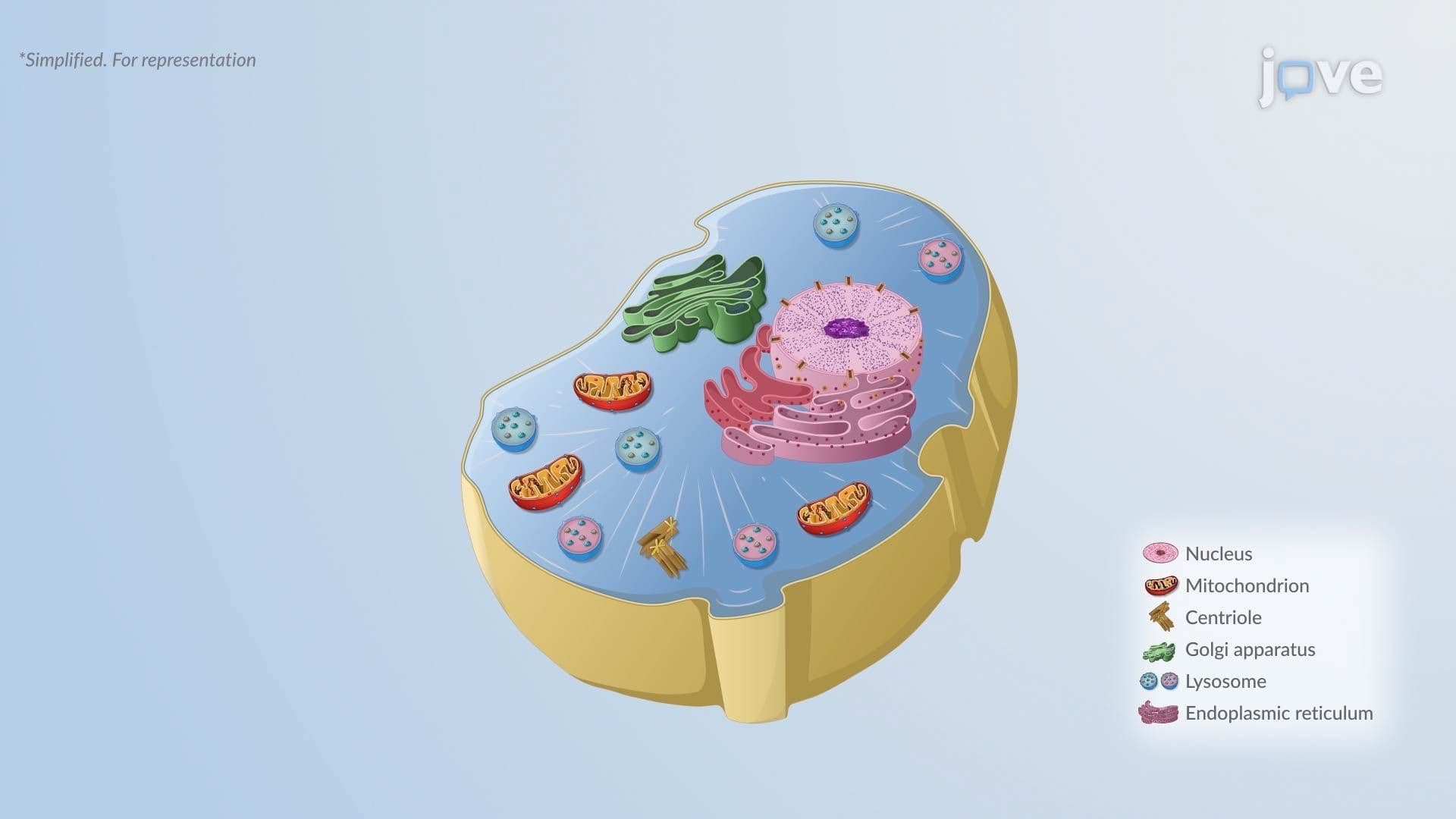
3. Cellular Adaptation: Atrophy and Hypertrophy Cells continuously adapt to changing demands. *Atrophy*, a decrease in cell size, occurs physiologically as the thymus shrinks after childhood, and pathologically when prolonged cerebral ischemia associated with atherosclerosis causes brain atrophy. Atrophic cells contain fewer mitochondria and less rough endoplasmic reticulum, reducing metabolic output. *Hypertrophy*, an increase in cell size, occurs in skeletal muscle with consistent resistance training (physiologic) through IGF-1/Akt/mTOR signaling, increasing contractile protein production. Pathologic hypertrophy is illustrated by the heart enlarging in response to chronic hypertension; while initially compensatory, prolonged cardiac hypertrophy ultimately impairs function and can progress to heart failure.
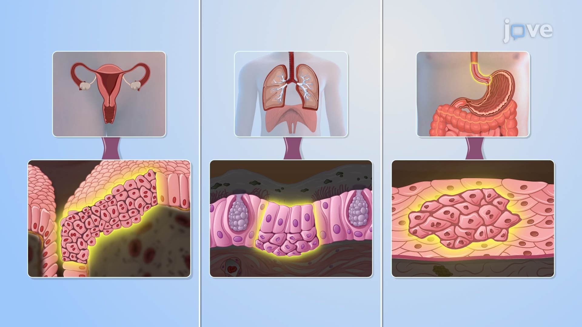
4. Cellular Adaptation: Hyperplasia, Dysplasia, and Metaplasia *Hyperplasia* increases cell number, driven by growth factors or stem cell activation. Physiologic examples include endometrial proliferation during the menstrual cycle; a pathologic example is thyroid hyperplasia from chronic TSH elevation in iodine-deficient individuals. *Dysplasia* is not a true adaptation, it represents disorganized, abnormal cell growth, most often in epithelial tissue. When dysplasia penetrates the basement membrane, it becomes invasive carcinoma. *Metaplasia* replaces one mature cell type with another better suited to a hostile environment. In long-term smokers, normal ciliated columnar epithelium in the airways is replaced by stratified squamous epithelium, cells that cannot produce mucus or move debris, dangerously impairing airway protection.
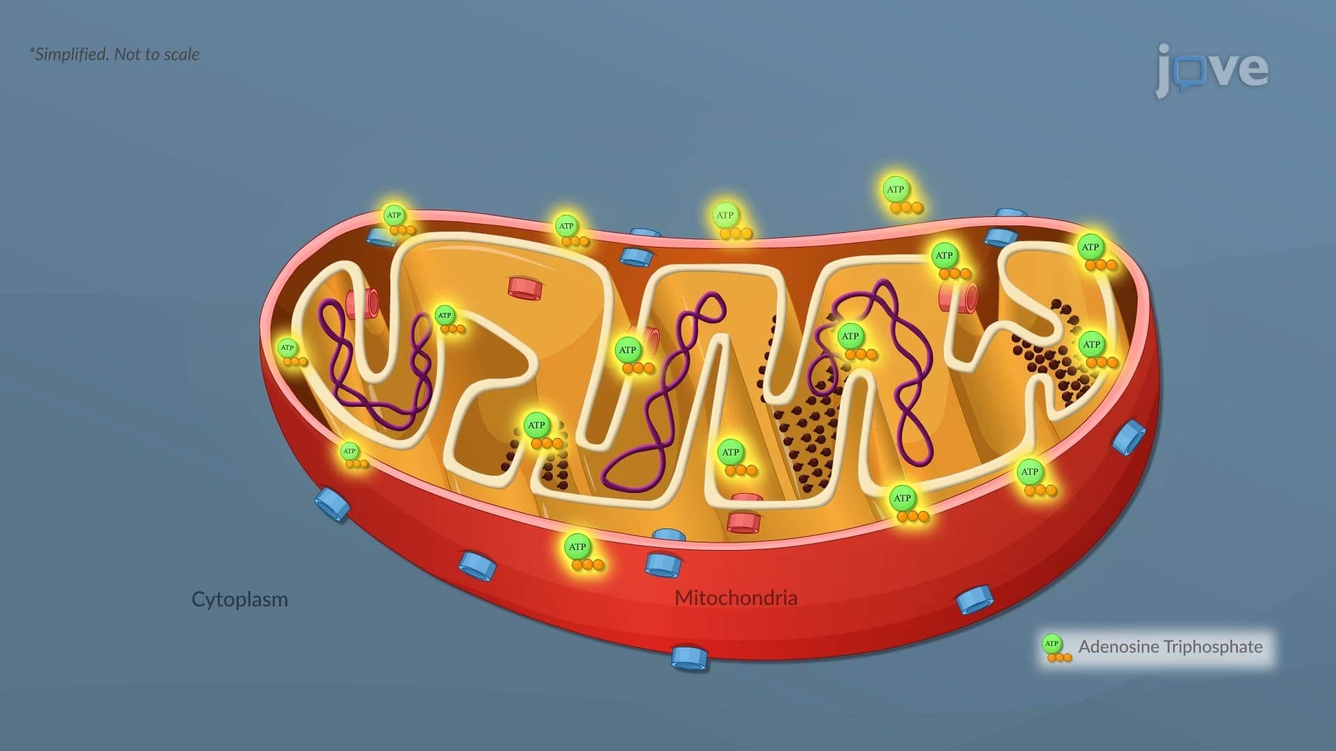
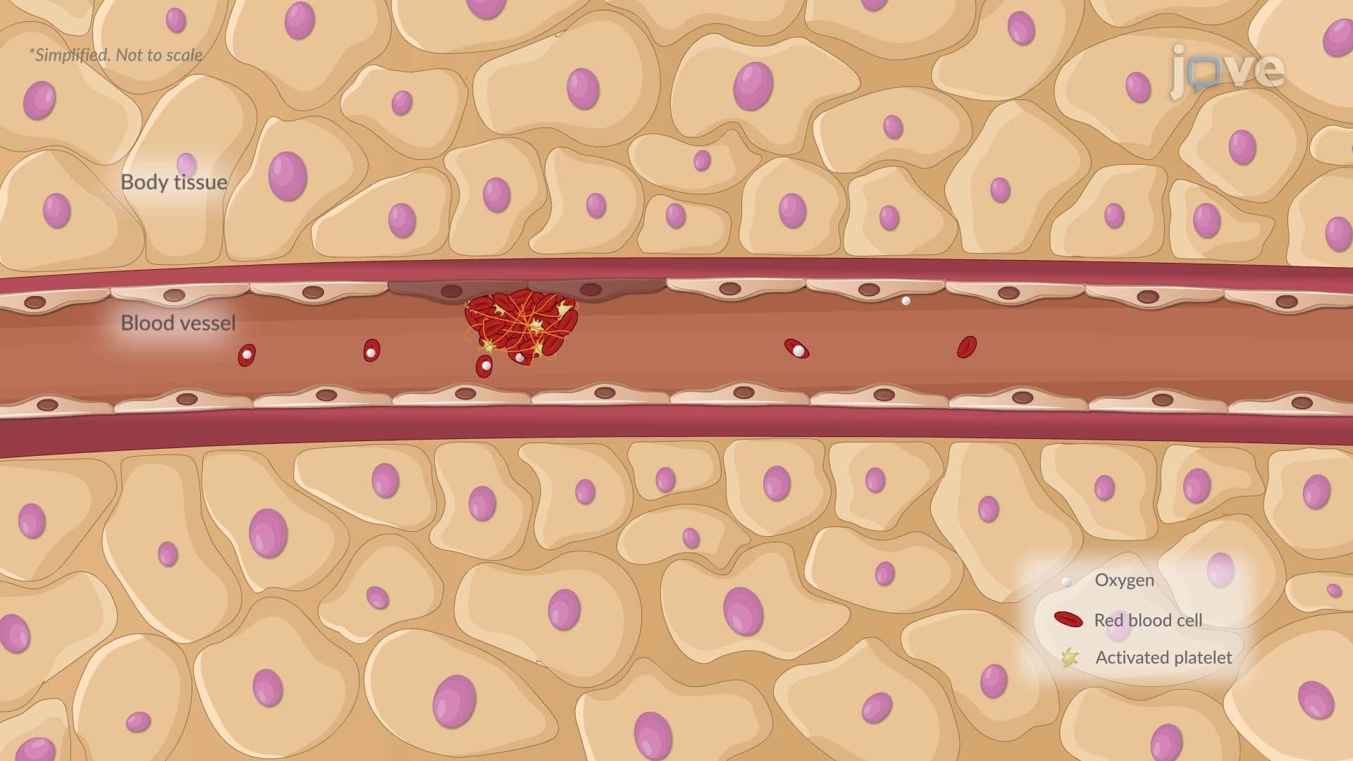
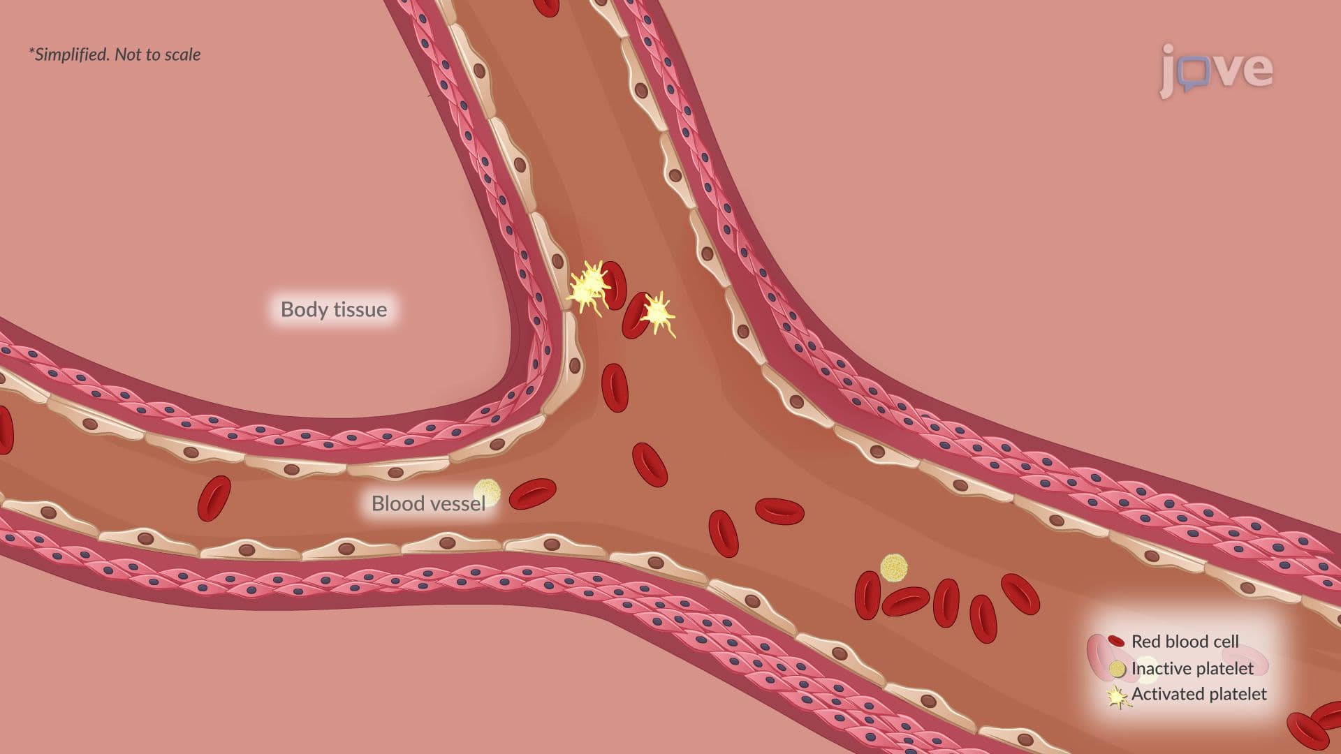
5. Mechanisms of Cellular Injury Cellular injury occurs when a cell cannot maintain homeostasis under stress. The most common cause is *hypoxic injury*, where reduced oxygen delivery, as in coronary artery thrombosis leading to myocardial infarction, depletes ATP. Without ATP, the sodium-potassium ATPase pump fails, sodium floods the cell, and water follows, causing swelling. *Chemical injury* may be direct (mercury or strong acids) or indirect through toxic metabolites, as in acetaminophen overdose damaging hepatocytes. *Physical injury* includes trauma, radiation, and extreme temperatures. *Infectious injury* involves direct cell damage by pathogens or collateral damage from the immune response. *Immunologic injury* results from abnormal immune activation, as in lupus erythematosus.
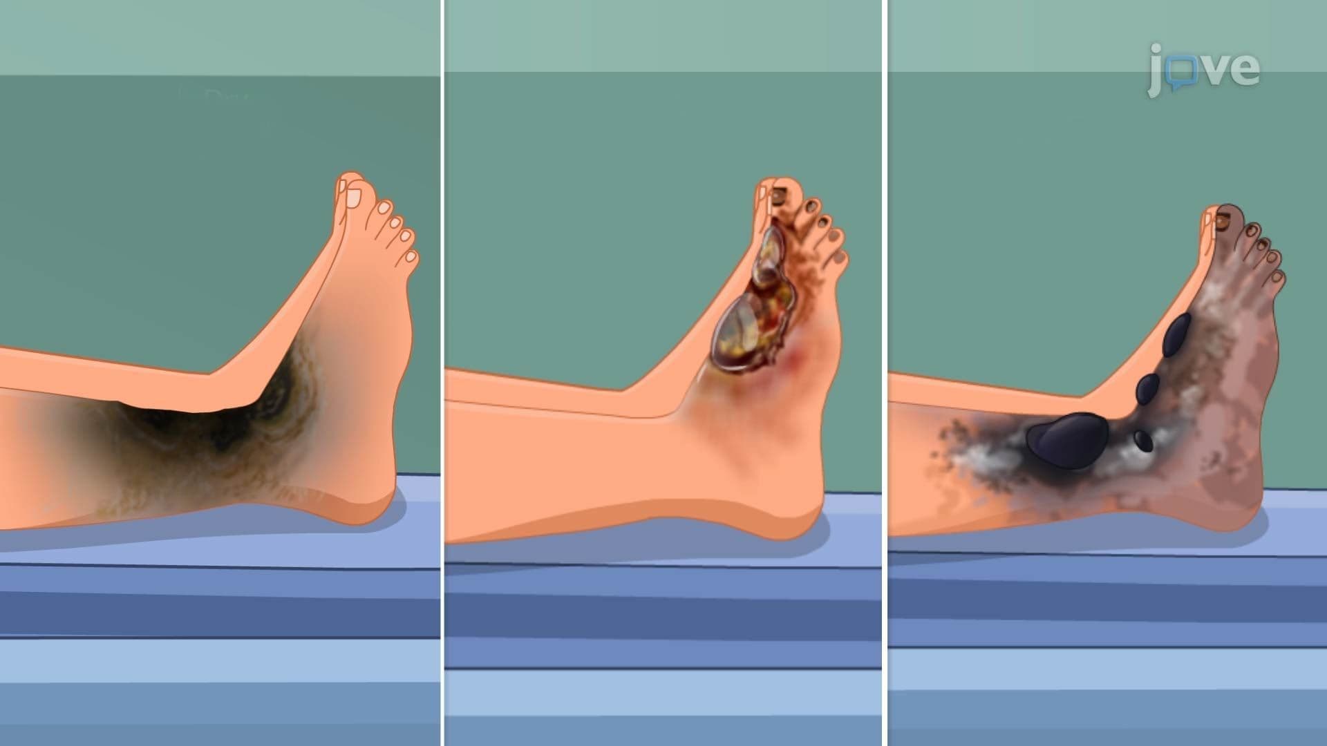
6. Cellular Death: Necrosis and Apoptosis When injury is irreversible, cells die through one of two pathways. *Necrosis* is uncontrolled cell death triggered by trauma or ischemia. Lysosomal enzymes leak out, digesting the cell from within, and nuclear changes follow in sequence: pyknosis (chromatin condensation and nuclear shrinkage), karyorrhexis (nuclear fragmentation), and karyolysis (complete nuclear dissolution). Cell rupture spills contents into surrounding tissue, triggering inflammation. Necrosis types include coagulative (firm, pale tissue, common in infarcts), liquefactive (fluid-filled softening, typical in brain infarcts), and gangrenous necrosis (affecting large tissue areas). *Apoptosis*, by contrast, is a programmed, orderly process: cells shrink, chromatin condenses, and membrane-bound apoptotic bodies form, cleared quietly by macrophages without inflammation.
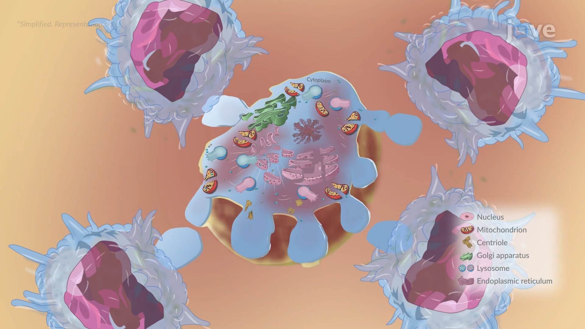
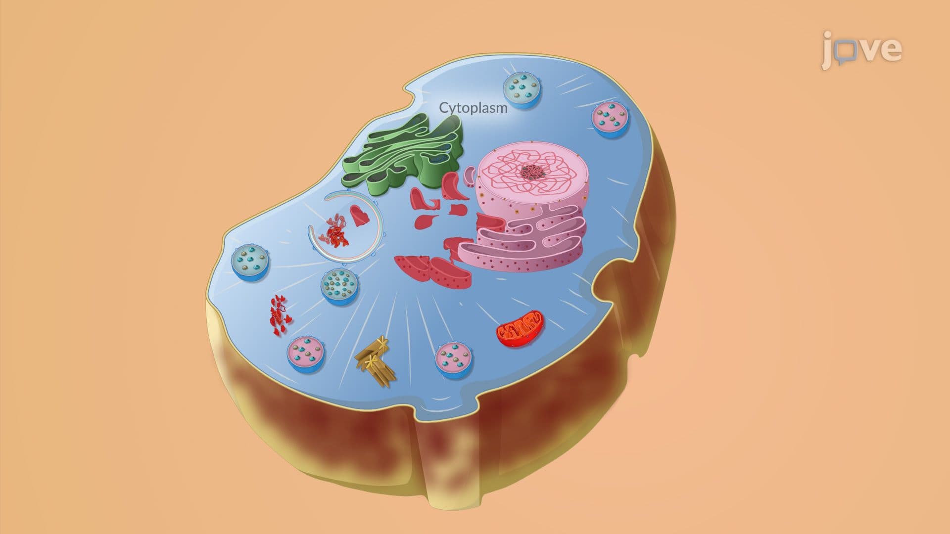
7. Autophagy *Autophagy* is the cell's internal recycling program, operating at baseline to maintain quality control and upregulated during nutrient deprivation or cellular stress. The process begins with a *phagophore*, a membrane structure that engulfs damaged organelles or misfolded proteins and develops into an *autophagosome*. This vesicle then fuses with a lysosome, where contents are enzymatically degraded and recycled for energy. Unlike necrosis, autophagy is a survival mechanism, not a death pathway. It helps cells endure starvation, clears dysfunctional mitochondria, and supports metabolic continuity, making it physiologically distinct from both necrosis and apoptosis.
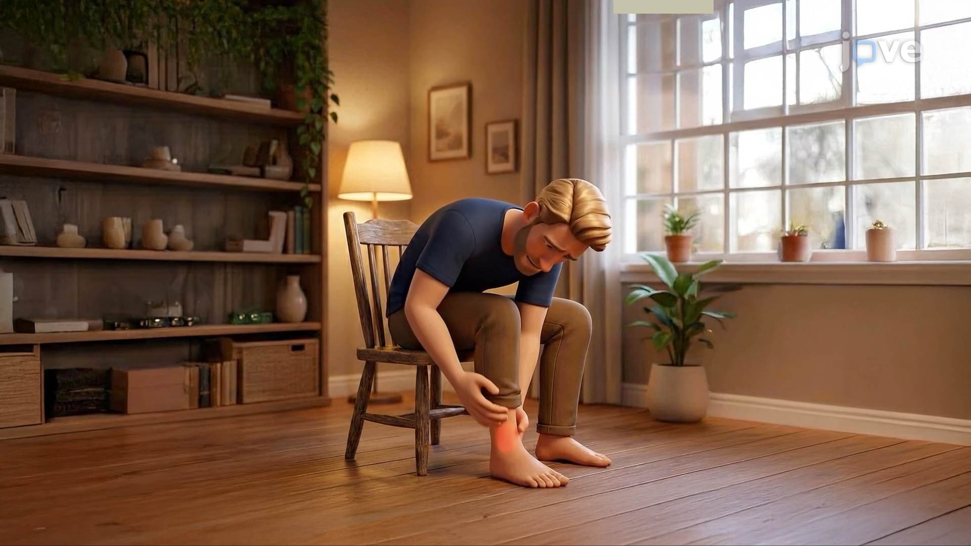
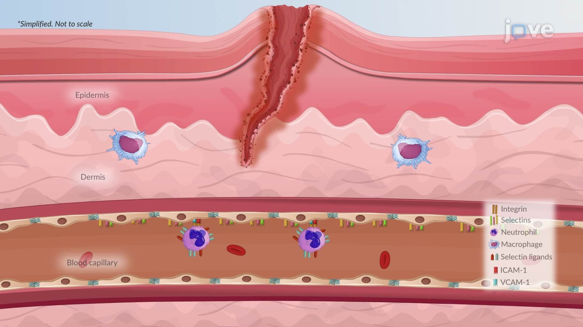
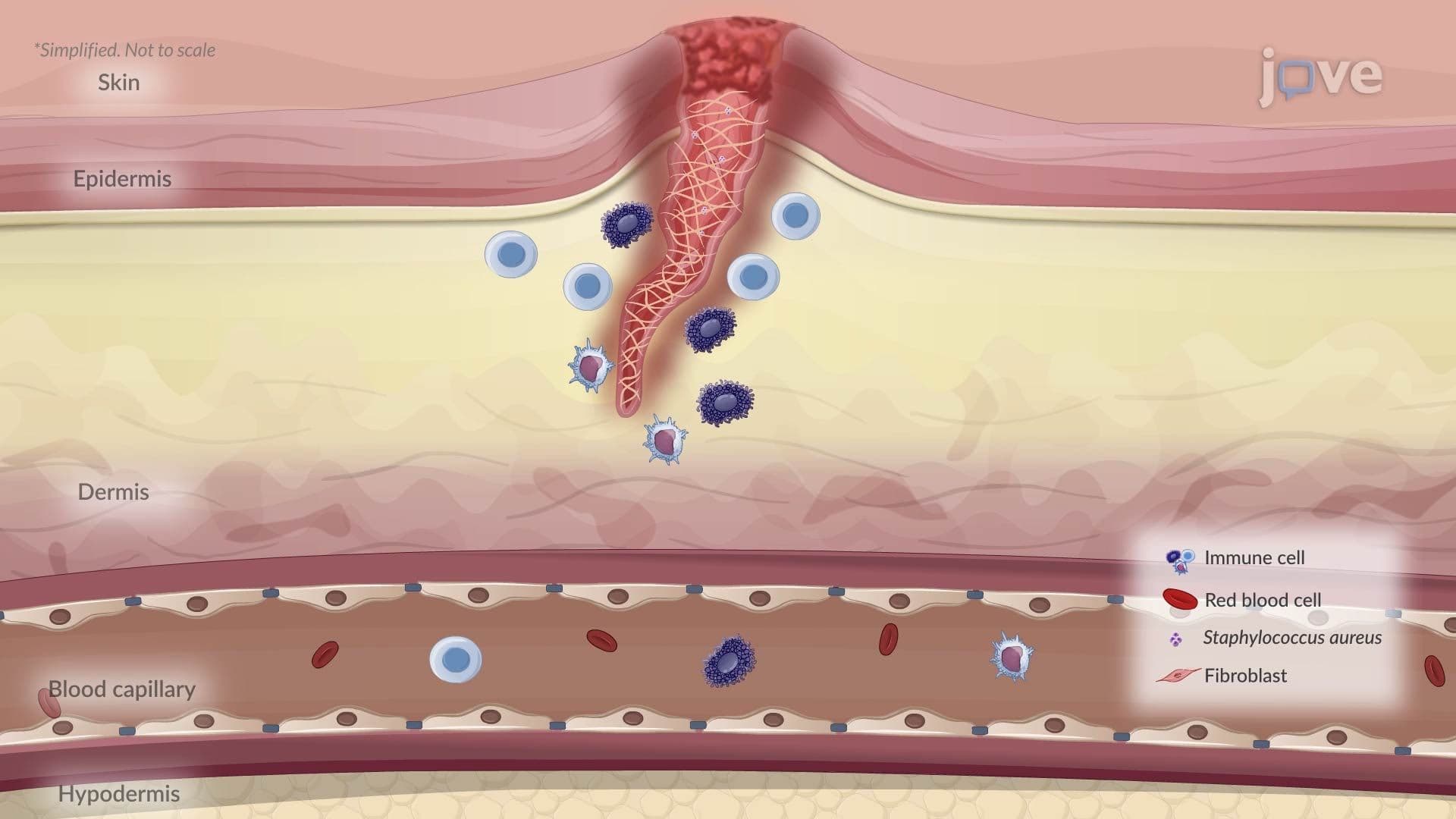
8. Inflammation: Acute Response Inflammation is the body's first-line defensive response to injury, infection, or harmful stimuli. *Acute inflammation* is rapid, lasting minutes to days. Resident immune cells, mast cells, macrophages, and dendritic cells, detect pathogen- and damage-associated molecular patterns via pattern-recognition receptors. Activated cells release *histamine* and *prostaglandins*, causing vasodilation and increased blood flow, producing the classic signs: redness, heat, swelling, and pain. The cellular phase follows: leukocytes undergo margination, rolling on endothelial *selectins*, firm adhesion via integrins binding ICAM-1 and VCAM-1, then *diapedesis* into tissue. Inside phagolysosomes, pathogens are destroyed by reactive oxygen species and proteolytic enzymes. Systemic effects, fever, leukocytosis, tachycardia, emerge in severe infection or sepsis.
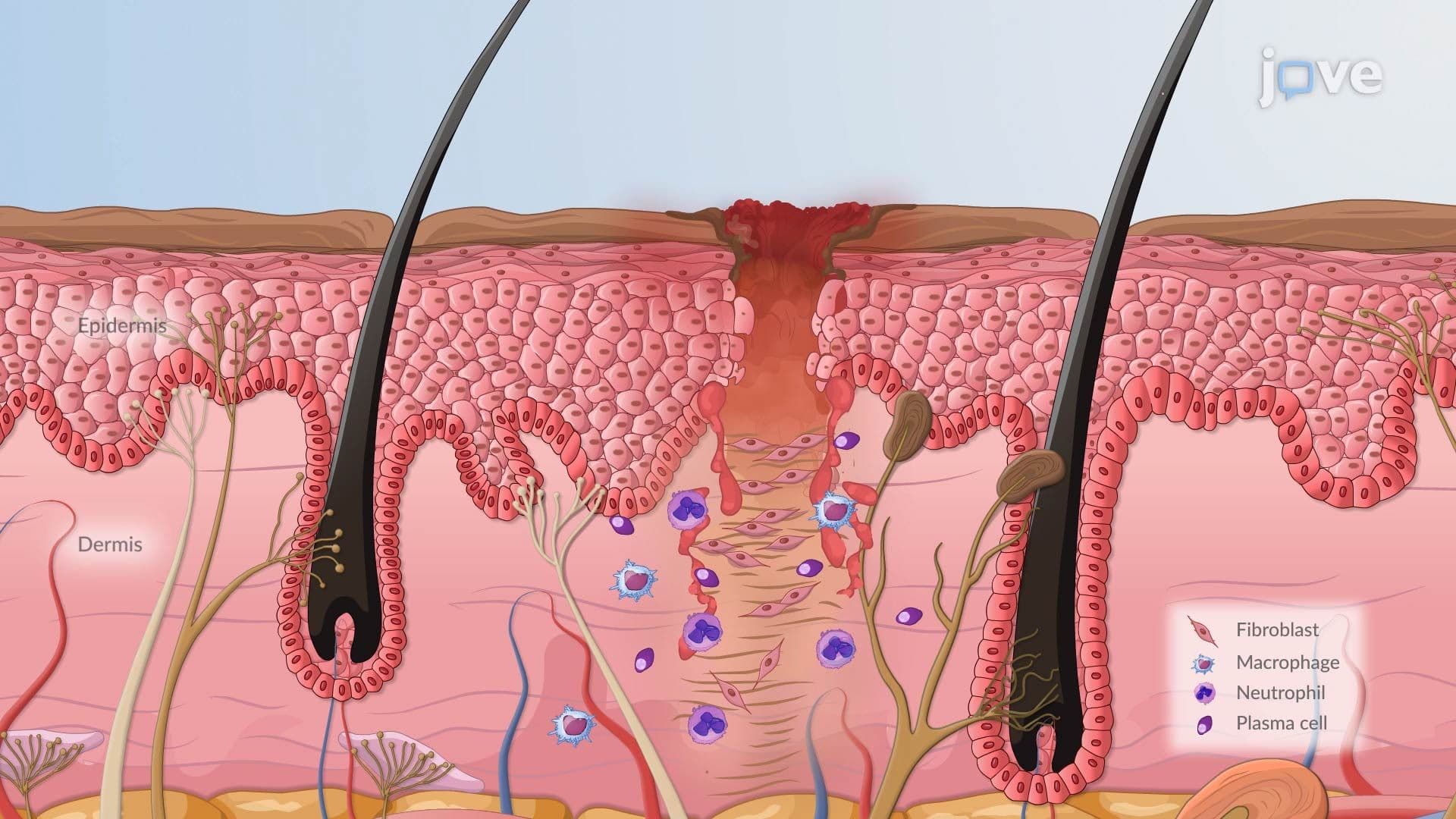
9. Chronic Inflammation and Healing When acute inflammation is unresolved, *chronic inflammation* develops, a prolonged response involving lymphocytes, macrophages, and fibroblasts that can last months to years. Progressive tissue destruction occurs as enzymes and reactive molecules damage surrounding healthy tissue. Fibroblasts respond by depositing collagen, causing *fibrosis* (scarring). In some diseases, such as tuberculosis, the immune system forms a *granuloma*, a compact cluster of activated macrophages surrounded by lymphocytes, designed to wall off persistent pathogens. *Healing* then proceeds through resolution (full recovery with no scar, as in mild sunburn), regeneration (functional replacement, as in liver hepatocyte regrowth), or replacement by fibrous scar tissue (as in post-myocardial infarction remodeling), each with distinct clinical implications.
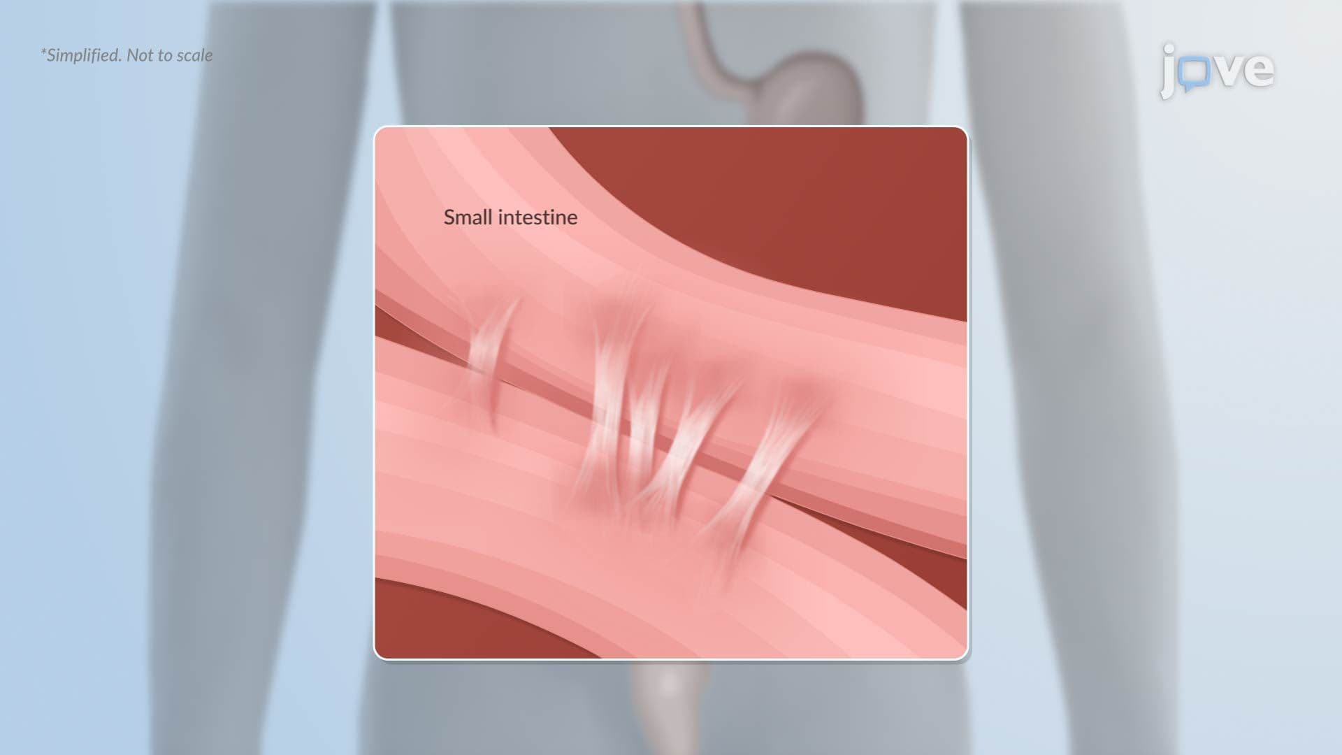
10. Complications of Healing Healing does not always proceed cleanly. *Contractures* form when scar tissue tightens around a wound, restricting movement, a serious complication following severe burns, particularly at mobile joints like fingers and elbows. *Adhesions* are bands of internal scar tissue; post-surgical abdominal adhesions can bind loops of intestine and cause bowel obstruction, a common complication seen in US emergency departments. *Hypertrophic scars* are raised, thick, and confined to the wound boundary, caused by excess collagen deposition. *Keloids* extend beyond the original wound edges and are more prevalent in certain populations. Dense scar tissue can also compress local blood vessels, reducing oxygen delivery and causing chronic ulceration or tissue breakdown.
