Loading concepts…

 35 Concepts
 5.1K Views
 Popular topics
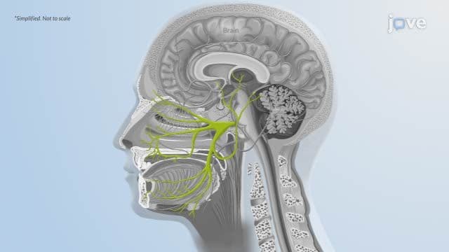

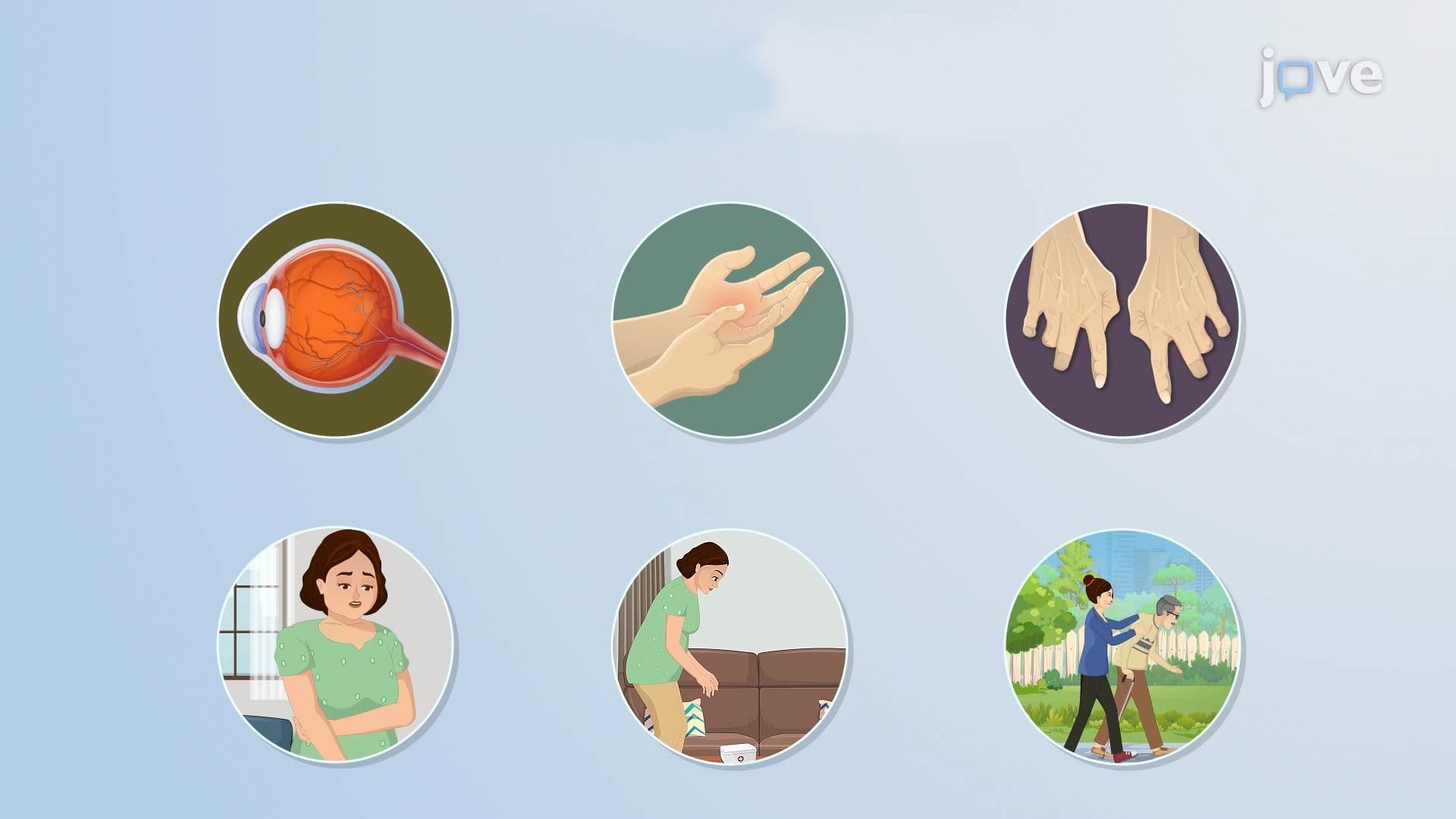
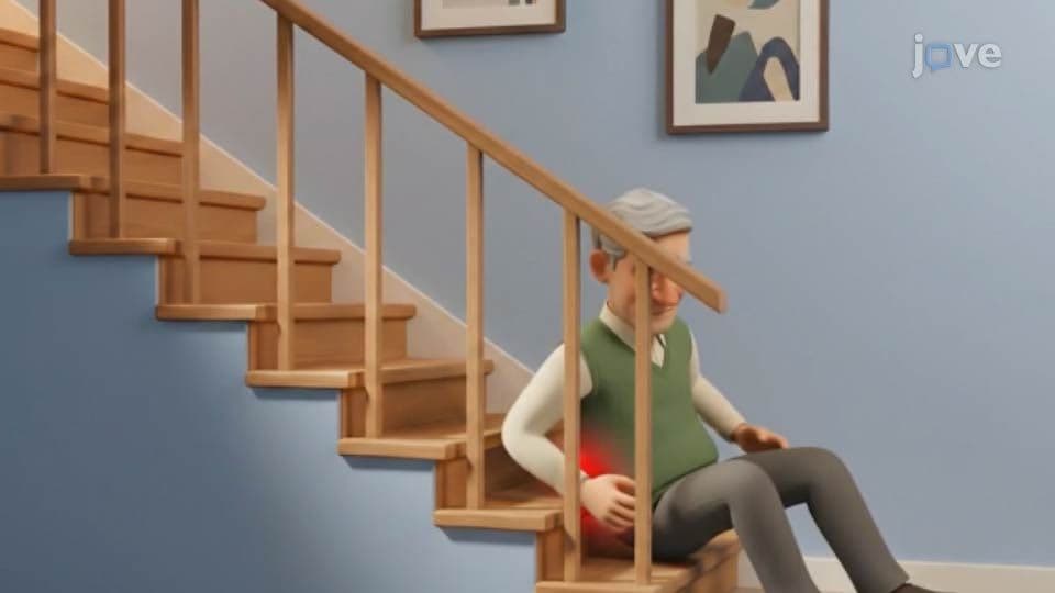
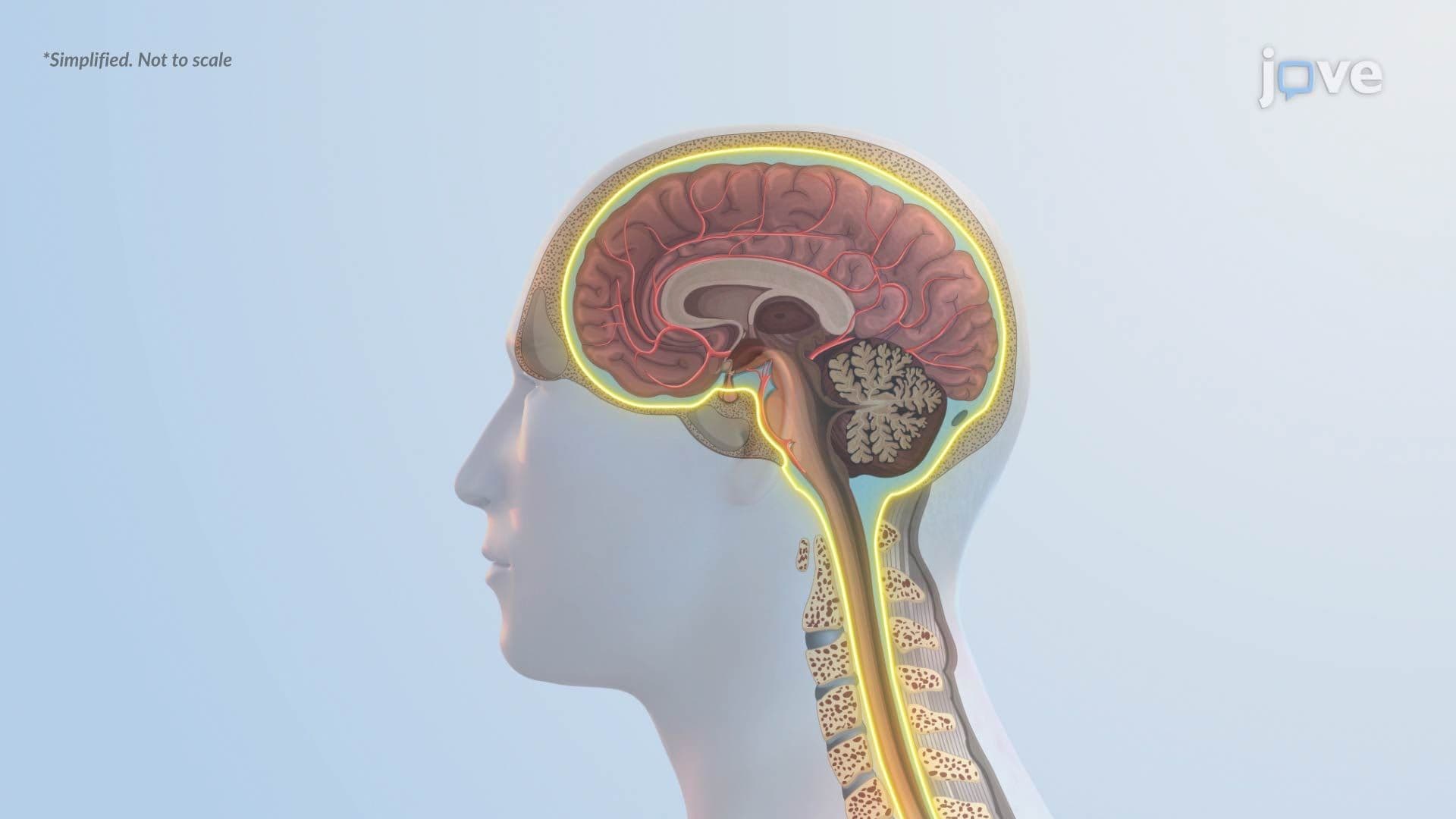



Nervous System Disorders Fundamentals covers the essential mechanisms, classifications, and clinical consequences of major CNS and PNS conditions-from stroke and traumatic brain injury to Alzheimer's disease and seizure disorders. JoVE Coach guides students through vascular, inflammatory, degenerative, and traumatic pathologies, connecting cellular-level dysfunction to real-world neurological outcomes critical for US healthcare and science education.
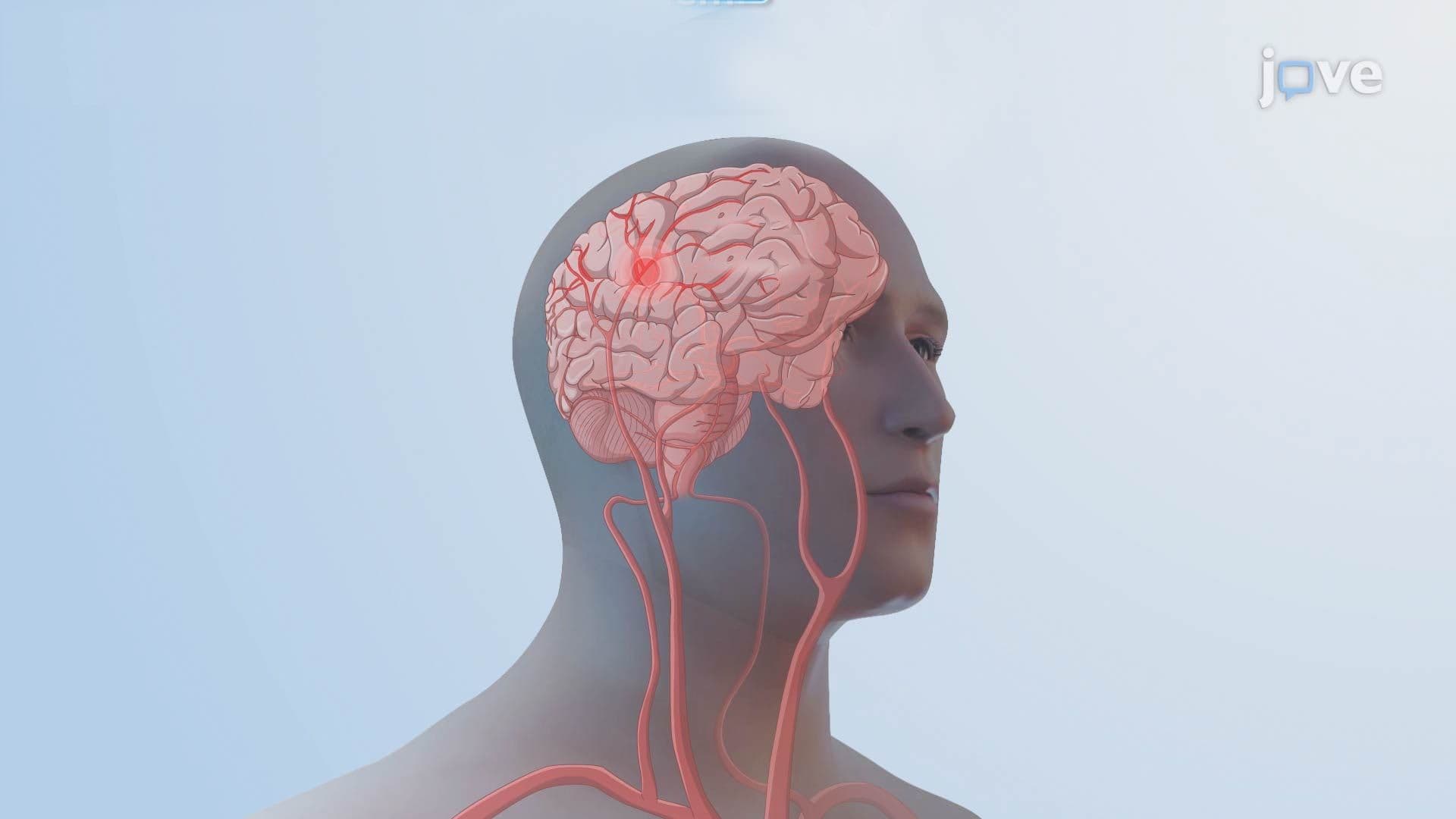
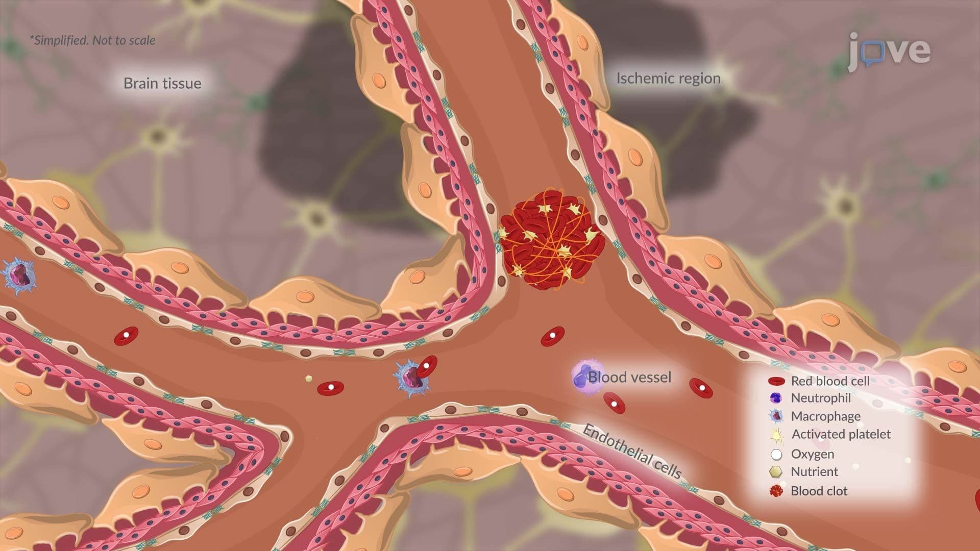
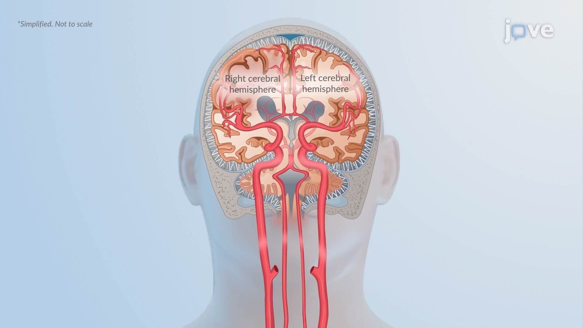
1. Stroke: Ischemic and Hemorrhagic Types Stroke is a sudden disruption of cerebral blood flow resulting in neurological dysfunction. Ischemic strokes-the most common type-occur when a cerebral artery is blocked by a thrombus (often from atherosclerotic plaque rupture) or an embolus (frequently from atrial fibrillation). Hemorrhagic strokes result from vessel rupture, causing either intracerebral hemorrhage directly into brain tissue or subarachnoid hemorrhage into the space around the brain, often from a ruptured berry aneurysm. Both types can be life-threatening, and distinguishing them is essential for clinical management in US emergency settings.
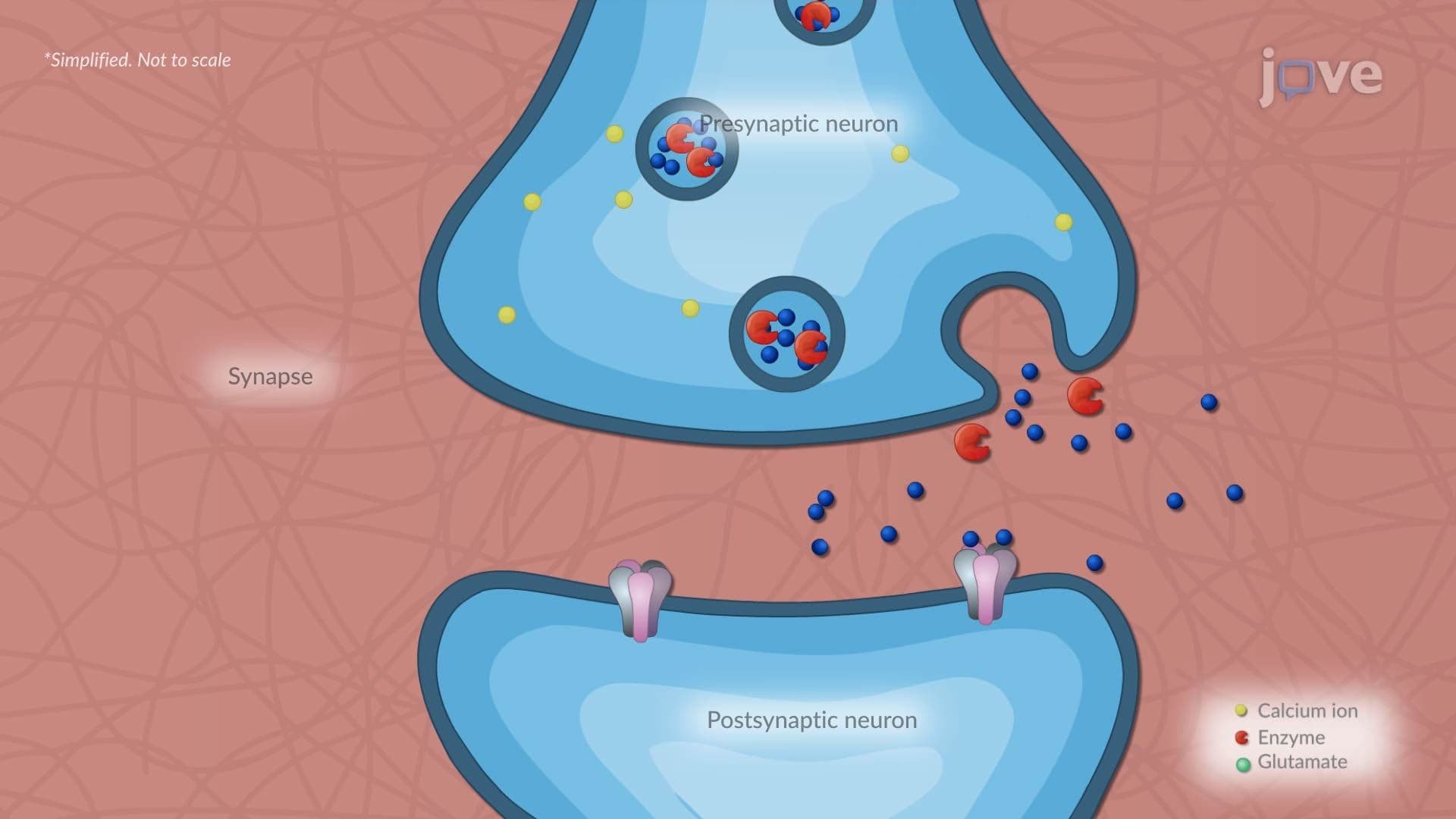
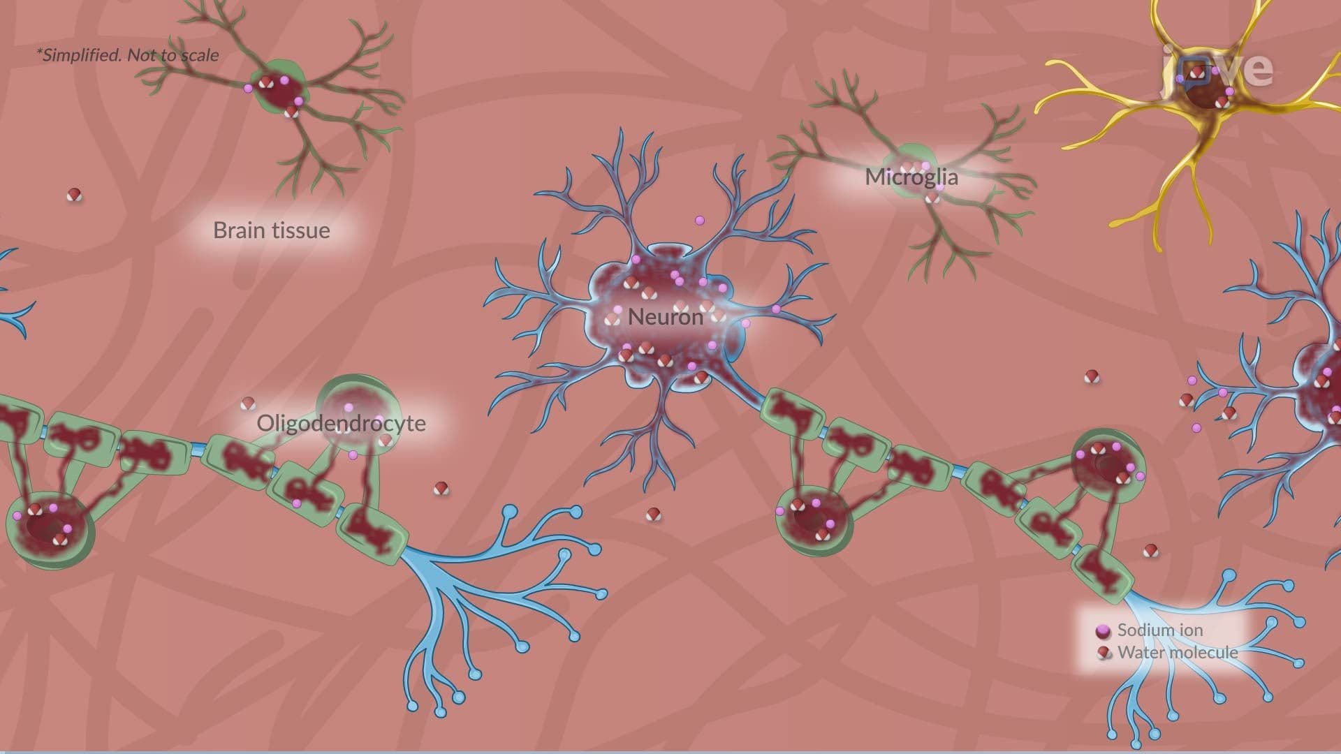
2. Ischemic Stroke Pathophysiology and the Ischemic Penumbra When cerebral blood flow is blocked, oxygen and glucose delivery fail within minutes. An infarct core of irreversibly dead cells forms at the center, surrounded by the penumbra-metabolically stressed tissue that may still be salvageable. Ion pump failure allows sodium and calcium to flood cells, triggering cytotoxic edema. Excess calcium drives glutamate release and excitotoxicity, damaging neurons further. Inflammatory cells worsen injury and contribute to vasogenic edema. The longer the blockage persists, the larger the infarct core grows, reinforcing why rapid intervention-such as tPA administration in US stroke centers-is time-critical.
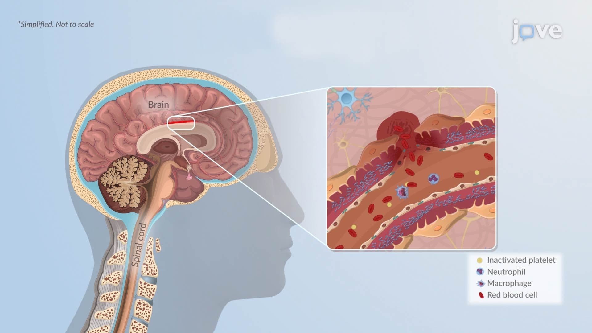
3. Hemorrhagic Stroke Pathophysiology After a vessel ruptures, blood accumulates as a hematoma, compressing surrounding brain tissue and raising intracranial pressure. Secondary injury follows as hemoglobin and iron leak into tissue, triggering oxidative stress and microglial-mediated inflammation that compromises the blood-brain barrier. In subarachnoid hemorrhage, vasospasm can cause delayed cerebral ischemia days after the initial bleed, while blood in the cerebrospinal fluid can obstruct drainage, causing hydrocephalus. These cascading secondary events often determine long-term neurological outcomes and are the focus of intensive care management in US neurocritical care units.
4. Transient Ischemic Attack (TIA) A transient ischemic attack-commonly called a "mini-stroke"-involves a temporary focal loss of blood flow that causes stroke-like symptoms resolving within hours without permanent tissue infarction. Mechanisms include large-artery atherosclerosis causing transient flow restriction, emboli that temporarily block then dissolve from vessels, and reduced perfusion distal to a high-grade carotid stenosis. TIA is a major warning sign: roughly 10-15% of patients in the US experience a full stroke within 90 days of a TIA, making rapid evaluation with tools like the ABCD2 score clinically essential.
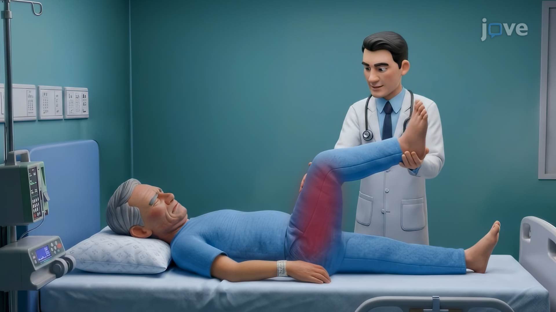
5. Bacterial Meningitis: Introduction and Pathophysiology Bacterial meningitis is an acute, life-threatening infection of the leptomeninges and cerebrospinal fluid. Common pathogens in US adults include *Streptococcus pneumoniae* and *Neisseria meningitidis*, while *Listeria monocytogenes* affects immunocompromised individuals and older adults. Bacteria colonizing the nasopharynx invade the bloodstream, penetrate the blood-CSF barrier, and multiply in the relatively immune-privileged subarachnoid space. This triggers intense inflammation-cytokine release, neutrophil recruitment, BBB disruption, and edema-raising intracranial pressure and reducing cerebral perfusion. Classic signs include fever, severe headache, neck stiffness, and positive Kernig and Brudzinski signs.
6. Encephalitis and Brain Abscess Encephalitis is inflammation of the brain parenchyma itself, most commonly caused by viruses such as herpes simplex virus type 1 (HSV-1), which has a tropism for the temporal lobes, or arboviruses like West Nile virus. Autoimmune encephalitis-such as anti-NMDA receptor encephalitis-involves antibody-mediated disruption of neural signaling. In contrast, a brain abscess is a localized pus-filled mass arising from bacterial invasion-often from sinus, dental, or cardiac sources-that acts as a space-occupying lesion, elevating intracranial pressure and causing focal neurological deficits, seizures, and papilledema. Both conditions require urgent diagnosis and treatment in US hospital settings.
7. Multiple Sclerosis (MS) Multiple sclerosis is a chronic autoimmune CNS disease in which the immune system attacks myelin sheaths, demyelinating axons in the brain and spinal cord. Over time, axonal damage accumulates, producing irreversible neurological disability. MS typically affects US adults between ages 20 and 40, occurring twice as often in women. Symptoms are diverse-optic neuritis, spasticity, paresthesias, bladder dysfunction, and cognitive impairment-and often follow a relapsing-remitting pattern where episodes of neurological worsening alternate with partial or full recovery. It is one of the most common causes of non-traumatic disability in young American adults.
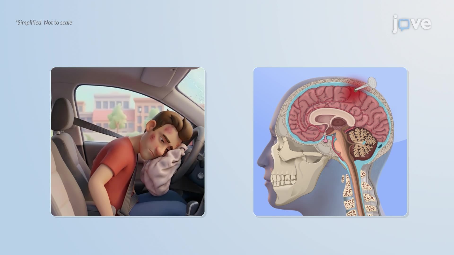
8. Traumatic Brain Injury (TBI) TBI results from external mechanical forces disrupting normal brain function, classified by severity using the Glasgow Coma Scale (GCS), loss of consciousness duration, and post-traumatic amnesia. Mild TBI (GCS 13-15), commonly called a concussion, frequently occurs in US contact sports. Moderate TBI (GCS 9-12) often results from motor vehicle crashes, while severe TBI (GCS 3-8) can follow major falls or high-impact collisions. Primary injury is immediate and mechanical; secondary injury unfolds over hours to days involving inflammation, excitotoxicity, and ischemia-processes that are the targets of US neurointensive care interventions.
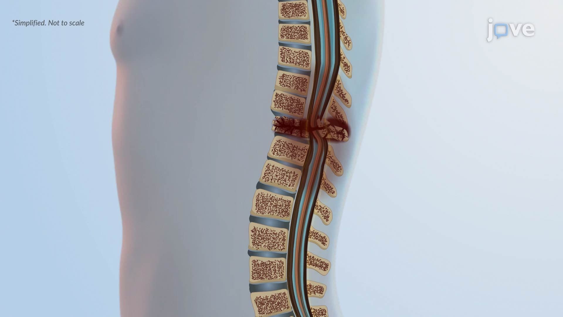
9. Spinal Cord Injury: Primary and Secondary Mechanisms Spinal cord injury involves an immediate primary phase-compression, contusion, laceration, or distraction of cord tissue-that disrupts neurons, supporting cells, and blood vessels. A secondary injury phase then unfolds over minutes to weeks, driven by ATP depletion, glutamate-mediated excitotoxicity, calcium overload, inflammatory cell infiltration, and reactive oxygen species. Oligodendrocyte loss strips axons of myelin, impairing signal conduction. In later stages, reactive astrocytes form a glial scar that limits but does not reverse injury. Understanding this two-phase model is foundational for rehabilitation medicine and spinal cord research programs at US institutions like the NIH.
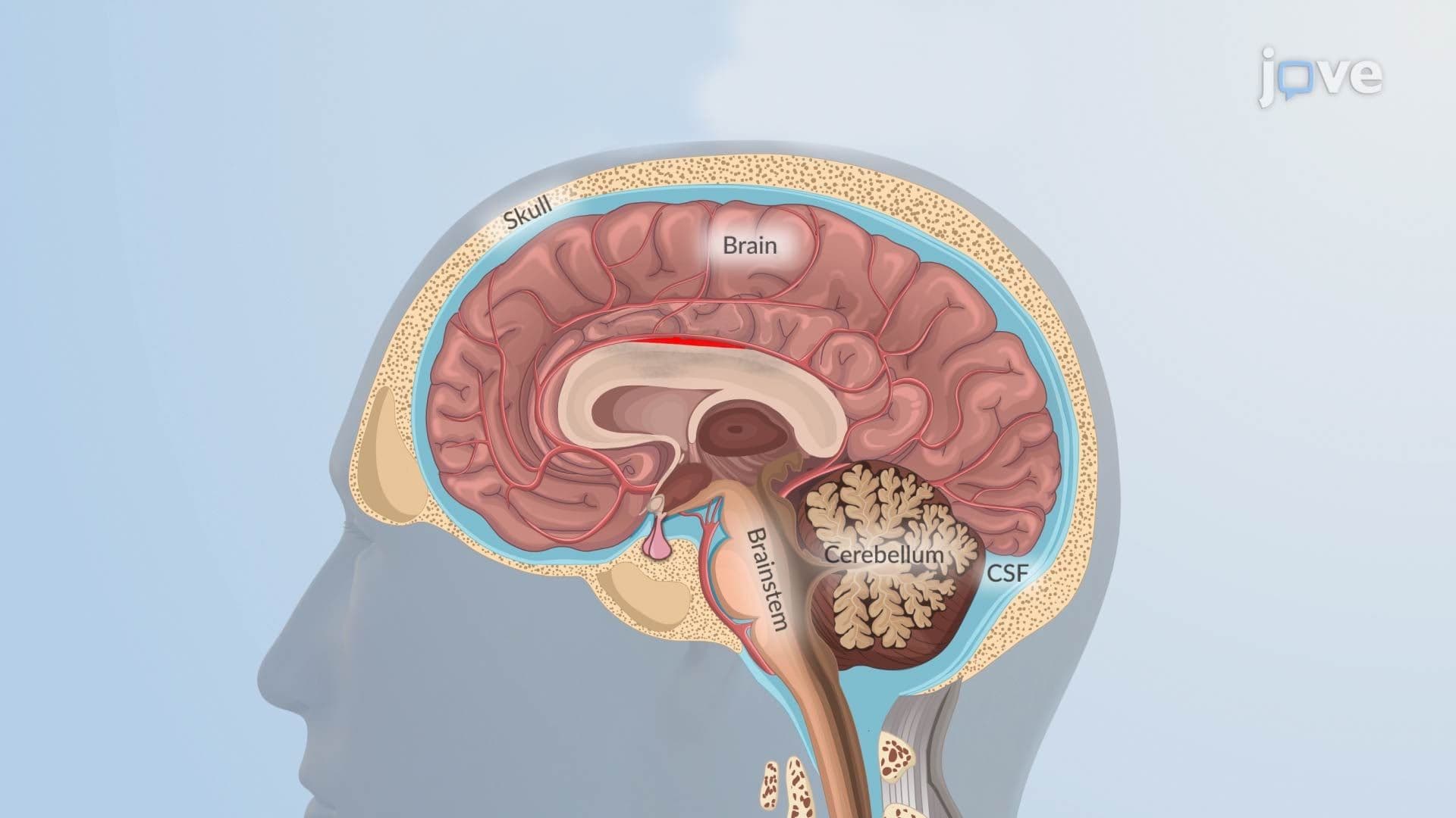
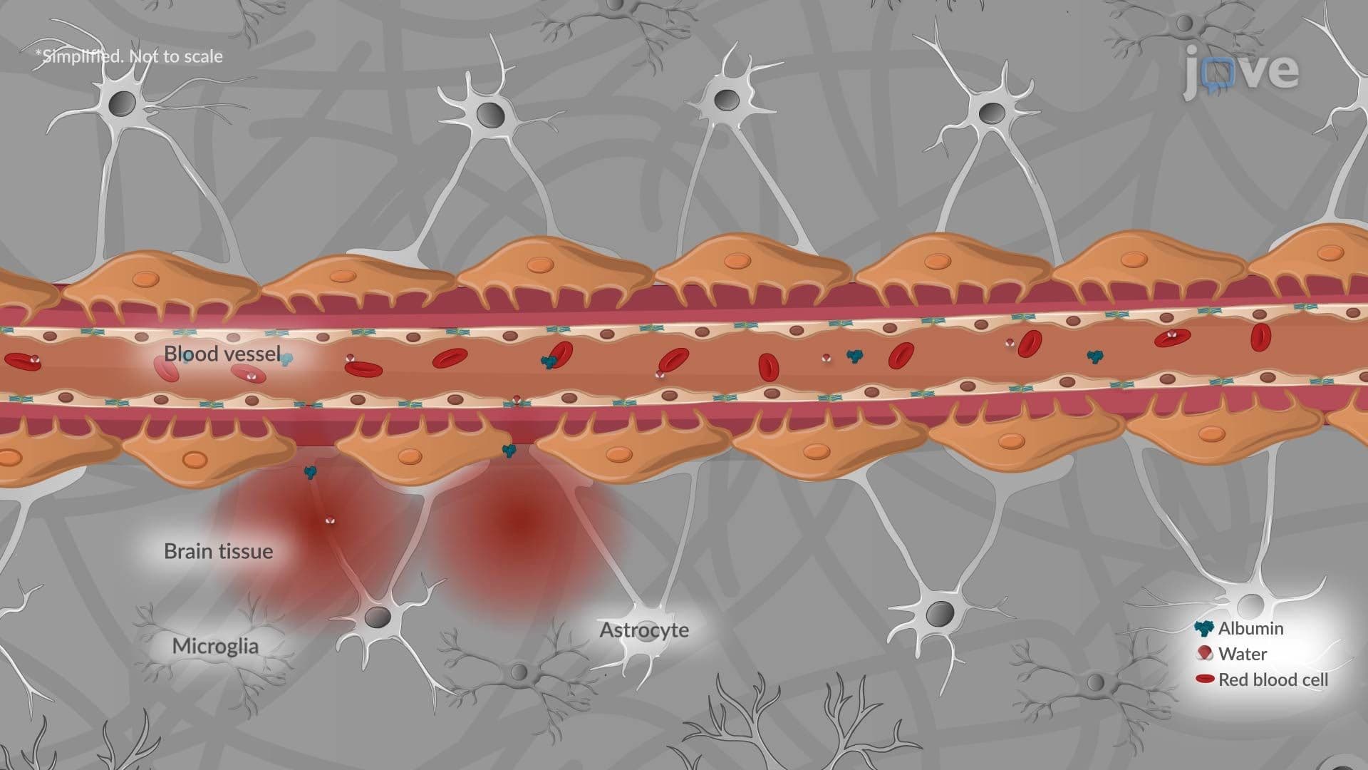
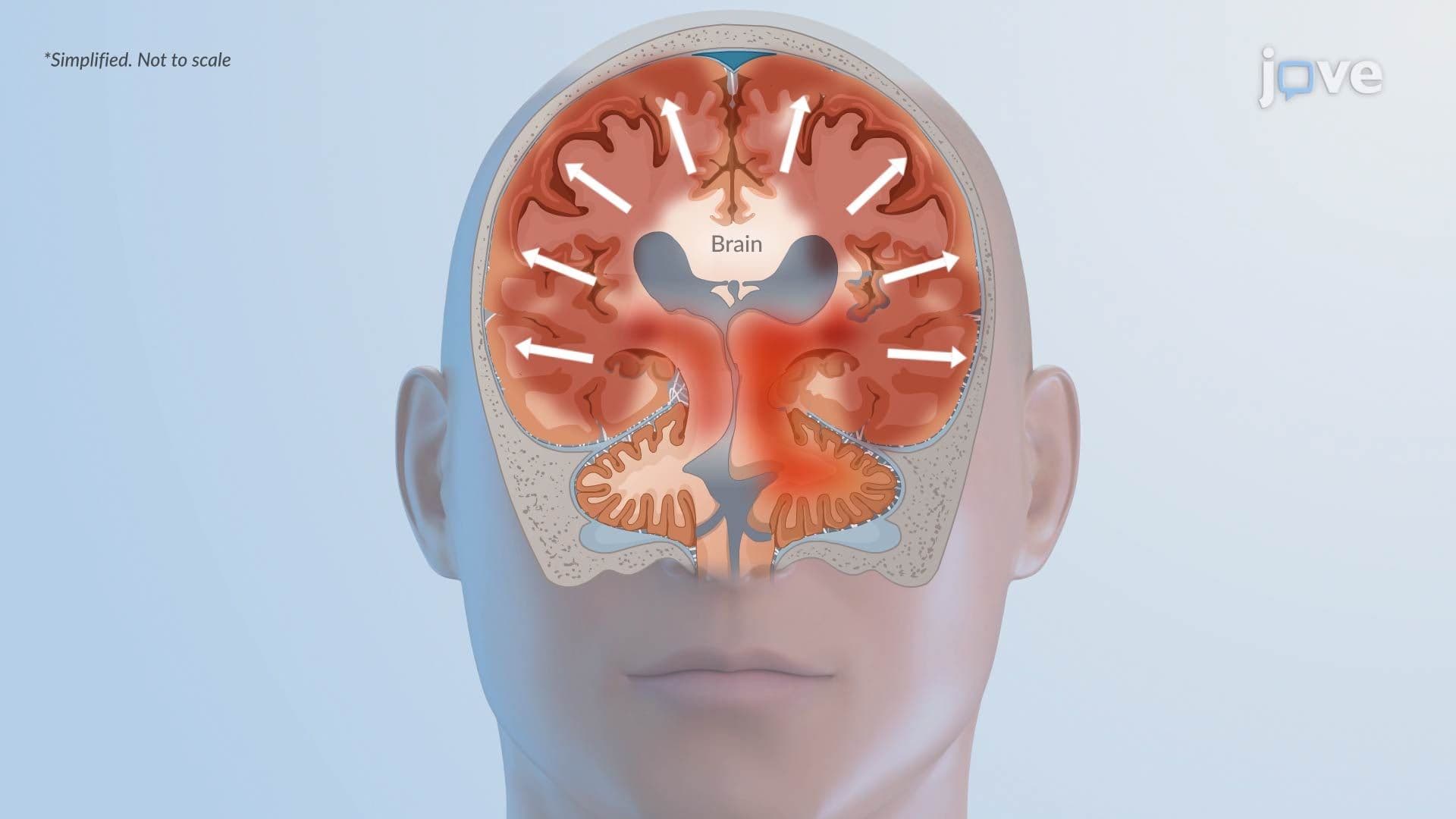
10. Increased Intracranial Pressure and Cerebral Edema The Monro-Kellie doctrine states that total intracranial volume is fixed; any increase in one component-brain tissue, CSF, or blood-must be offset by another or ICP rises. Causes include hemorrhage, large infarcts, tumors, and infections. Cerebral edema is a major driver: vasogenic edema arises from blood-brain barrier breakdown allowing protein-rich fluid into extracellular space; cytotoxic edema results from cellular energy failure and intracellular water accumulation; interstitial edema occurs with hydrocephalus; and ionic edema develops via osmotic gradients without protein leakage. Uncontrolled ICP elevation leads to herniation syndromes, brainstem compression, and death.
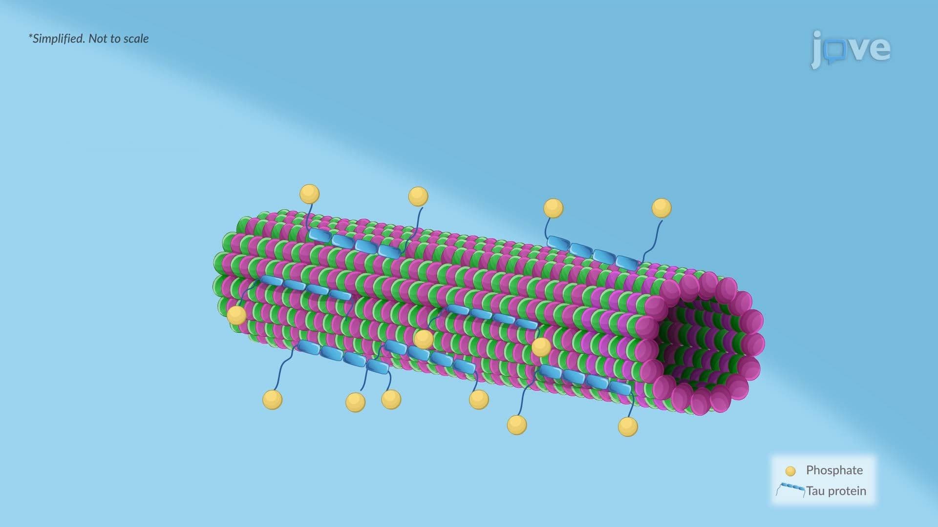
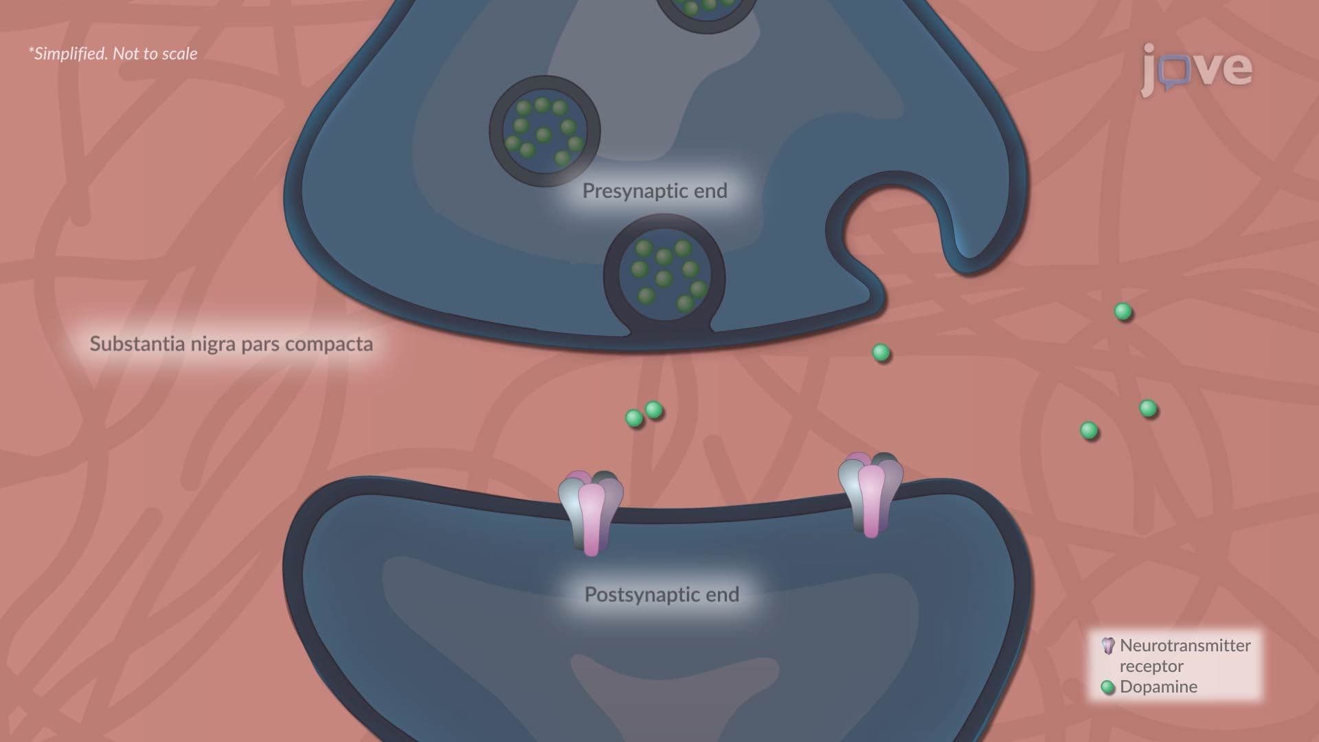
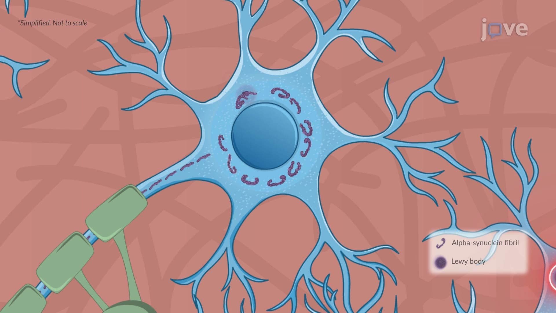
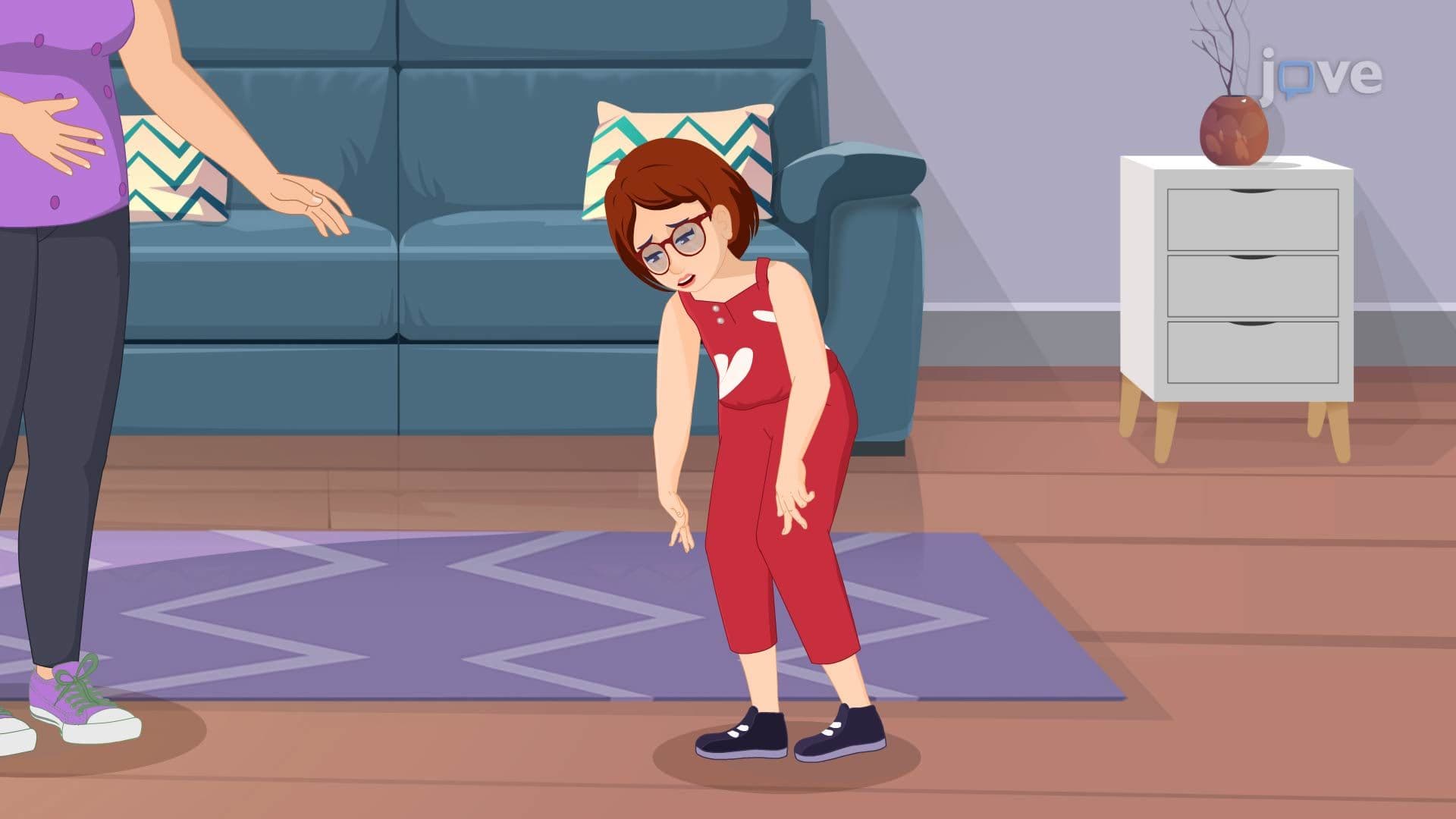
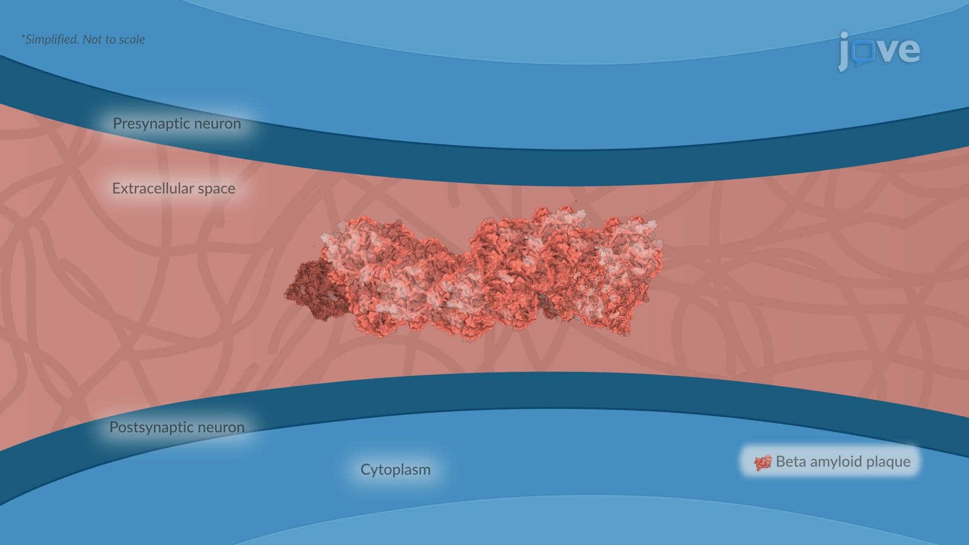
11. Neurodegenerative Disorders: Alzheimer's, Parkinson's, and Huntington's Disease Alzheimer's disease, the leading cause of US dementia, features extracellular beta-amyloid plaques and intracellular neurofibrillary tangles from hyperphosphorylated tau, causing progressive hippocampal and cortical atrophy. Parkinson's disease involves degeneration of dopaminergic neurons in the substantia nigra, producing the classic triad of resting tremor, rigidity, and bradykinesia, alongside Lewy body accumulation of misfolded alpha-synuclein. Huntington's disease is caused by a CAG repeat expansion in the HTT gene, leading to striatal neuronal death. Together, these disorders represent the major focus of neurodegeneration research and clinical neurology across US academic medical centers.
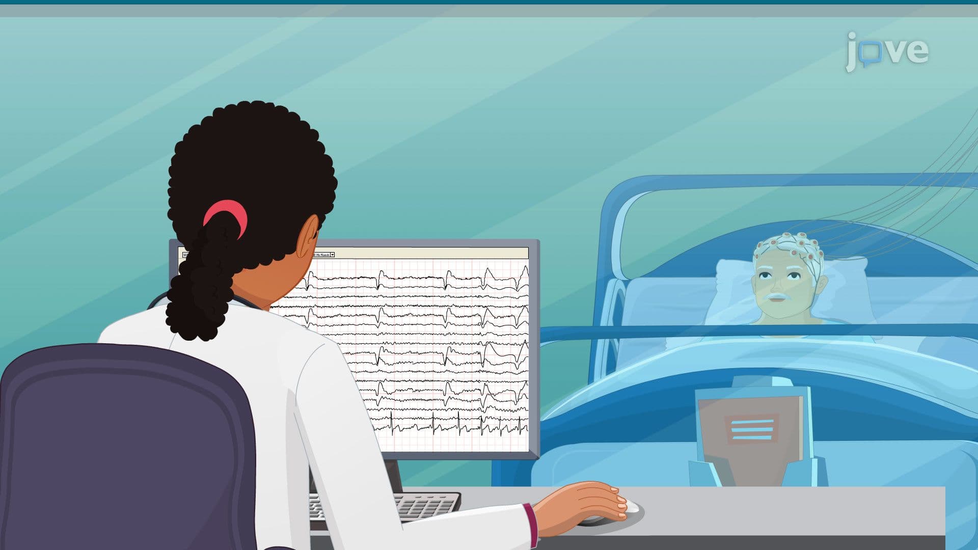
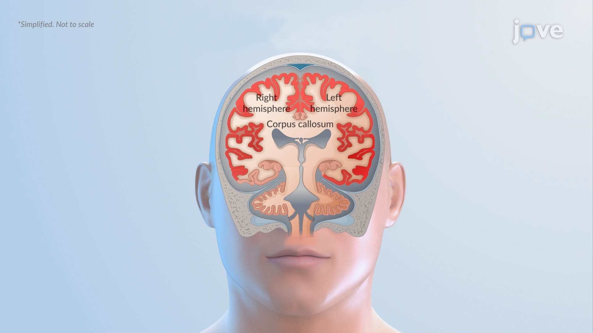
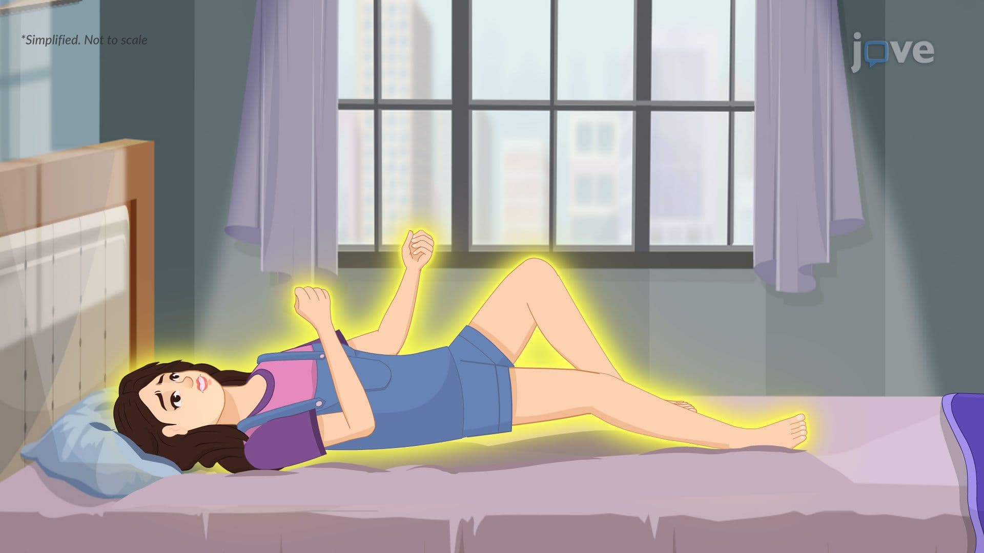
12. Seizures, Epilepsy, and Myasthenia Gravis Seizures result from abnormal, excessive, or synchronous neuronal activity in the brain and are classified as focal (originating in one hemisphere) or generalized (involving both hemispheres simultaneously). Epilepsy is a chronic disorder defined by recurrent unprovoked seizures. Myasthenia gravis is an autoimmune neuromuscular junction disorder in which antibodies target acetylcholine receptors at the neuromuscular junction, causing fluctuating muscle weakness that worsens with activity-particularly affecting extraocular and bulbar muscles. All three conditions are heavily represented on US licensing exams including the USMLE and NCLEX, and require a strong mechanistic understanding for clinical reasoning.
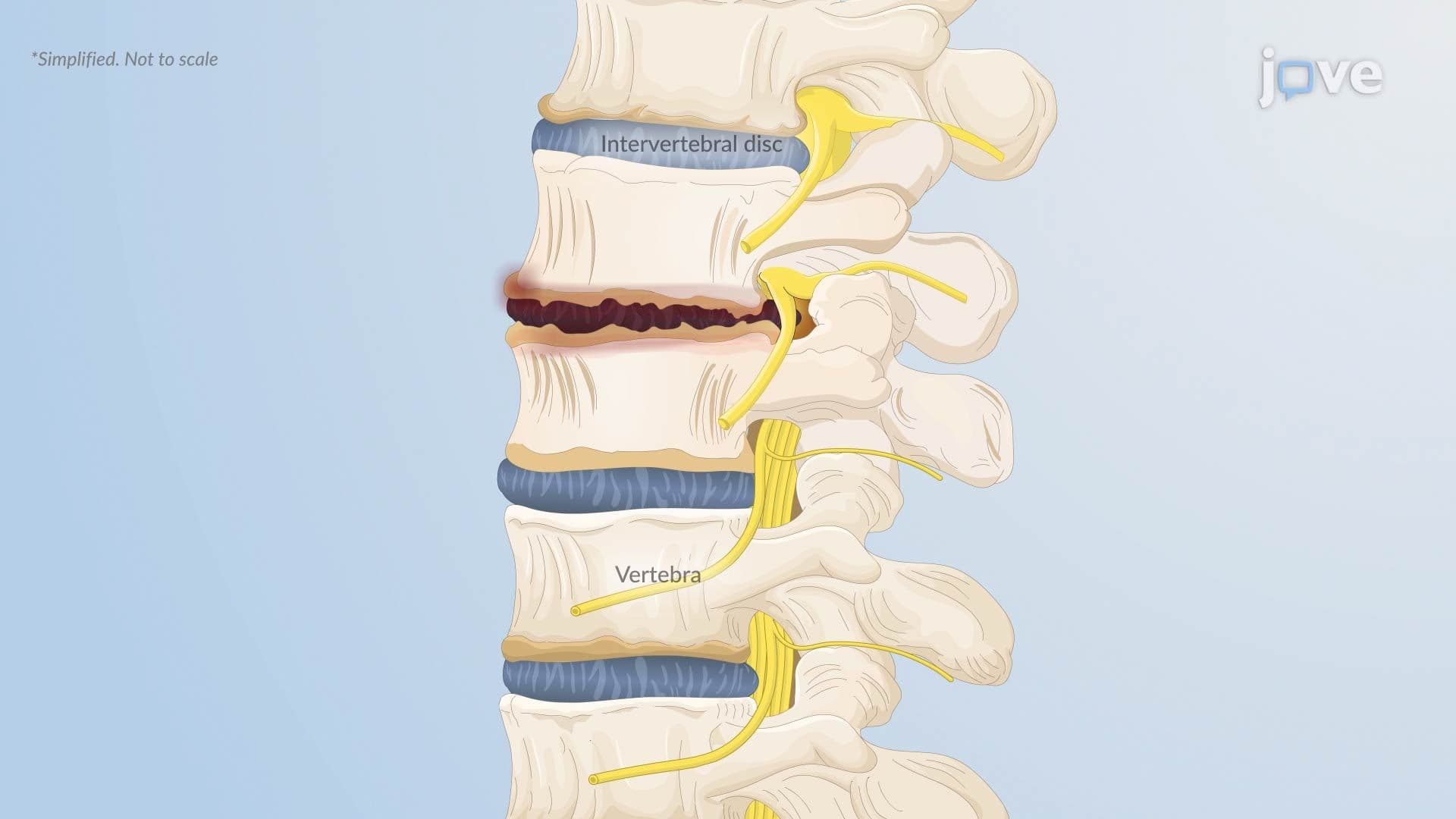
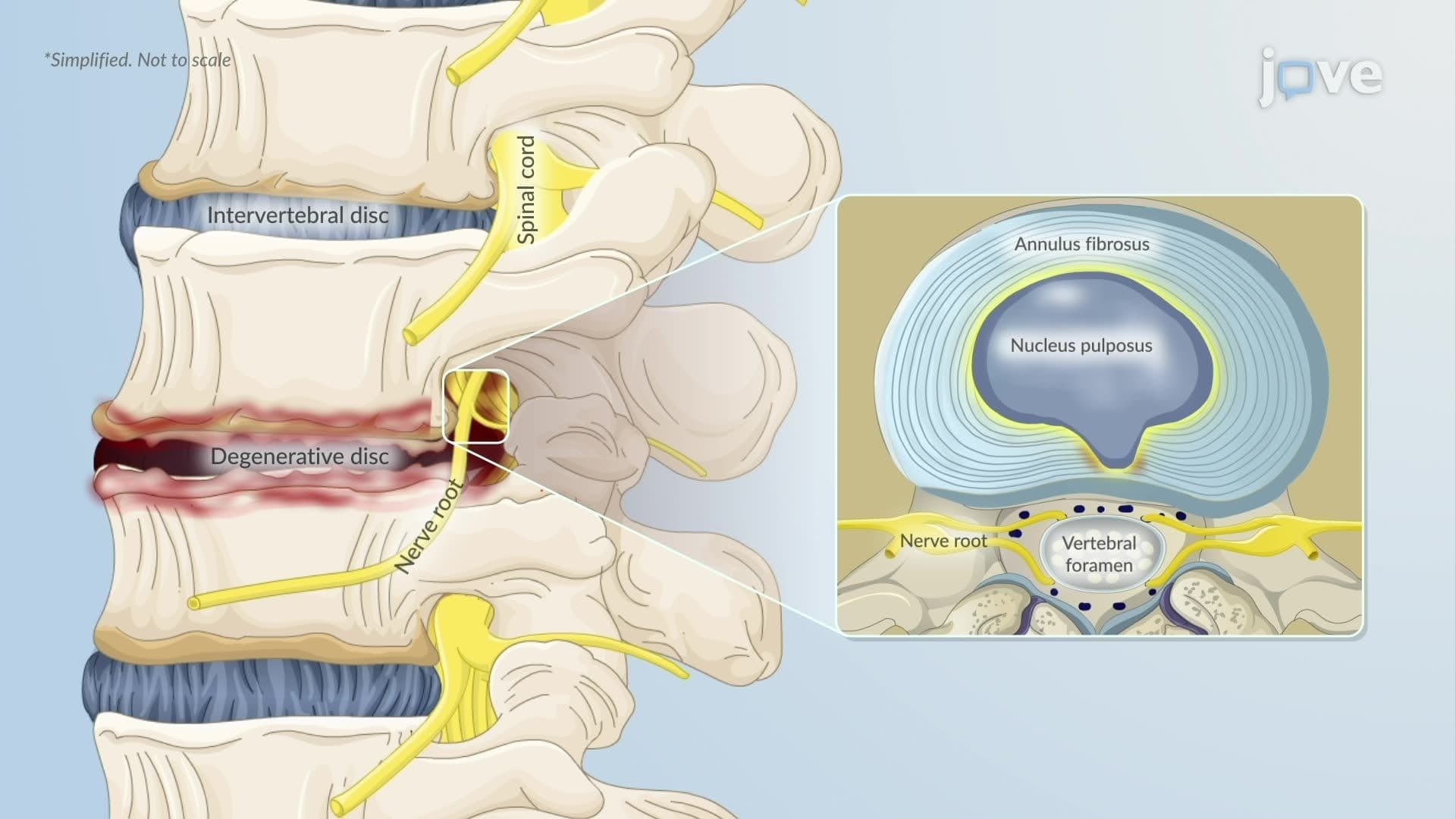
13. Disc Disease and Alterations in Muscle Tone Herniated intervertebral disc occurs when the nucleus pulposus protrudes through the annulus fibrosus, compressing spinal nerve roots and causing radiculopathy-pain, weakness, or numbness in the distribution of the affected nerve. Degenerative disc disease involves age-related dehydration and structural breakdown of disc tissue, reducing shock absorption and narrowing the spinal canal. Alterations in muscle tone-including spasticity (increased tone from upper motor neuron lesions) and flaccidity (decreased tone from lower motor neuron damage)-reflect the level and extent of nervous system injury and guide clinical assessment in US physical and occupational therapy settings.
