Loading video…

 35 Concepts
 5.1K views

Video Summary: What Is Brain Abscess L
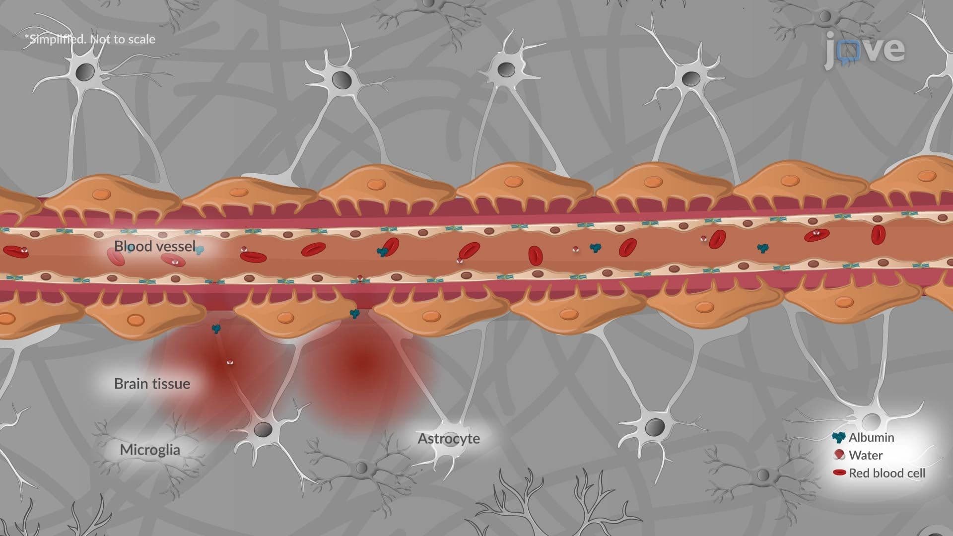
Did you know a simple ear or sinus infection can travel to your brain and become life-threatening? Brain Abscess L describes a dangerous, pus-filled mass that forms inside brain tissue when bacteria like *Staphylococcus aureus* breach the blood-brain barrier. In the US, this condition is seen in neurosurgical and emergency medicine settings, where rapid diagnosis can mean the difference between recovery and death. Watch the full video on JoVE Coach to master this concept with expert-led visuals and step-by-step explanations.
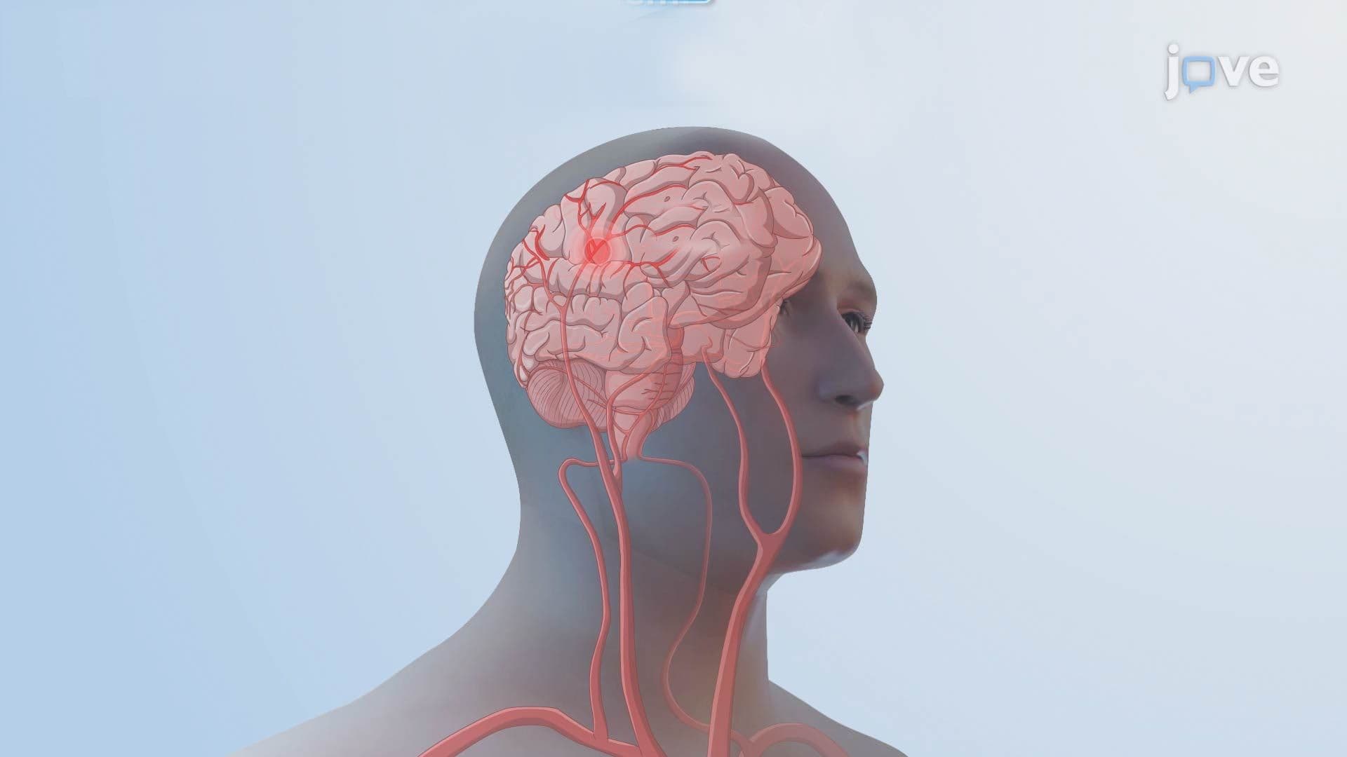
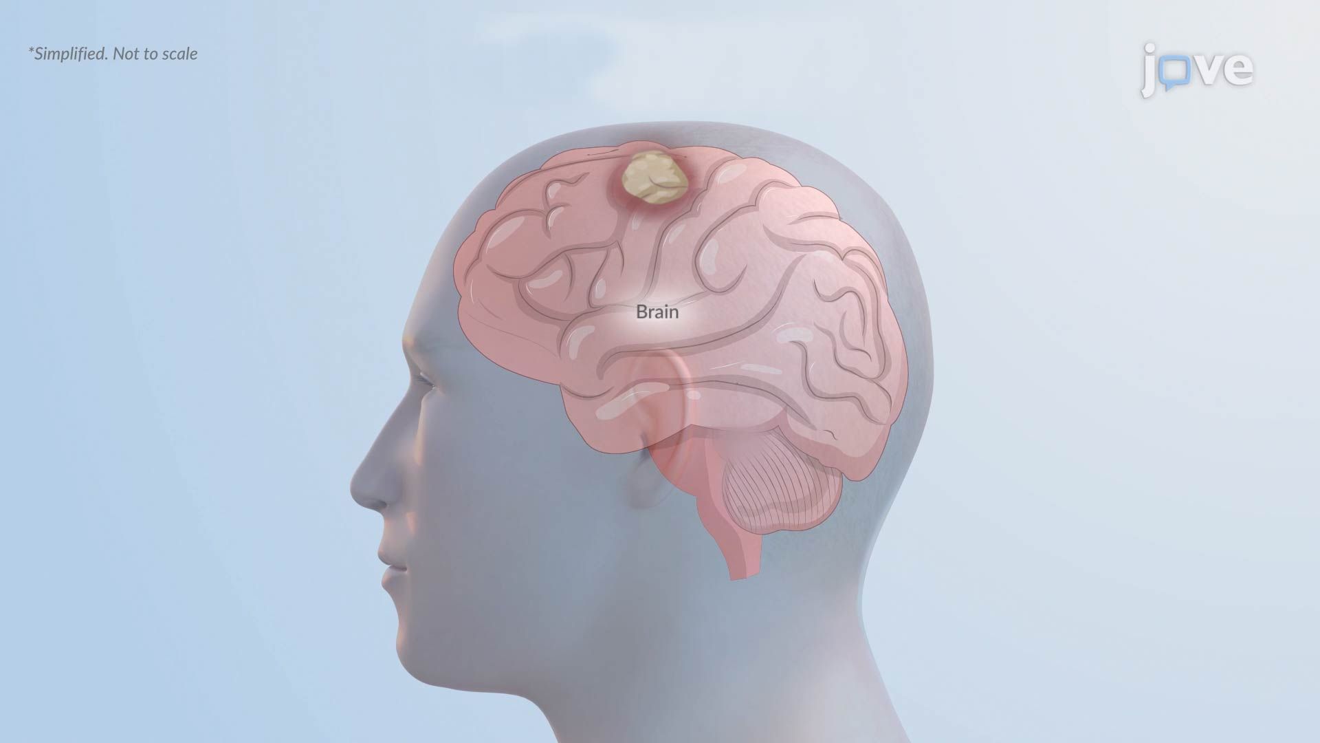
A brain abscess is one of the most serious infections the human body can experience. Classified as a focal, pus-filled mass within brain tissue, it develops when pathogens invade and infect the brain, triggering a cascade of inflammation and tissue death known as necrosis. Unlike chronic neurodegenerative diseases such as Alzheimer's disease or Parkinson's disease, a brain abscess is an acute infectious condition, one that can escalate from a manageable infection to a fatal emergency within days if left untreated. Understanding brain abscess basics is essential for students studying neurology, microbiology, and human physiology.
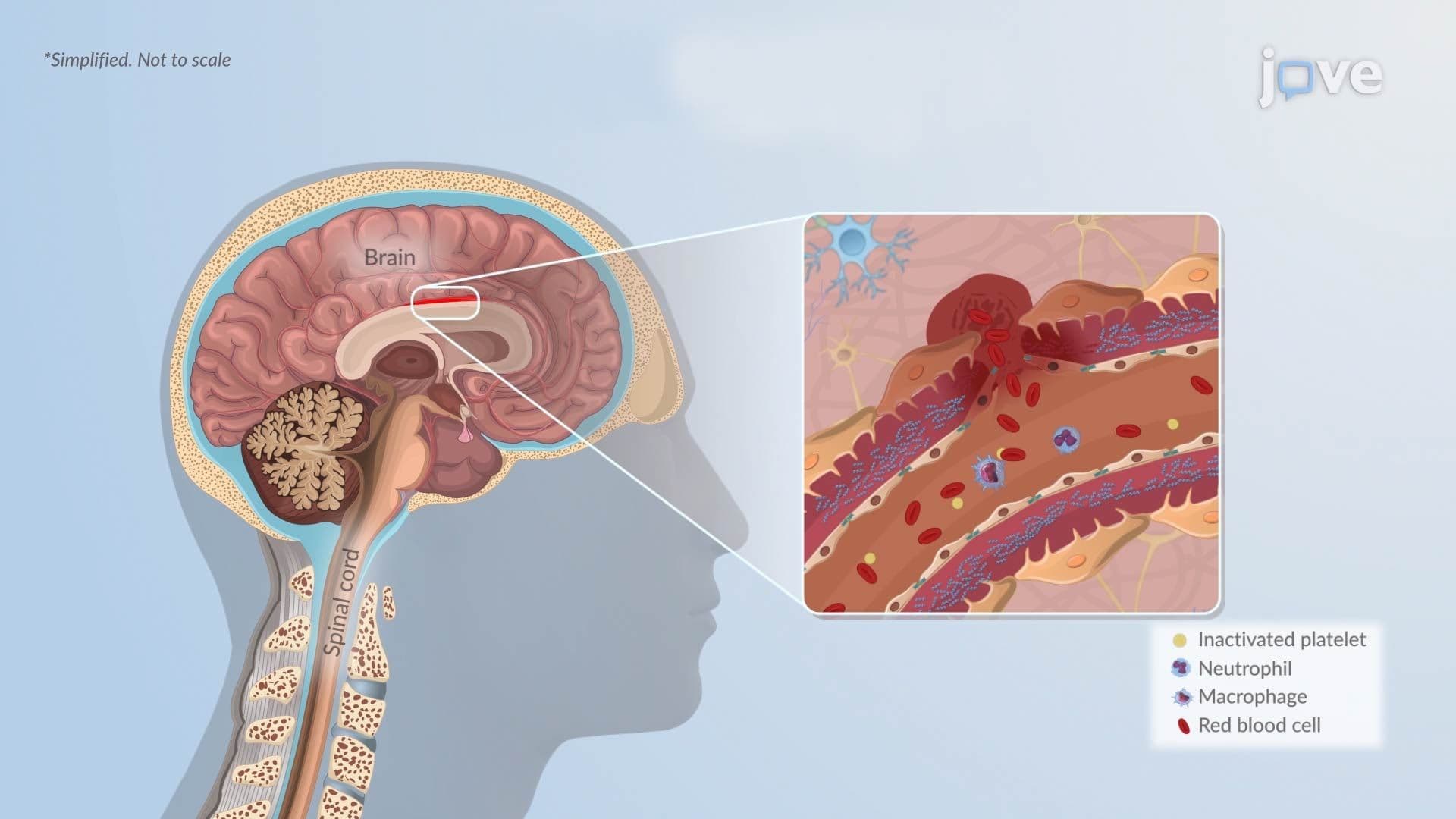
Several distinct pathways allow pathogens to reach the brain. The most common involve the spread of existing infections in nearby structures. Ear infections (otitis media), chronic sinusitis, and dental abscesses can erode tissue boundaries and allow bacteria to migrate directly into adjacent brain regions. Lung infections such as bacterial pneumonia and heart conditions like infective endocarditis can seed the brain through the bloodstream, a process called hematogenous spread.
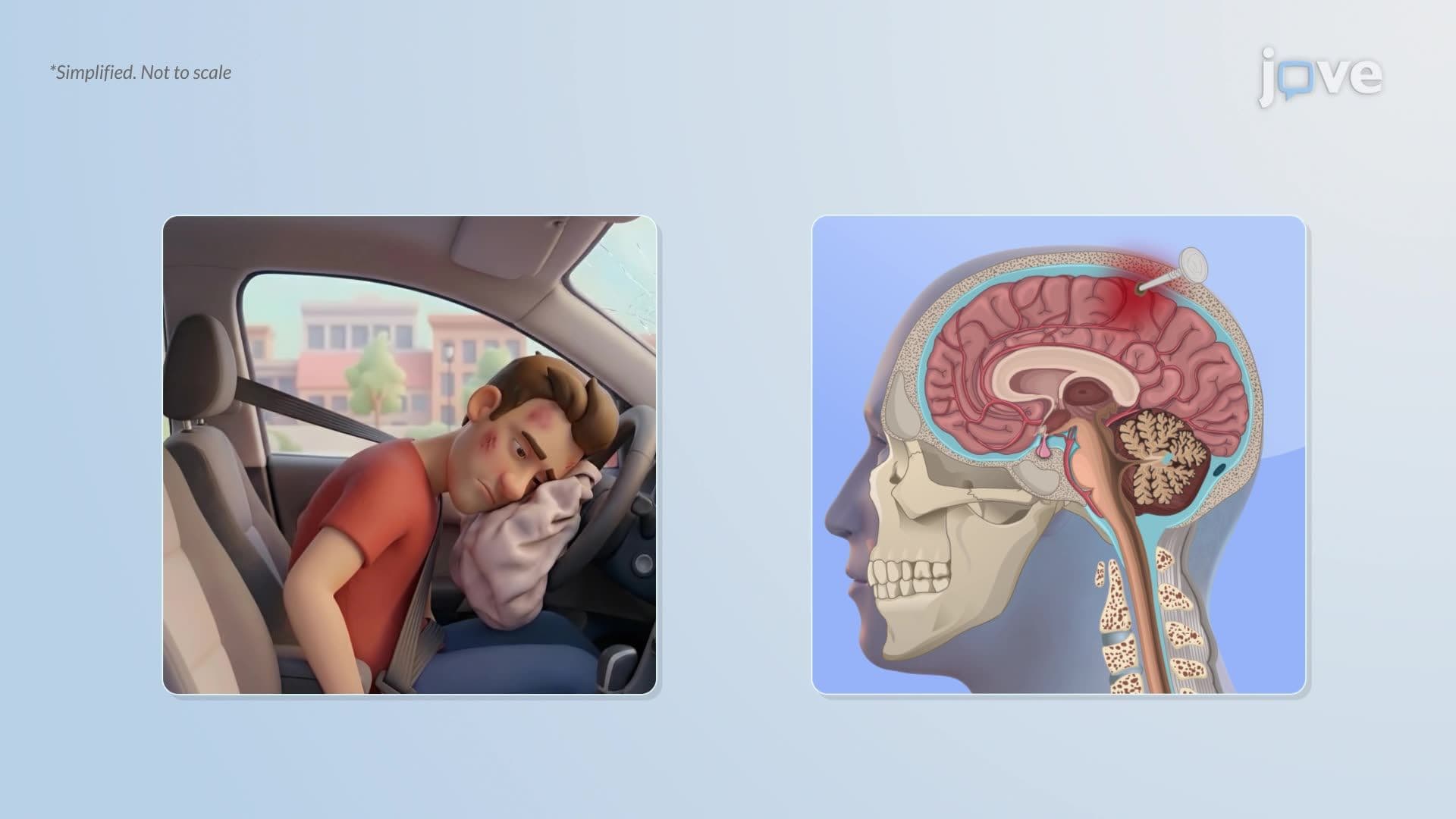
Physical breaches of the skull are another major route. Penetrating head trauma, compound skull fractures, and neurosurgical procedures can introduce bacteria directly into brain tissue. In US trauma centers and neurosurgical units, post-procedural infection protocols exist specifically to minimize this risk. The primary bacteria implicated include *Staphylococcus aureus*, commonly associated with skin and post-surgical infections, and viridans group streptococci, which are frequently linked to dental and oral sources.
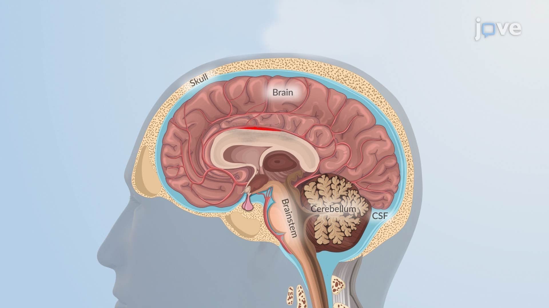
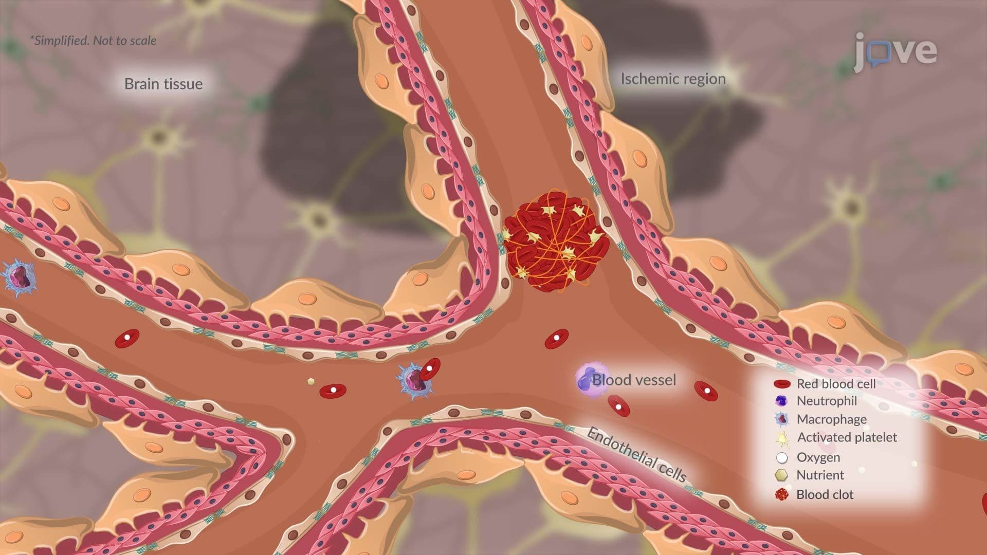
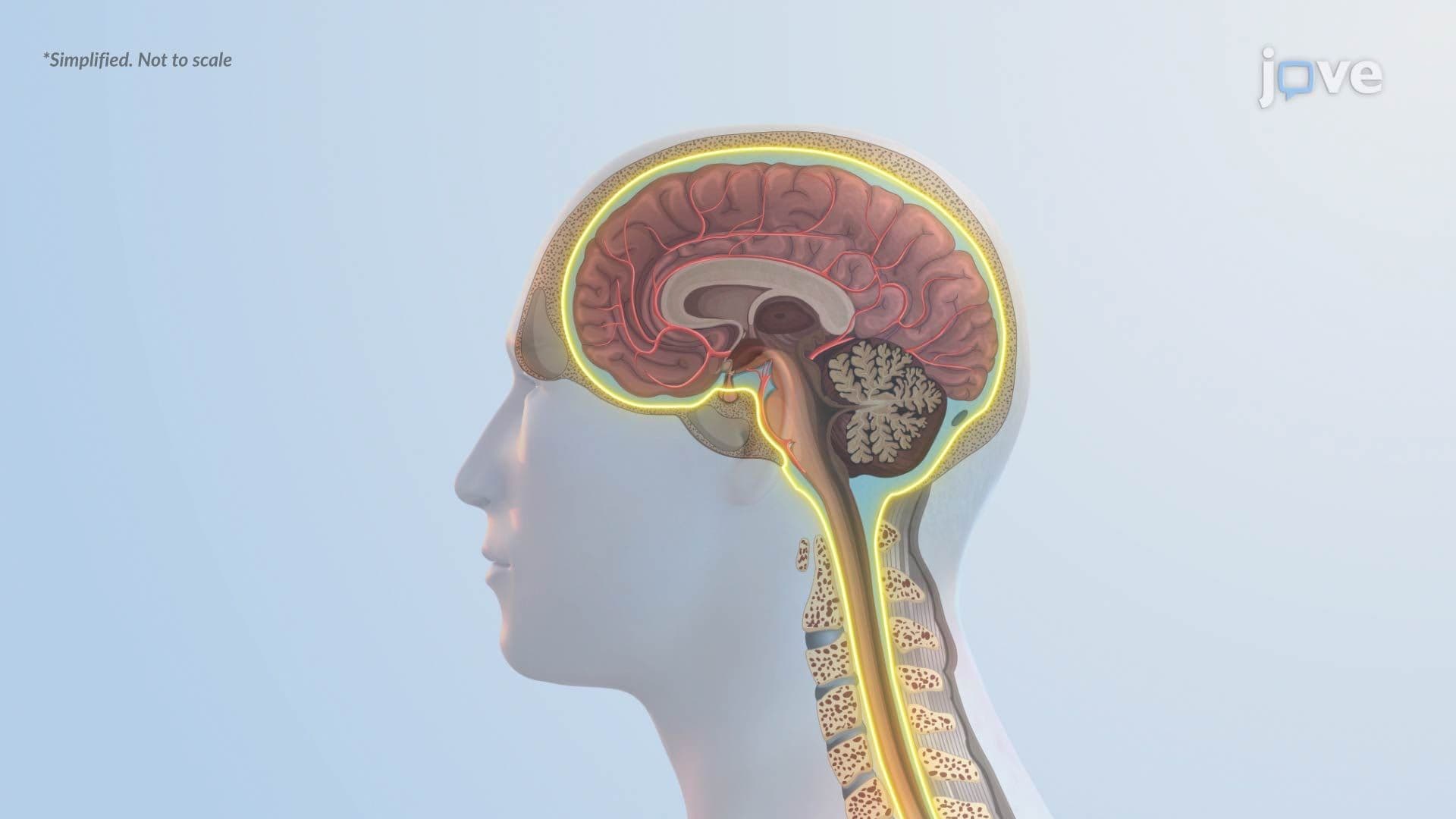
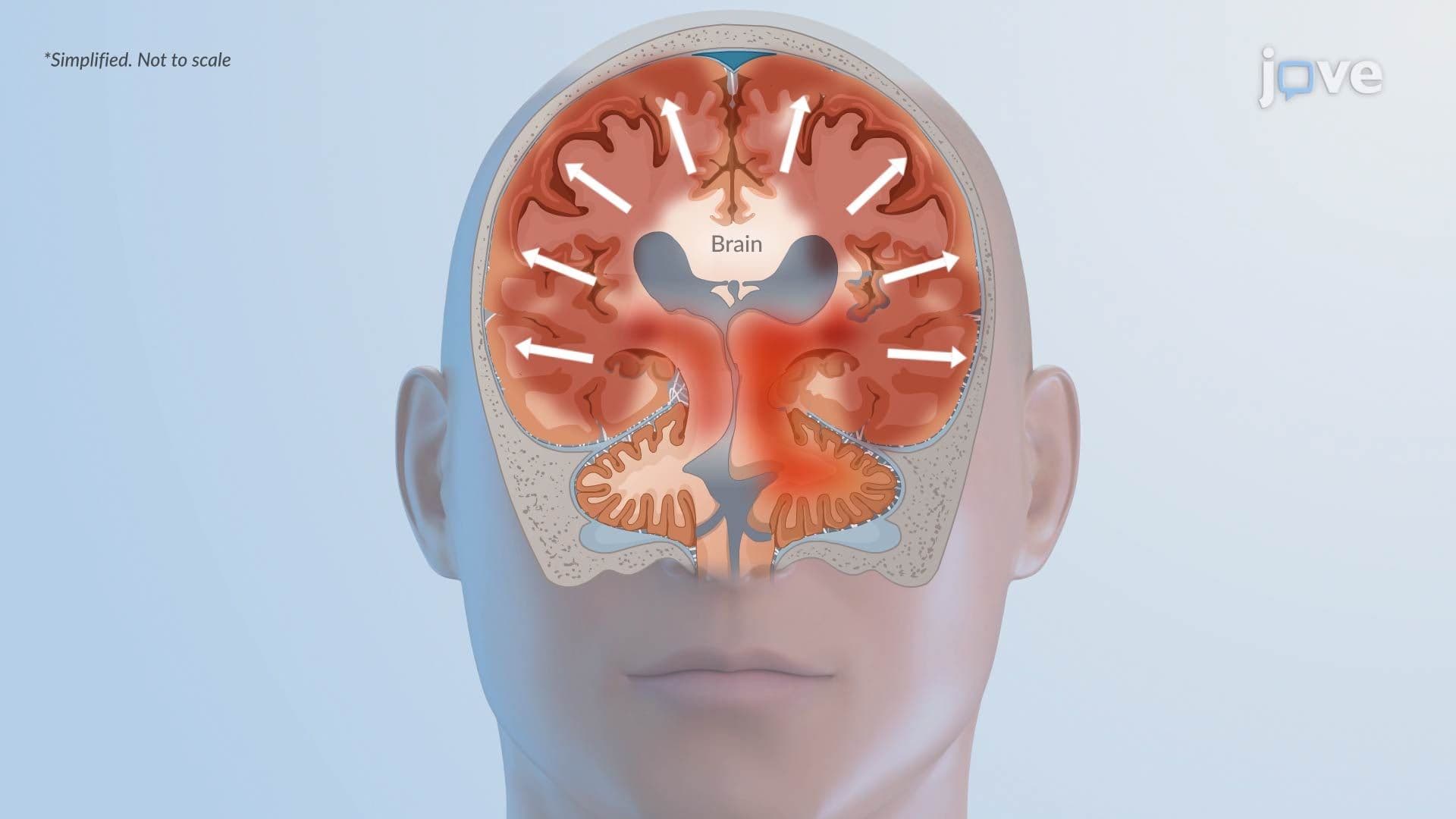
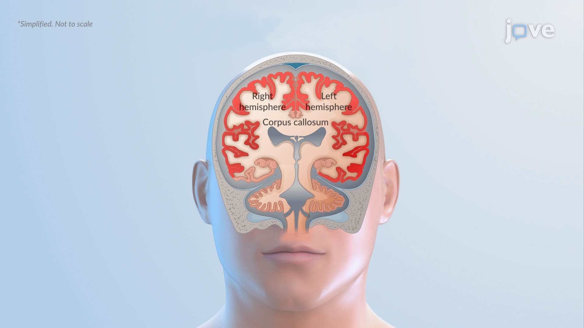
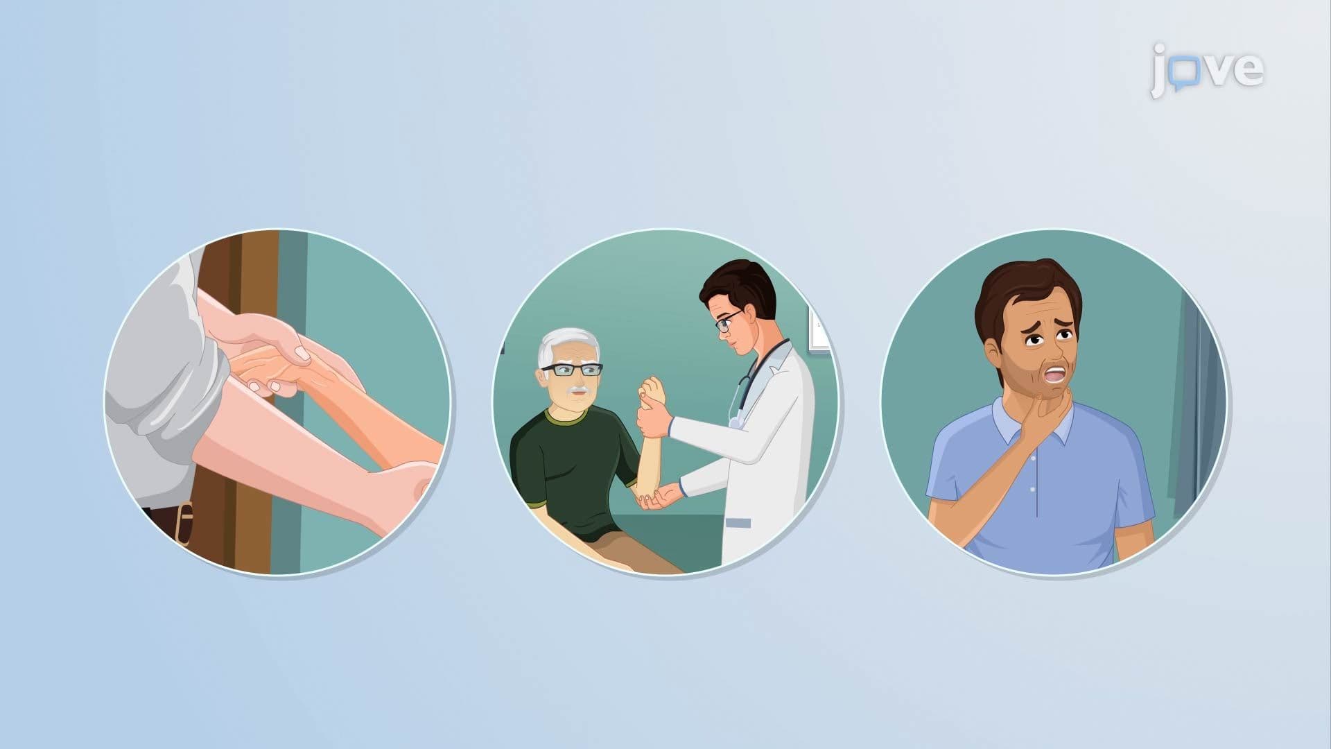
As the abscess grows, it behaves like any expanding mass inside the skull, a fixed, rigid structure. The brain has no room to accommodate extra volume, so an enlarging abscess compresses surrounding tissue and raises intracranial pressure. This is why brain abscesses are classified as space-occupying lesions, a term also applied to tumors and large hematomas. Rising intracranial pressure explains many of the classic symptoms: persistent, worsening headache, nausea and vomiting, and papilledema (swelling of the optic nerve visible on eye examination). Visual field defects and other focal neurological deficits occur when specific brain regions are compressed or damaged.
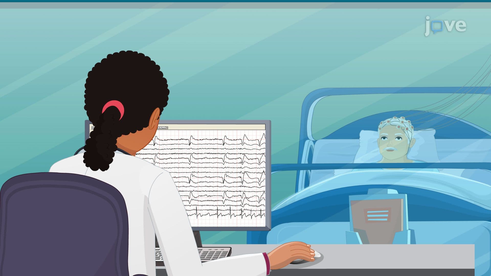
The classic triad of brain abscess, headache, fever, and focal neurological deficits, is a high-yield concept for students preparing for the MCAT, USMLE Step 1, and NCLEX. However, this full triad is only present in a minority of patients, making diagnosis challenging. In US emergency departments, contrast-enhanced MRI or CT imaging is the standard diagnostic tool, revealing the characteristic ring-enhancing lesion that distinguishes an abscess from other brain pathology such as a tumor or stroke.
Untreated cases carry a significant mortality risk. With prompt neurosurgical drainage and targeted antibiotic therapy, outcomes improve dramatically. This real-world urgency is why brain abscess is taught alongside other critical neurological emergencies in college-level anatomy, physiology, and pathophysiology courses. For students asking, *"How are neurological disorders diagnosed?"*, brain abscess serves as an excellent model case, it combines microbiology, anatomy, and clinical reasoning into a single, compelling example.
Related Micro-courses