Loading video…

 35 Concepts
 5.1K views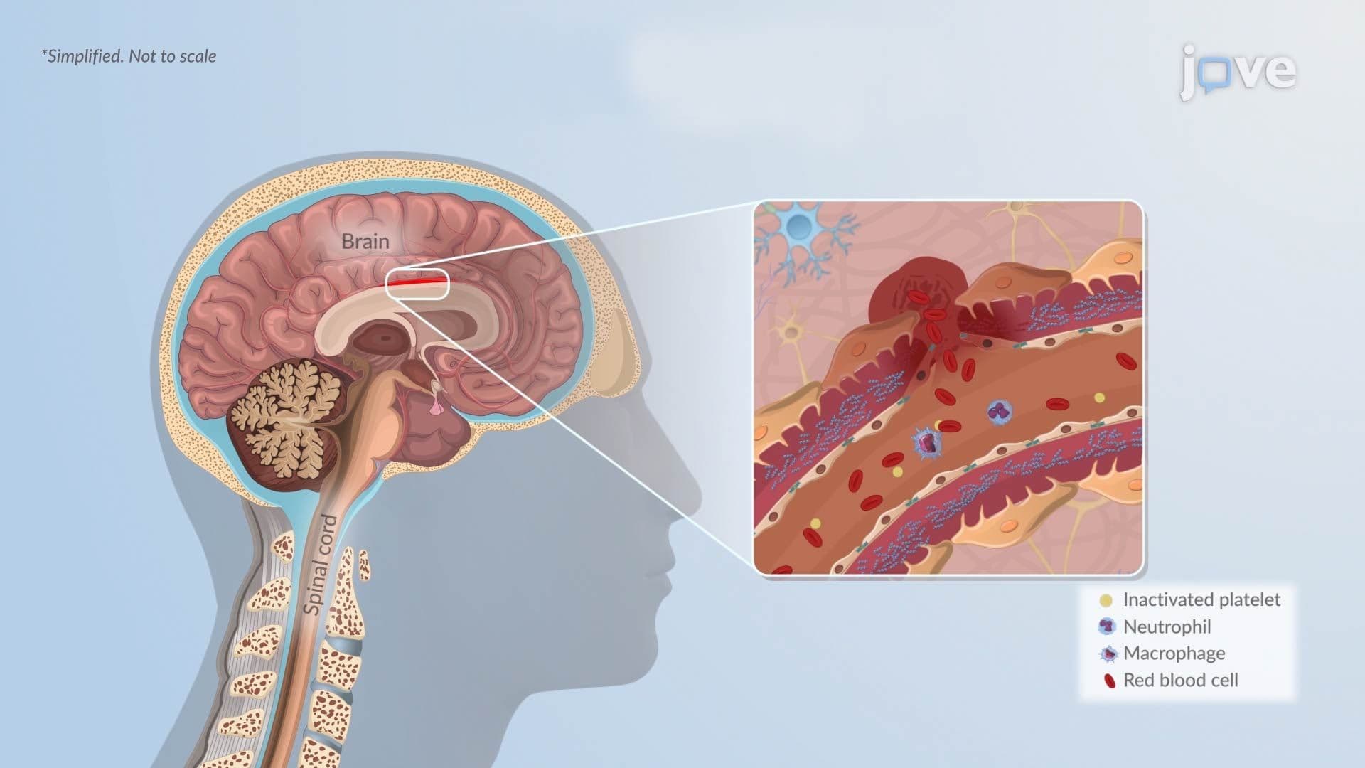

Video Summary: What Is Hemorrhagic Stroke L
Every year, nearly 800,000 Americans experience a stroke, and the bleeding kind can be the most devastating. Hemorrhagic Stroke L occurs when a weakened brain blood vessel ruptures, flooding surrounding tissue with blood and triggering dangerous swelling or compression. At major US trauma centers like Johns Hopkins, rapid identification of hemorrhagic stroke type can mean the difference between life and death. Watch the full video on JoVE Coach to master this concept with expert-led visuals and step-by-step explanations.
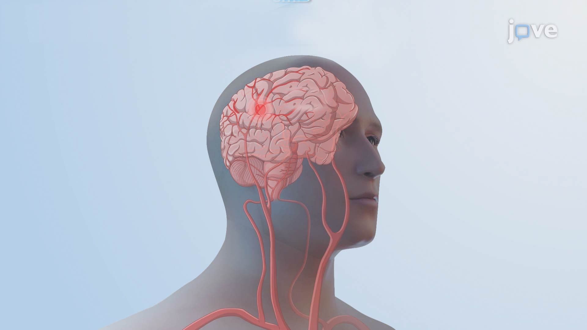
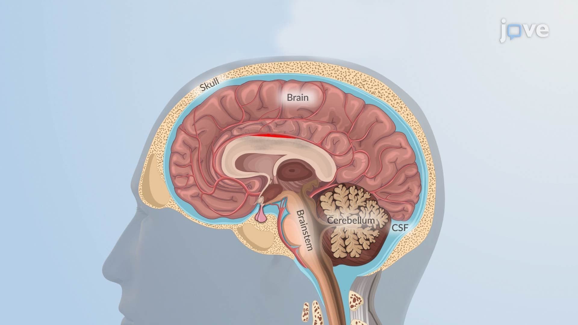
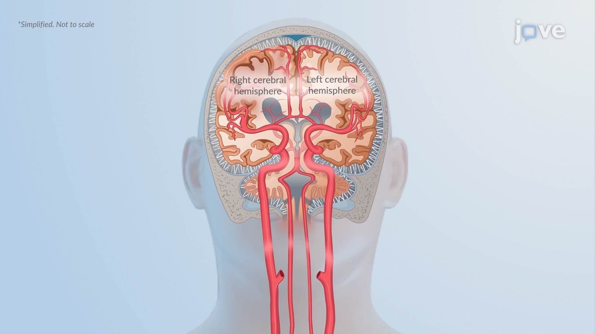
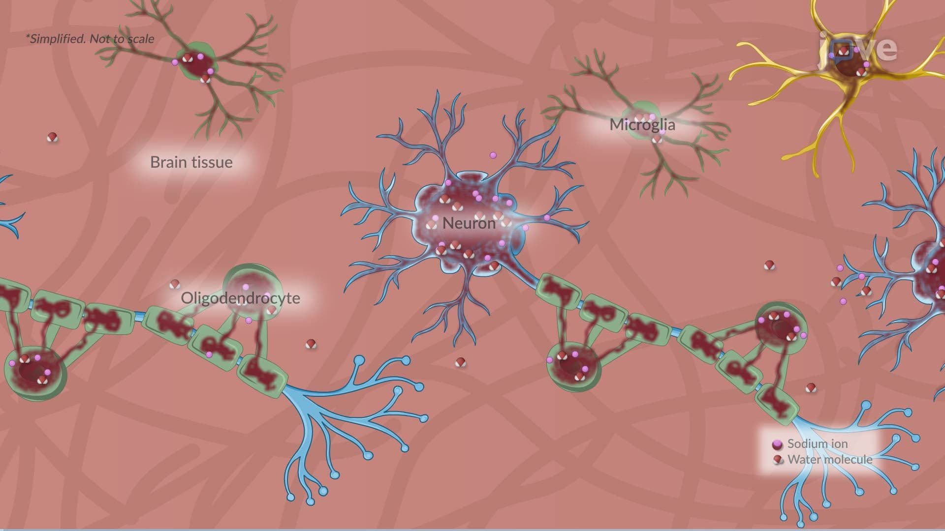
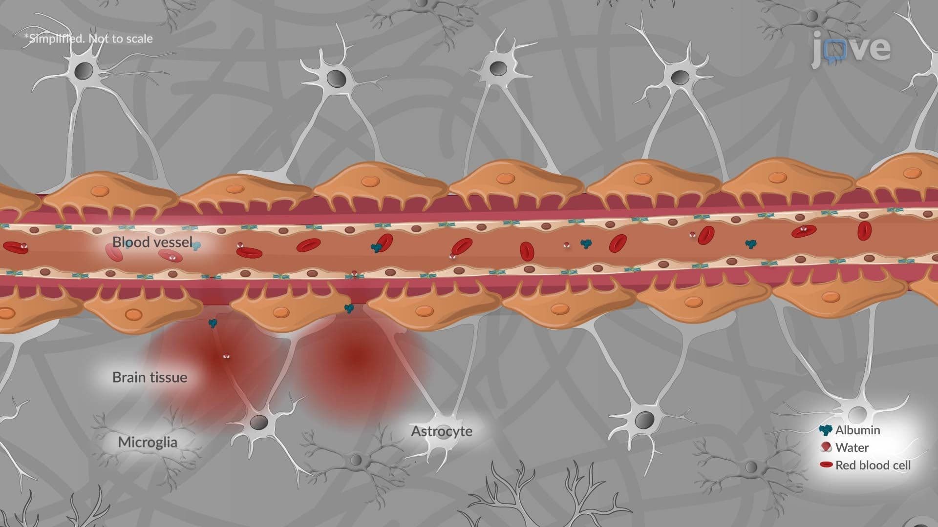
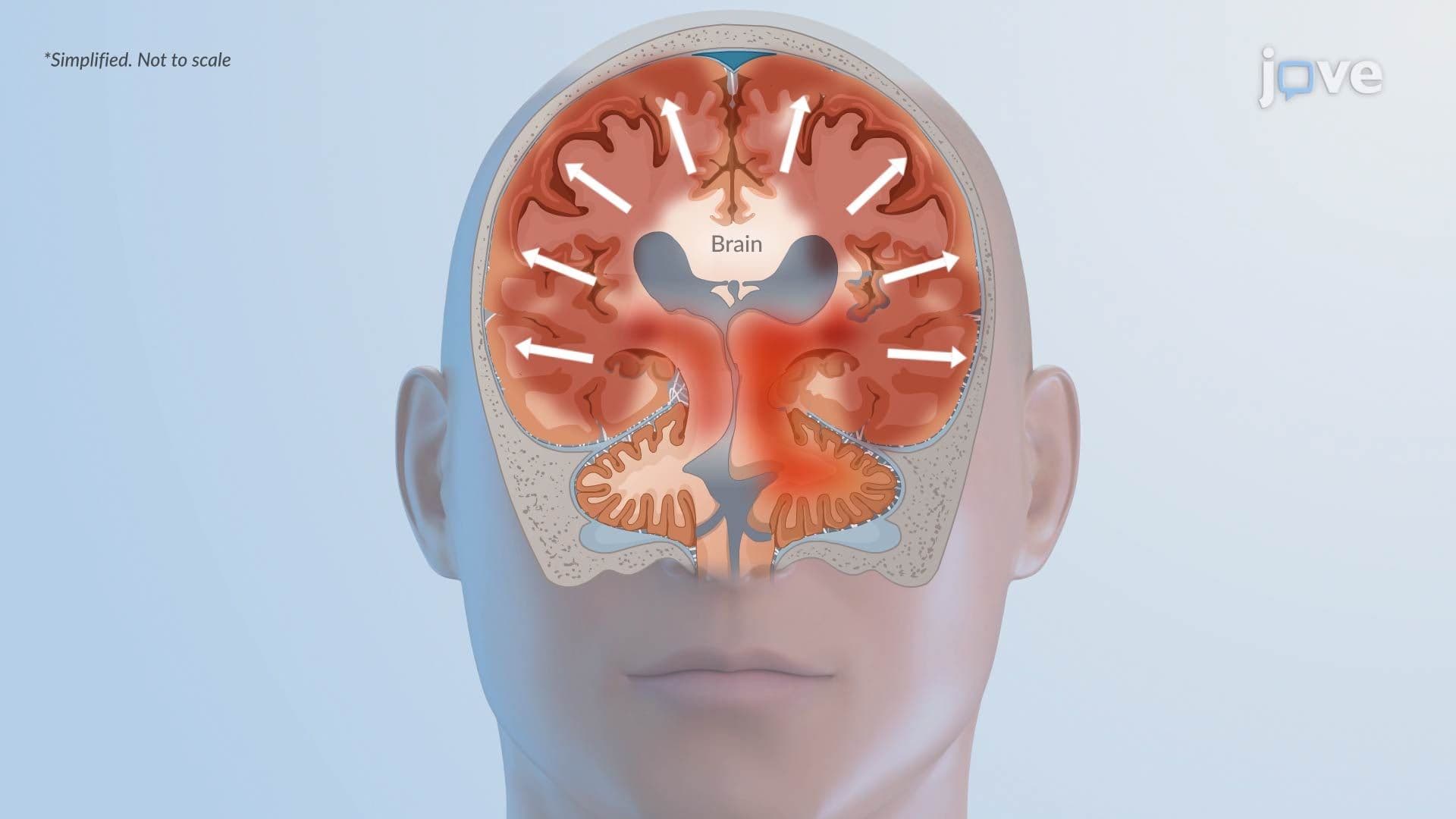
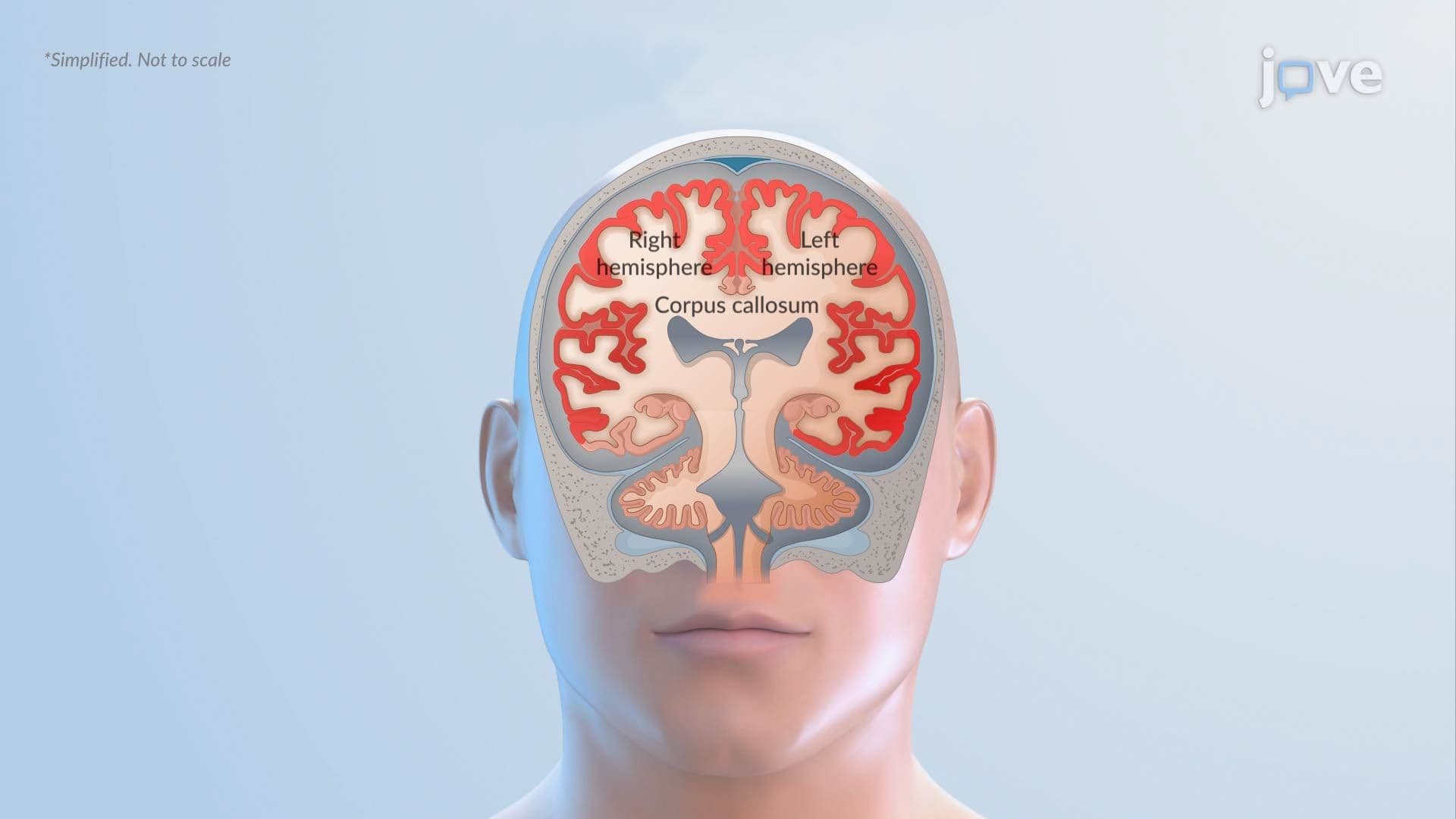
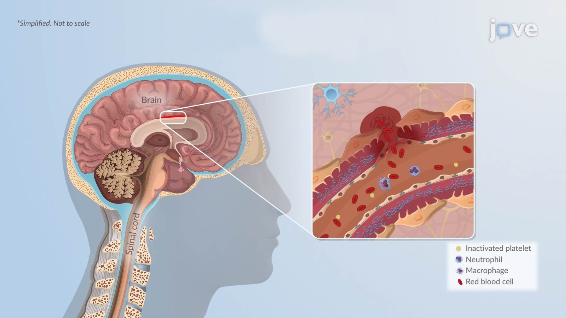
Hemorrhagic Stroke L is a life-threatening neurological event that occurs when a blood vessel inside or around the brain ruptures and bleeds. Unlike ischemic stroke, which results from a blocked artery, hemorrhagic stroke involves active bleeding that creates a focal hematoma, a localized pool of blood that compresses and damages surrounding brain tissue. This compression can trigger a cascade of complications including brain swelling (cerebral edema), increased intracranial pressure, and in severe cases, intraventricular hemorrhage, where blood floods the brain's internal fluid-filled chambers. Understanding this mechanism is foundational for any student studying neurology, anatomy, or pathophysiology.
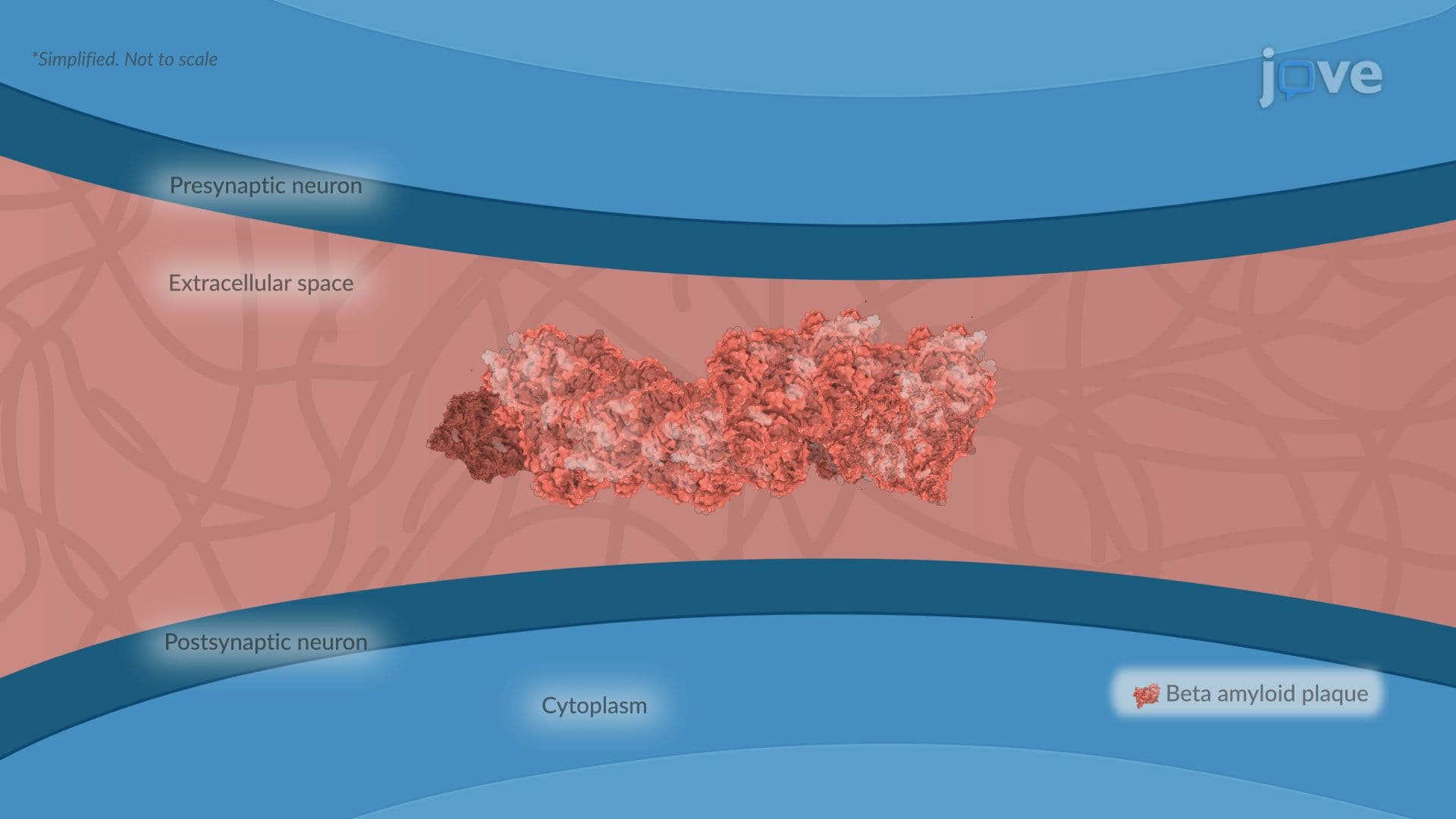
Intracerebral hemorrhage (ICH) is the most common form of hemorrhagic stroke and occurs when a blood vessel ruptures directly into the brain parenchyma, the functional tissue of the brain itself. The most frequent cause is chronic hypertension, which over time weakens the walls of small cerebral blood vessels, making them prone to rupture. A second major cause is cerebral amyloid angiopathy (CAA), a condition where abnormal amyloid protein deposits accumulate in vessel walls, stiffening and weakening them. CAA is notably associated with Alzheimer's disease, making it a critical bridge concept between neurodegenerative diseases and stroke. Arteriovenous malformations (AVMs), tangled webs of abnormal blood vessels that bypass normal capillary networks, represent a less common but serious cause, particularly in younger patients. In US emergency departments, non-contrast CT scans are the first-line imaging tool used to confirm ICH diagnosis quickly.
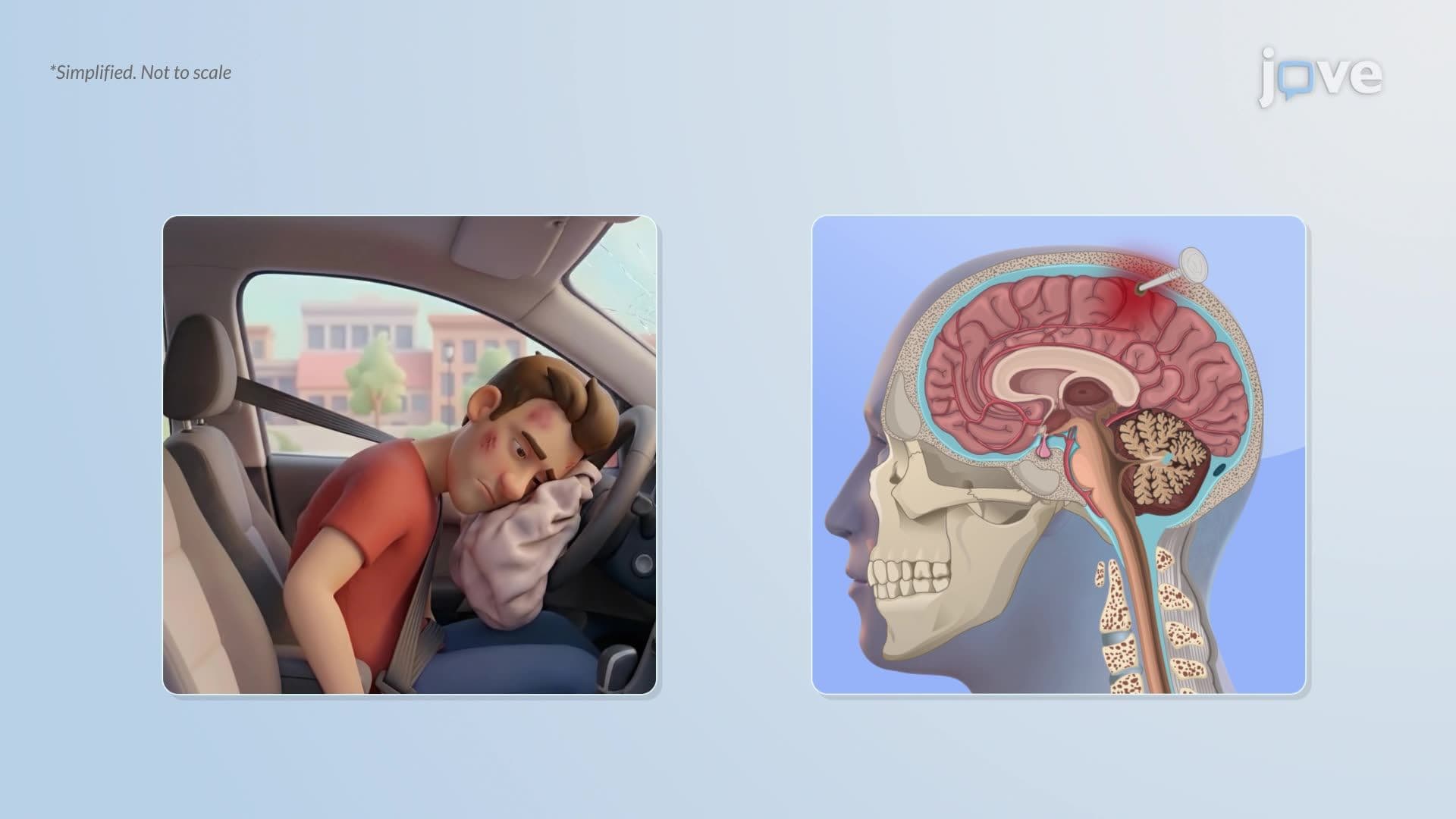
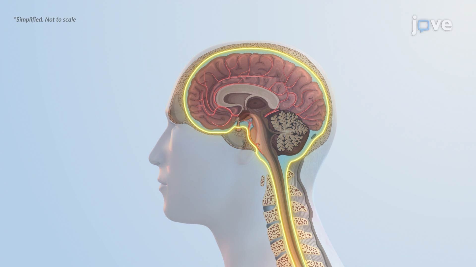
Subarachnoid hemorrhage (SAH) involves bleeding into the subarachnoid space, the fluid-filled gap between the arachnoid mater and the pia mater, two of the brain's three protective meningeal layers. The most common cause is the rupture of a berry aneurysm, a small, balloon-like bulging of an arterial wall that forms at vessel branch points in the circle of Willis. When a berry aneurysm bursts, blood rapidly fills the subarachnoid space, causing a sudden, severe "thunderclap headache", often described by patients as the worst headache of their life. Head trauma is another significant cause of SAH and is commonly seen in sports-related injuries or motor vehicle accidents across the US. Diagnosis typically involves CT imaging followed by lumbar puncture if imaging is inconclusive.
For high school and college students, hemorrhagic stroke is a high-yield concept that appears across multiple disciplines. In AP Biology and AP Psychology, understanding how nervous system disorders disrupt normal brain function provides essential context. On the MCAT, questions frequently test the distinction between stroke types, vascular anatomy, and the pathophysiology of neurological conditions. In college-level neuroscience or anatomy and physiology courses, hemorrhagic stroke serves as a concrete example of how structural brain damage leads to functional deficits. Clinically, nurses preparing for the NCLEX must understand hemorrhagic stroke assessment, while pre-med students studying for the USMLE need to differentiate ICH from SAH by cause, location, and imaging findings. Connecting hemorrhagic stroke to broader nervous system disorders, including epilepsy, multiple sclerosis, and Parkinson's disease, strengthens your overall understanding of how the brain responds to injury and disease.
Related Micro-courses