Loading video…

 35 Concepts
 5.1K views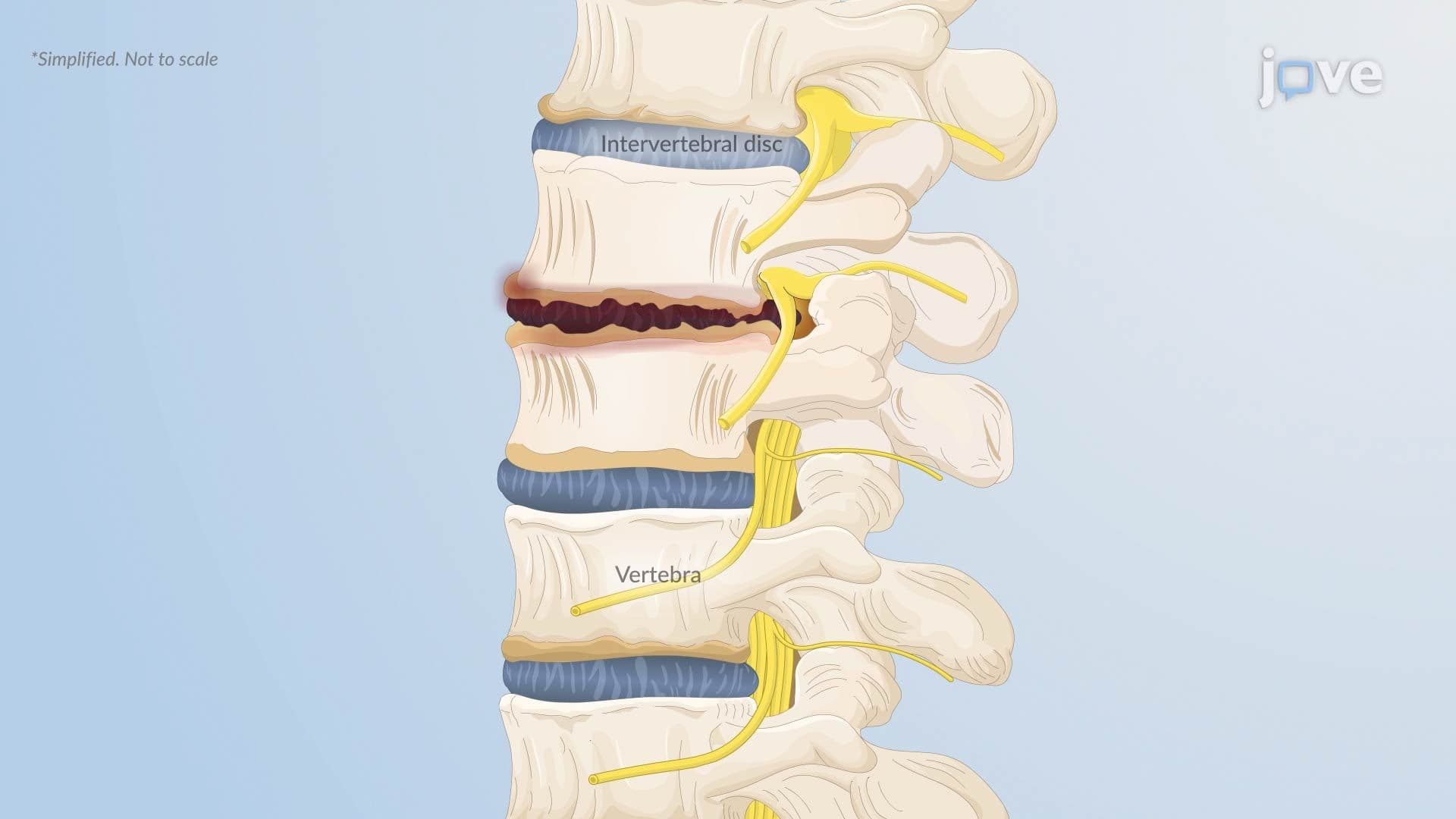

Video Summary: What Is Herniated Intervertebral Disc L
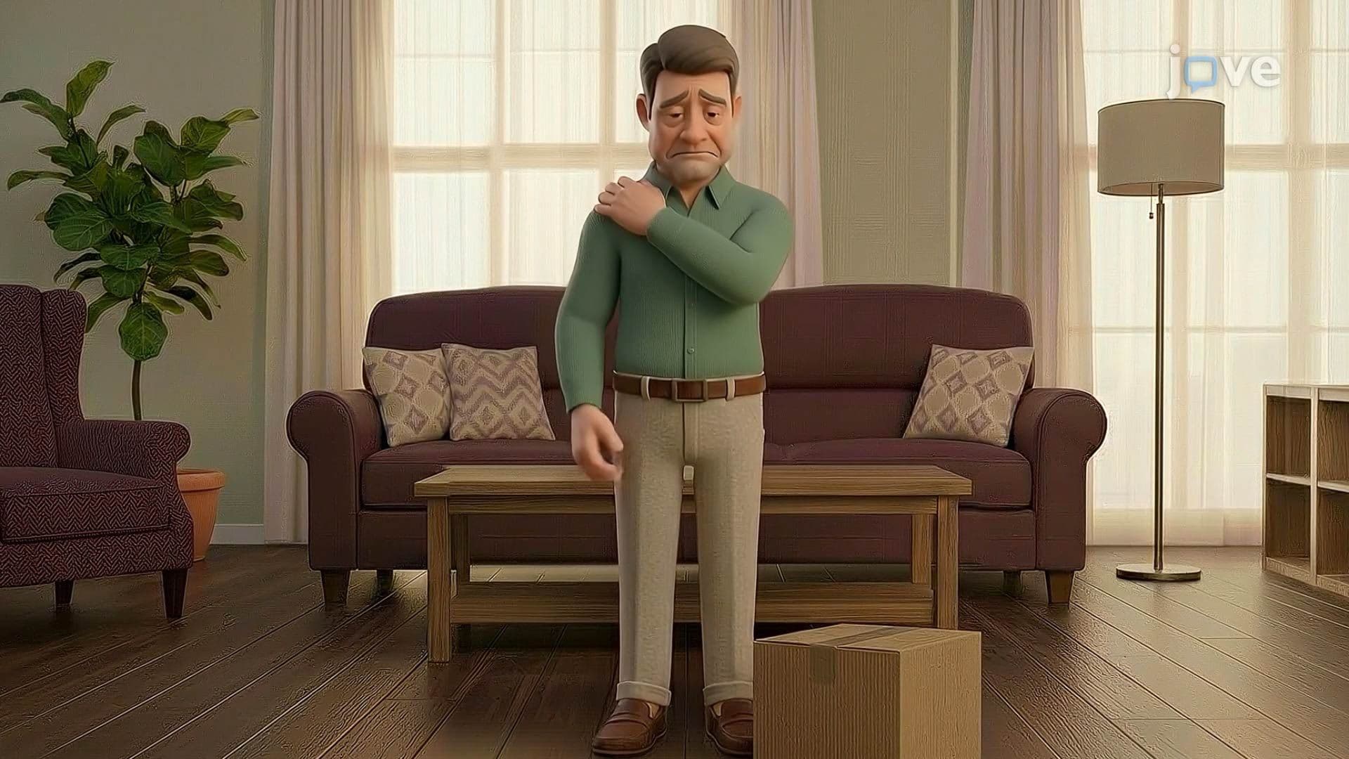
Ever wonder why a simple sneeze or awkward lift can send shooting pain down your entire leg? Herniated Intervertebral Disc L, a condition affecting the lumbar spine, occurs when the soft inner core of a spinal disc ruptures and presses on nearby nerves. NFL athletes and warehouse workers across the US commonly experience this injury. Watch the full video on JoVE Coach to master this concept with expert-led visuals and step-by-step explanations.
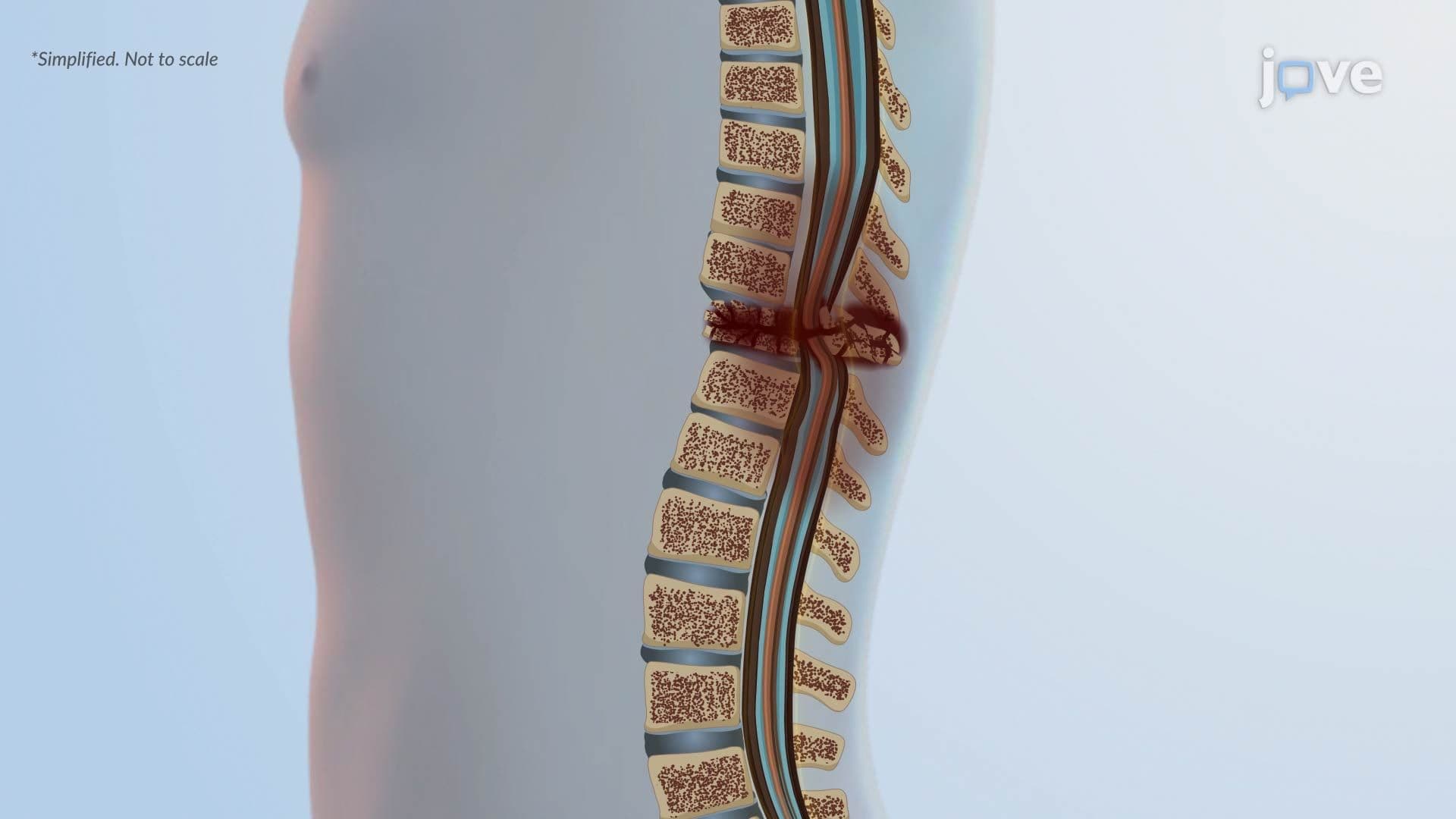
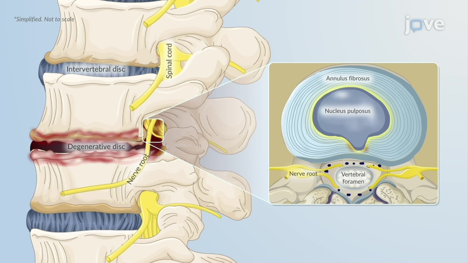
The human spine is an engineering marvel, flexible enough to allow movement yet strong enough to protect the spinal cord. Sandwiched between each pair of vertebrae is an intervertebral disc, a shock-absorbing structure with two distinct parts: the nucleus pulposus (a gel-like inner core) and the annulus fibrosus (a tough, fibrous outer ring). A herniated intervertebral disc L occurs when the nucleus pulposus is forced through a crack or weakness in the annulus fibrosus, causing disc material to bulge or fully rupture beyond its normal boundary. When that displaced material contacts a nearby spinal nerve root, pain and neurological symptoms follow.
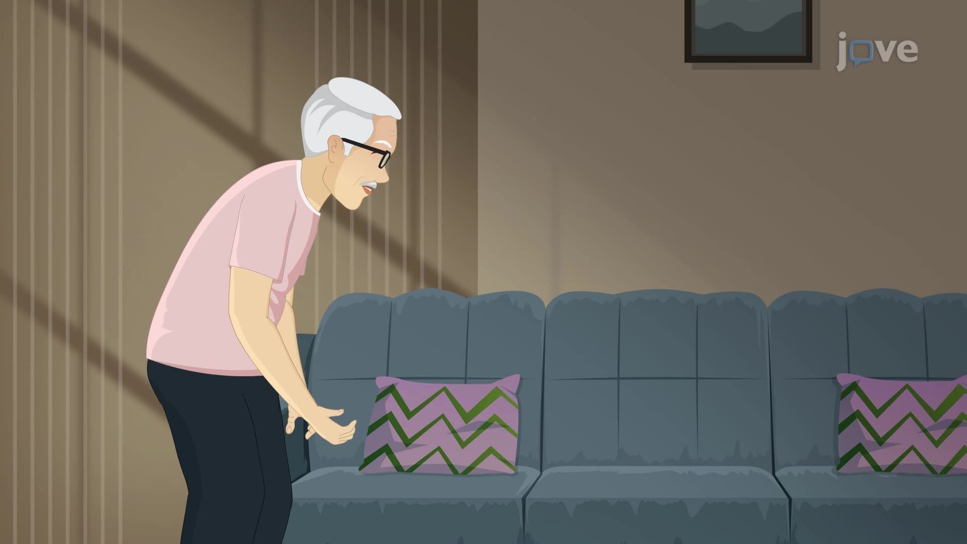
Not all spinal levels herniate equally. The lumbar region, specifically the L4-L5 and L5-S1 segments, absorbs the majority of the body's compressive and shear forces during everyday activities like walking, lifting, and sitting. This mechanical burden, combined with the natural reduction in disc hydration that comes with aging, makes the lower lumbar spine the most common site for herniation. In the US, lumbar disc herniation is one of the leading causes of missed workdays and is frequently seen in occupations requiring repetitive bending, twisting, or heavy lifting, such as construction, nursing, and warehouse logistics.
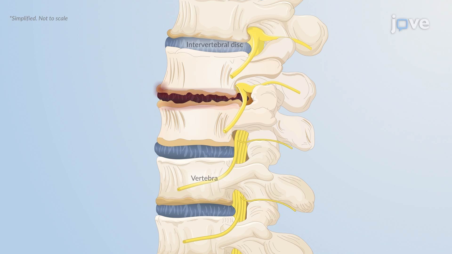
Disc herniation rarely happens in isolation. Most cases develop gradually through a process called degenerative disc disease, where discs lose water content and structural integrity over time. This degeneration creates micro-tears in the annulus fibrosus, setting the stage for herniation. Risk is amplified by:
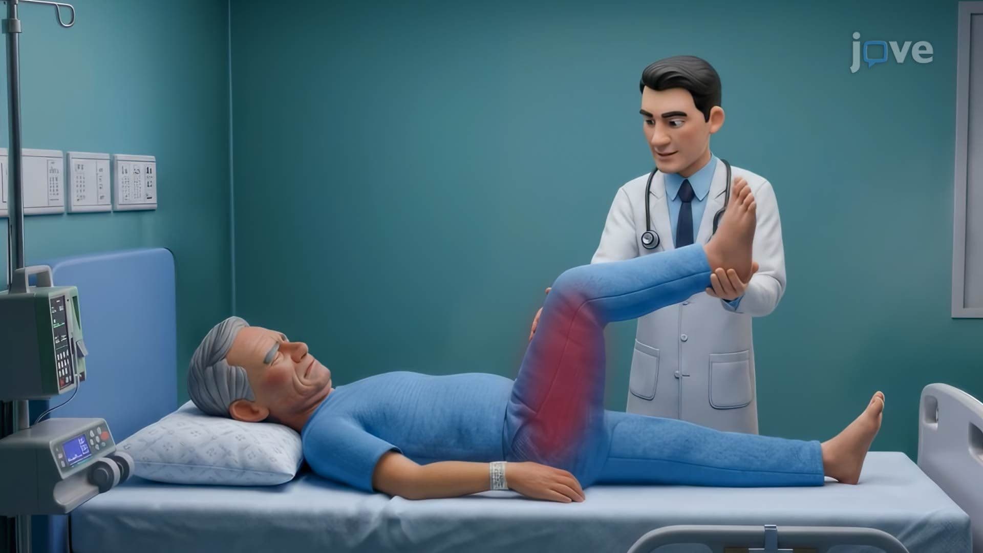
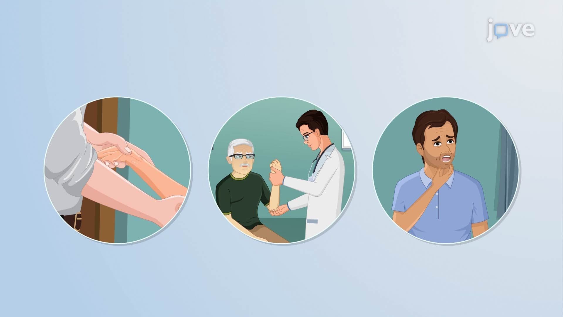
The defining clinical feature of lumbar disc herniation is radiculopathy, pain, tingling, or weakness that travels along the distribution of a compressed nerve root. When herniation at L4-L5 or L5-S1 compresses the sciatic nerve, patients experience sciatica: a sharp, shooting pain that radiates from the lower back through the buttock and down the leg, sometimes reaching the foot. The specific pattern of symptoms helps clinicians pinpoint which disc level is involved. For example, L5 nerve root compression often causes weakness in foot dorsiflexion, while S1 involvement typically produces diminished ankle reflex and pain along the lateral foot.
Herniated intervertebral disc L is a high-yield topic in US anatomy and physiology courses, pre-health programs, and standardized exams. On the MCAT, students are expected to understand spinal anatomy, nerve root distributions, and the pathophysiology of disc herniation in the context of musculoskeletal and nervous system function. In AP Biology and college-level A&P, herniated discs illustrate key concepts including connective tissue structure, nerve compression, and degenerative disease progression. Understanding this condition also bridges directly into clinical reasoning for students pursuing careers in physical therapy, medicine, or nursing.
Related Micro-courses