Loading video…

 35 Concepts
 5.1K views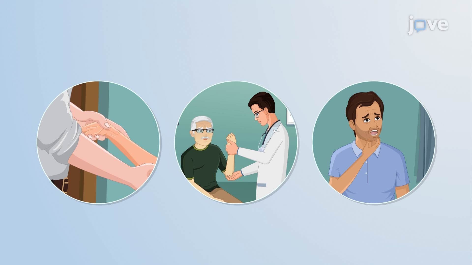

Video Summary: Alterations in Muscle Tone Ll Explained
Why does a stroke survivor's arm resist movement differently than someone with dementia? That puzzle sits at the heart of alterations in muscle tone ll. This concept covers three complex conditions, spasticity, paratonia, and dystonia, that disrupt normal muscle control through distinct neurological mechanisms. A US veteran recovering from stroke, for example, may experience velocity-dependent spasticity that makes physical therapy uniquely challenging. Watch the full video on JoVE Coach to master this concept with expert-led visuals and step-by-step explanations.
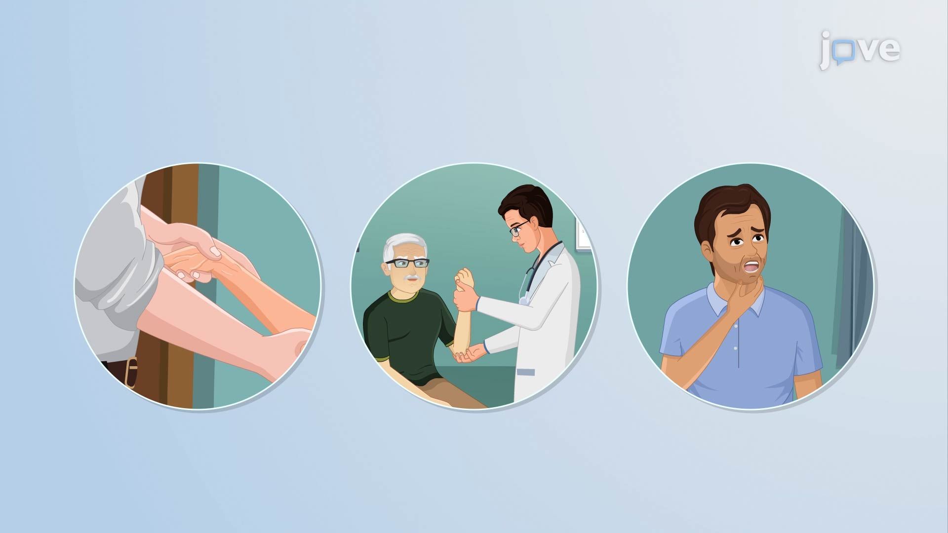
When muscle tone goes wrong in nuanced ways, the results can be disabling, confusing to diagnose, and deeply tied to specific areas of brain damage. Alterations in Muscle Tone ll moves beyond simple hypotonia or hypertonia to examine three clinically distinct conditions: spasticity, paratonia, and dystonia. Each one reflects a different breakdown in the nervous system's ability to regulate muscle activation, and each is associated with recognizable neurological diseases that appear frequently in US classrooms, clinics, and standardized exams.
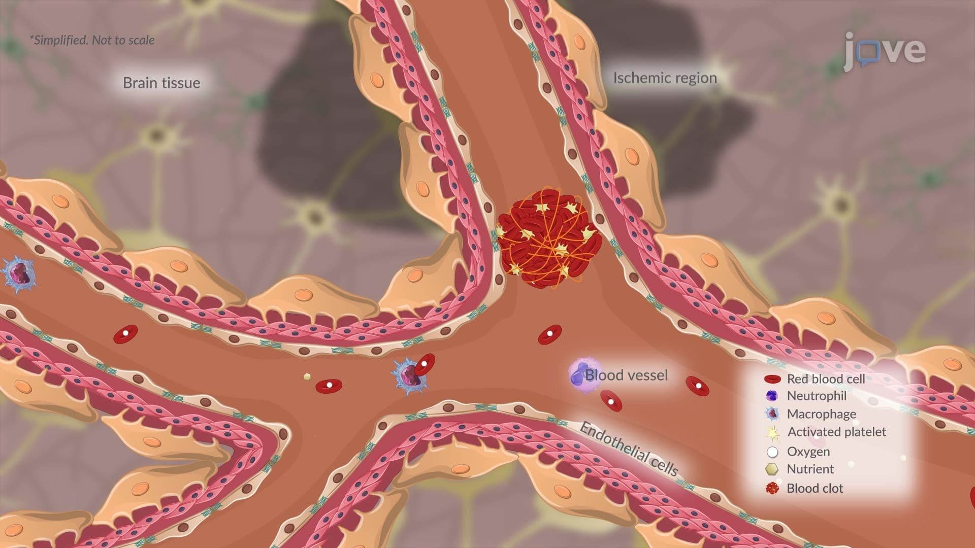
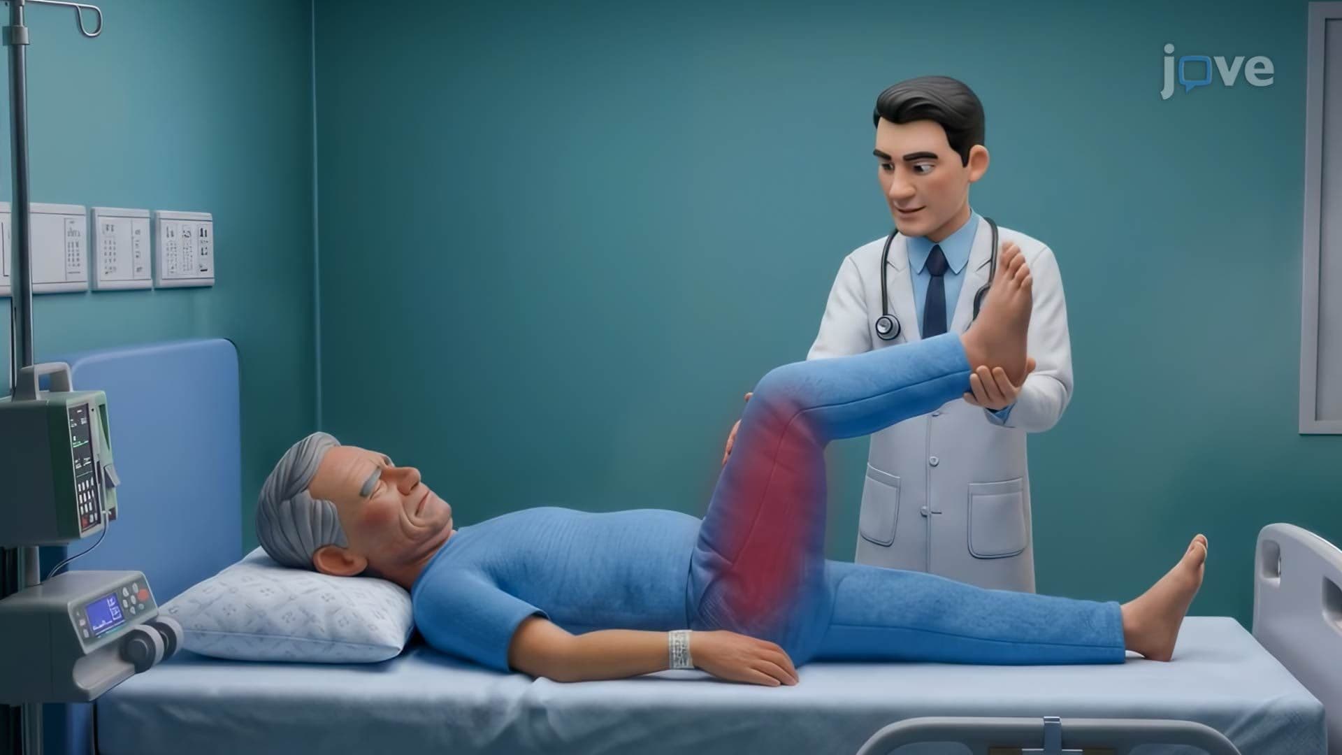
Spasticity is best understood through one defining characteristic, it is velocity dependent. This means the faster a clinician or examiner stretches a muscle, the more resistance they will feel. A slow, gentle movement may produce little pushback, while a rapid stretch triggers significant stiffness. This happens because upper motor neuron (UMN) damage disrupts the descending pathways that normally regulate spinal reflex arcs. Without proper inhibition from above, the stretch reflex becomes hyperactive.
Spasticity is commonly seen following stroke, in individuals with cerebral palsy, and in those diagnosed with multiple sclerosis (MS). In the United States, nearly 800,000 people suffer a stroke annually, and spasticity affects an estimated 30% of survivors, making it one of the most clinically significant muscle tone alterations in rehabilitation medicine. Physical therapists working in US hospitals routinely use passive range-of-motion assessments and the Modified Ashworth Scale to measure and track spasticity over time.
On AP Biology and college-level neuroscience exams, students are often asked to explain why UMN lesions produce hyperreflexia. The key insight is the loss of inhibitory control, not the neuron damage itself, but the downstream consequences for reflex regulation.
Paratonia, also called gegenhalten (German for "counter-hold"), presents very differently from spasticity. The resistance it generates is not velocity dependent. Instead, it tends to increase proportionally with the force the examiner applies, the harder you push, the harder the muscle pushes back. This involuntary, seemingly oppositional response can make neurological examinations challenging.
Paratonia is strongly associated with frontal lobe dysfunction. The frontal lobe plays a central role in voluntary motor inhibition and executive control. When it deteriorates, as it does in Alzheimer's disease and other dementias, patients lose the ability to consciously relax muscles during passive movement. In US memory care units, clinicians regularly observe paratonia as dementia progresses, making it a meaningful clinical marker of frontal lobe decline. NCLEX and USMLE prep materials often include paratonia as part of dementia-related neurological assessment questions.
Dystonia is a movement disorder rooted in abnormal signaling across motor control networks, specifically the motor cortex, basal ganglia, and cerebellum. Under normal conditions, these structures work together to initiate movement, refine it, and inhibit unnecessary muscle activation. In dystonia, this inhibition breaks down. The result is excessive, uncontrolled muscle activation that produces sustained contractions, twisting movements, and fixed abnormal postures.
Dystonia can be primary (genetic) or secondary (caused by another condition like Parkinson's disease, brain injury, or certain medications). In the US, dystonia affects an estimated 500,000 people, and it is frequently misdiagnosed early on. Cervical dystonia, involuntary neck twisting, is one of the most recognizable forms and is treated with botulinum toxin injections at major US neurology centers.
For college-level courses in neuroscience or pathophysiology, understanding dystonia requires grasping the basal ganglia's role in motor suppression. This is a high-yield topic on the MCAT, where questions may ask students to connect basal ganglia dysfunction to specific motor symptoms.
All three conditions discussed in Alterations in Muscle Tone ll are clinically relevant to major neurological disorders that students encounter across biology, health science, and pre-med curricula. Stroke, multiple sclerosis, Parkinson's disease, Alzheimer's disease, and epilepsy all involve disruptions to motor control pathways in ways that can produce spasticity, paratonia, or dystonia. Recognizing which condition fits which disease, and why, is exactly the kind of integrative thinking that separates strong performers on MCAT, USMLE Step 1, and college neuroscience midterms.
Related Micro-courses