Loading concepts…

 29 Concepts
 4.5K Views
 Popular topics
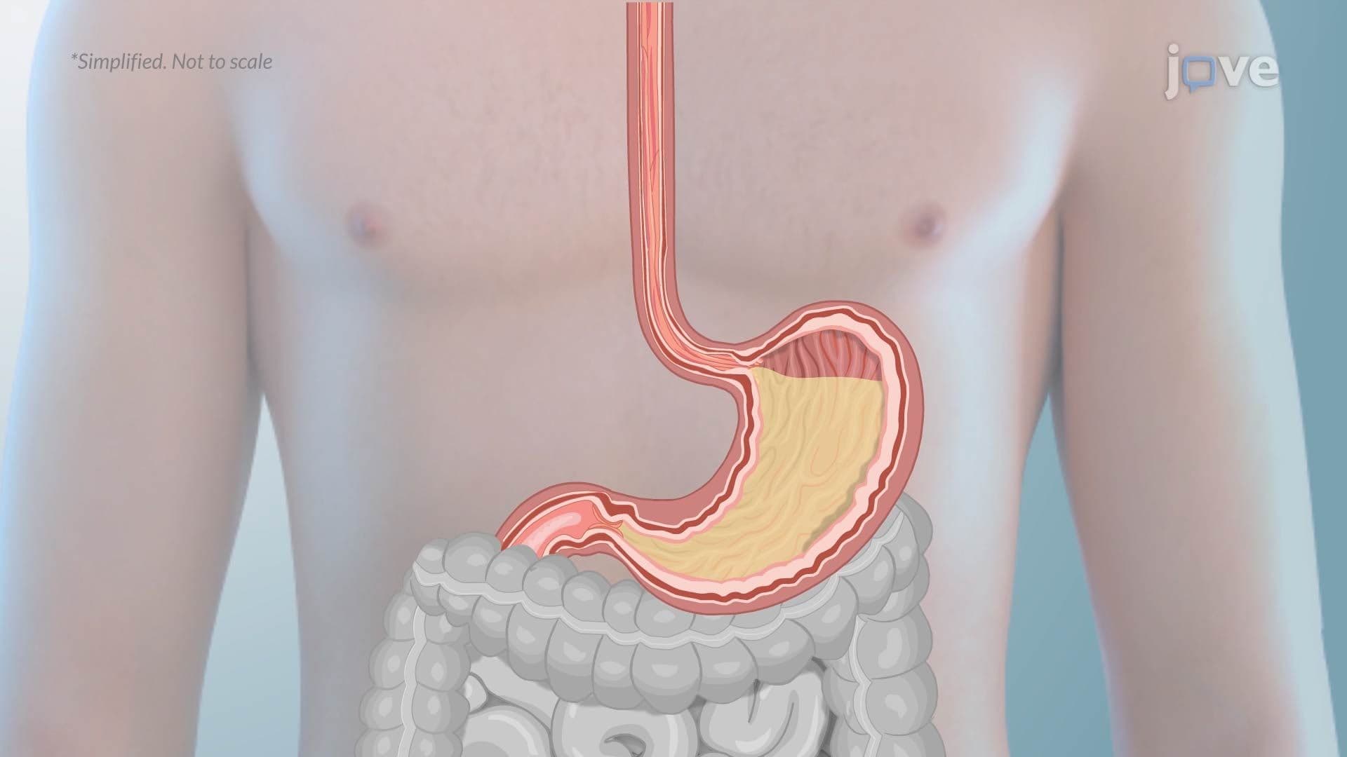



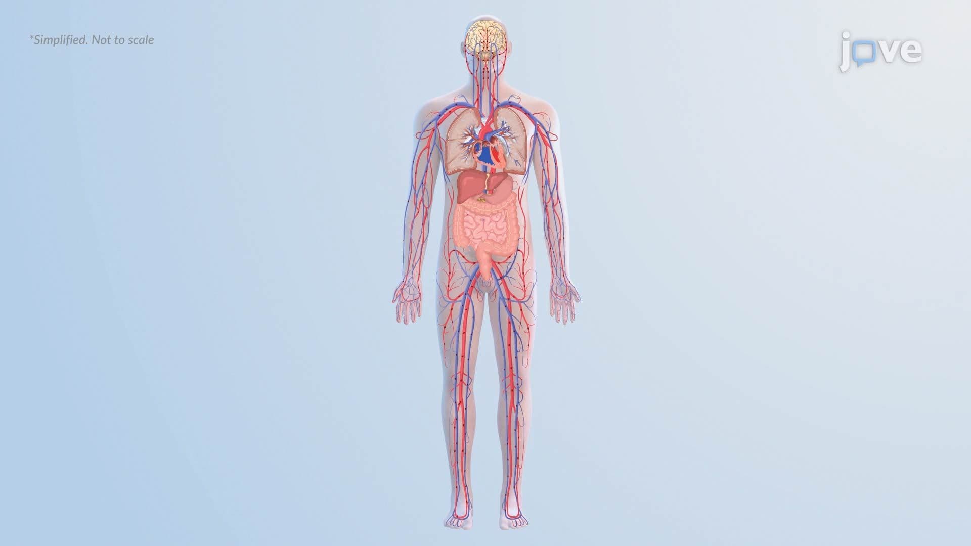

Gastrointestinal disorders represent some of the most clinically significant conditions covered in US health science education. This micro-course examines the full spectrum of common GI tract diseases, from esophageal achalasia and GERD to peptic ulcer disease, IBD, cirrhosis, and acute pancreatitis, building mechanistic understanding through pathophysiology, clinical manifestations, and complications. JoVE Coach supports learners in connecting cellular-level dysfunction to patient outcomes.
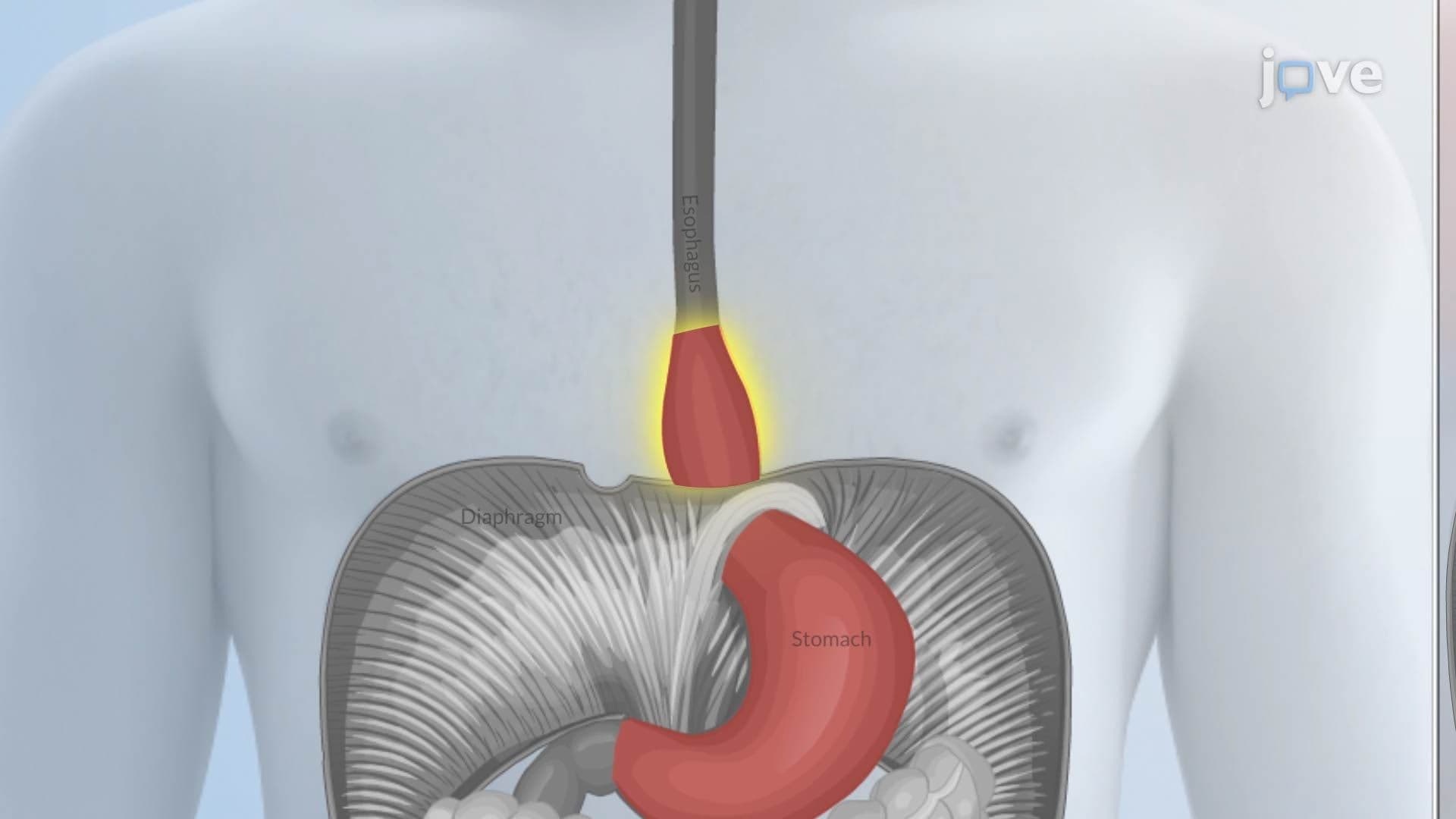
1. Esophageal Disorders: Achalasia, GERD, and Hiatal Hernia Esophageal achalasia involves failure of the lower esophageal sphincter (LES) to relax due to degeneration of inhibitory neurons in the myenteric plexus, impairing the nitric oxide signaling needed for normal LES relaxation. GERD occurs when the LES relaxes inappropriately, allowing acidic stomach contents to reflux into the esophagus, causing inflammation, Barrett esophagus, and chronic respiratory symptoms. Hiatal hernias, classified as sliding (Type I), paraesophageal (Type II), combined (Type III), or complex (Type IV), involve upward protrusion of stomach structures through the diaphragm's esophageal hiatus, disrupting the anatomical barriers that normally prevent reflux.
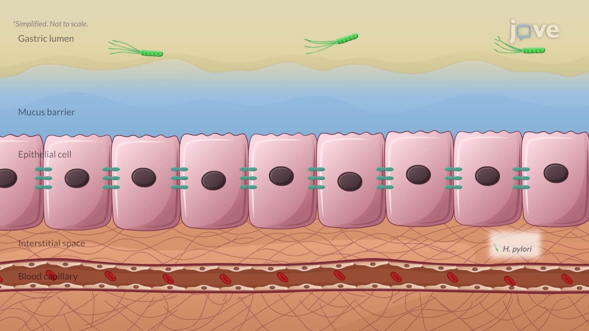
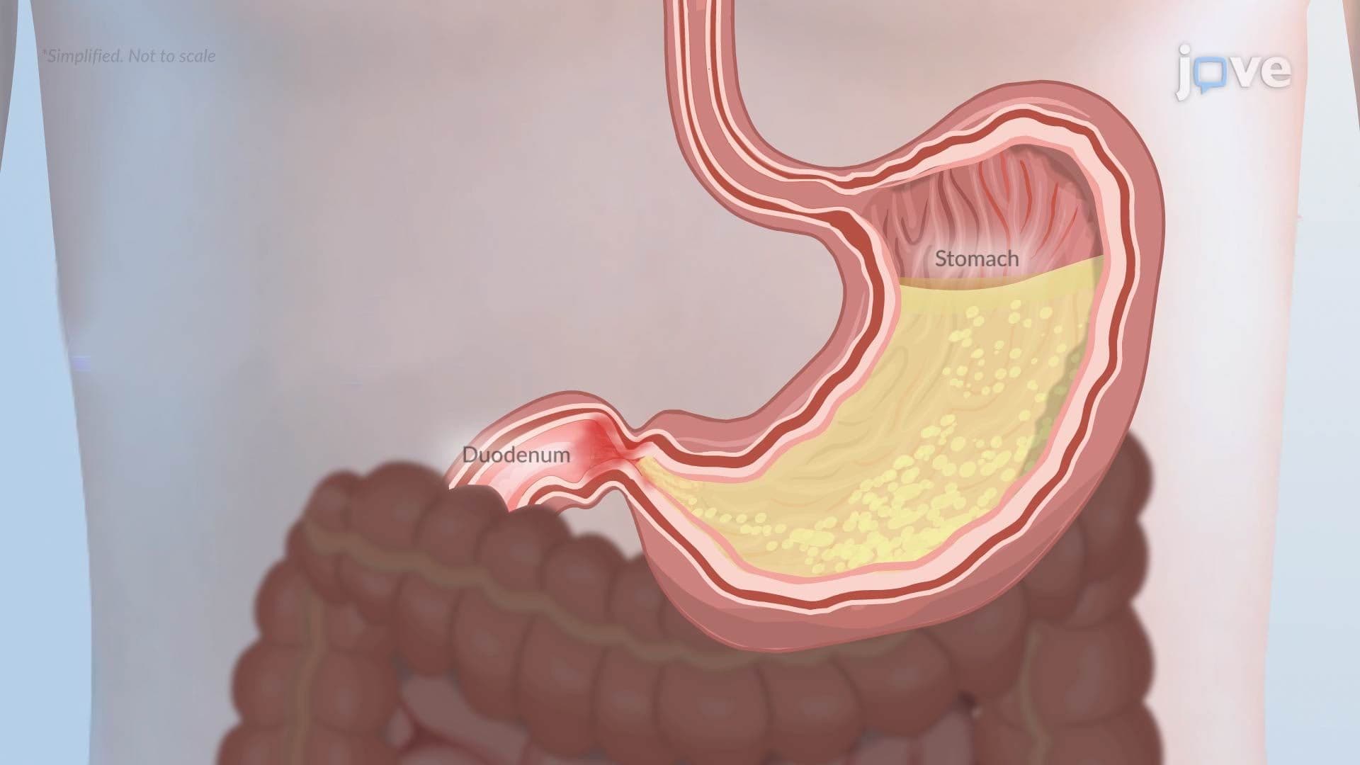
2. Gastritis and Pyloric Obstruction H. pylori-induced gastritis illustrates a critical relationship between bacterial virulence and immune evasion. The bacterium produces urease to neutralize gastric acid, then penetrates the mucus layer and injects virulence factors such as CagA, which disrupts tight junctions and cell signaling. It also triggers PD-L1 expression to suppress T-cell activity, allowing chronic infection. Pyloric obstruction, caused by scarring from chronic peptic ulcer disease or malignancy, blocks gastric emptying, leading to distension, vomiting of undigested food, and dangerous electrolyte imbalances including hypokalemia, hypochloremia, and metabolic alkalosis.
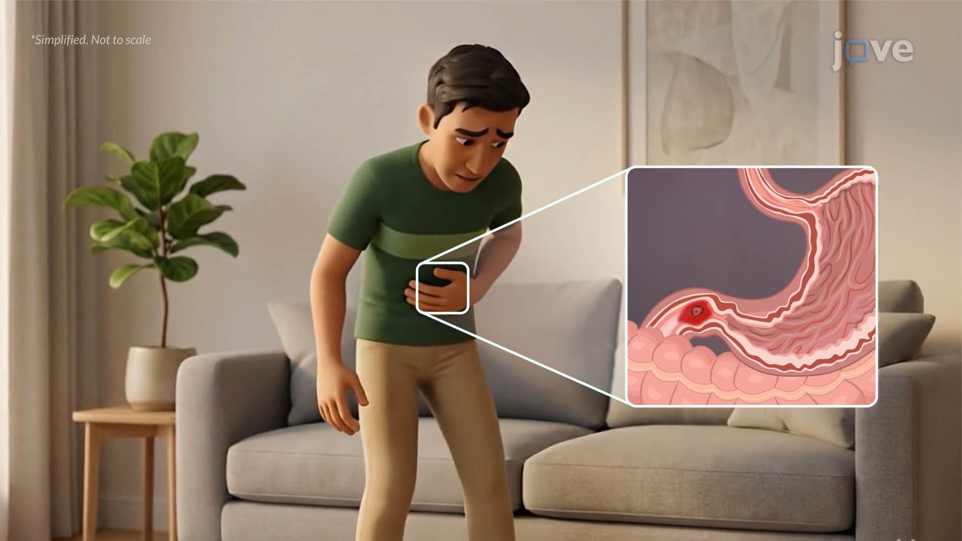
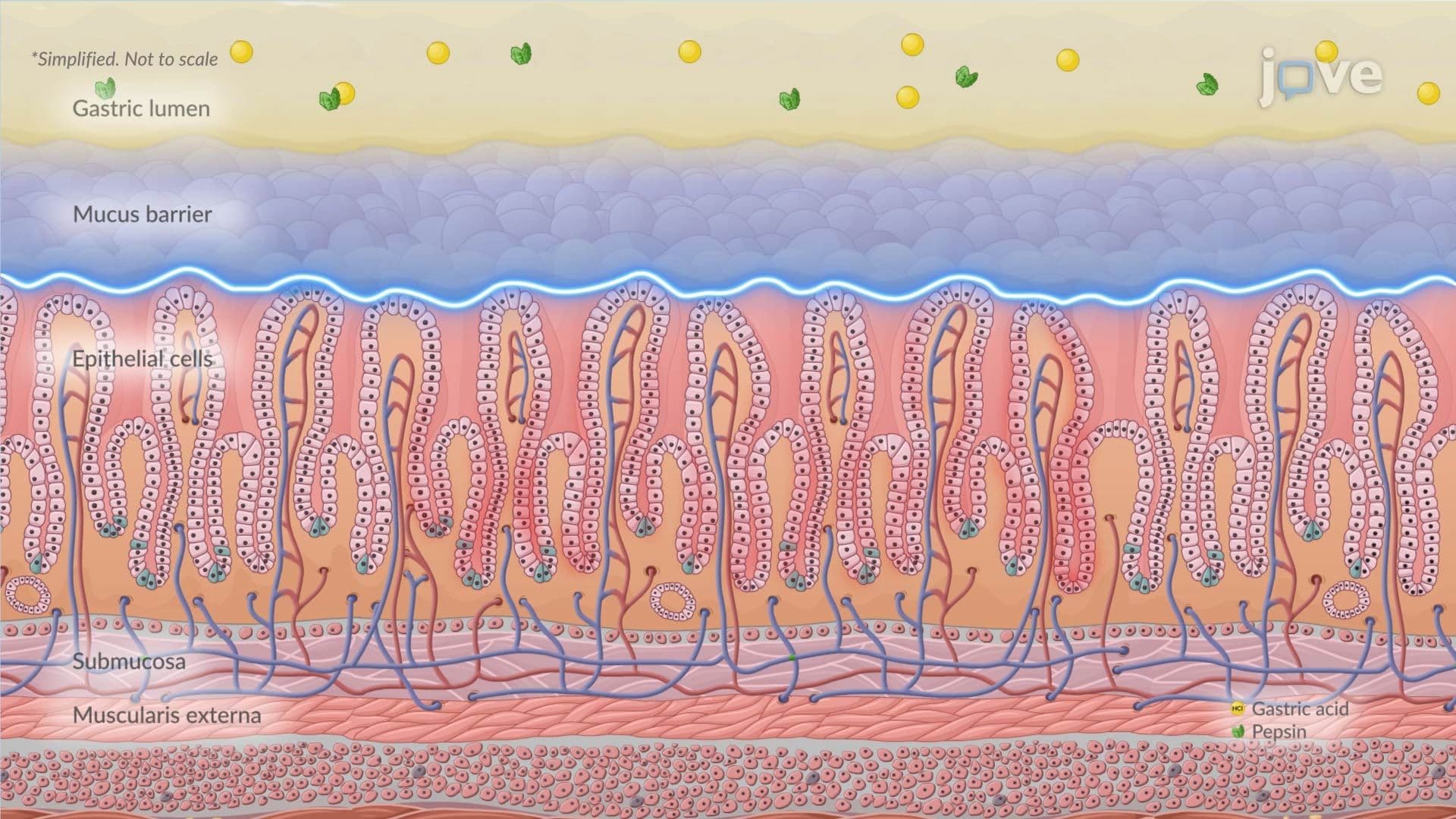
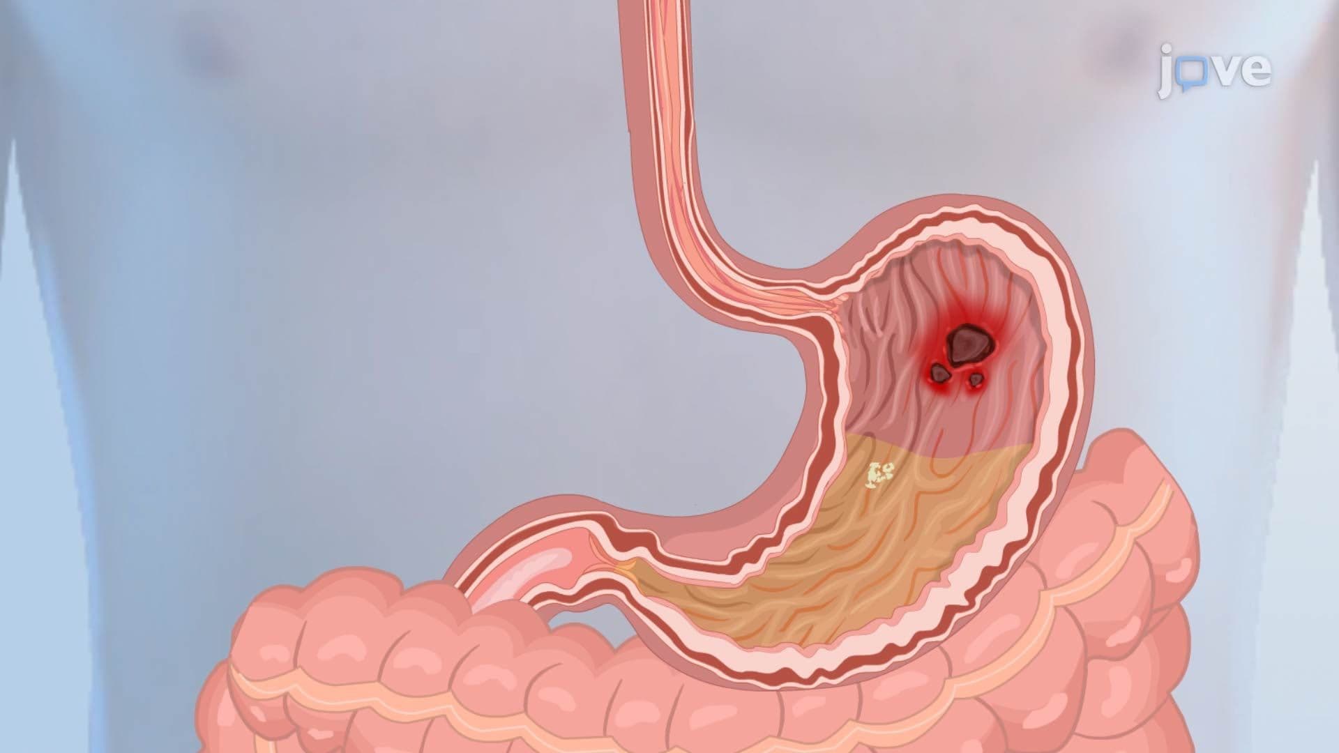
3. Peptic Ulcer Disease (PUD): Mechanisms, Presentation, and Complications Peptic ulcer disease involves erosion of the stomach or duodenal lining through the muscularis mucosae. The two dominant causes are H. pylori infection and NSAID use. H. pylori deploys urease, flagella, CagA, and VacA to breach mucosal defenses and sustain inflammation. NSAIDs inhibit COX-1 enzymes, reducing prostaglandins that normally maintain the protective mucus and bicarbonate layer. Gastric ulcers worsen with eating, while duodenal ulcers follow the classic "pain-food-relief" pattern. Major complications include gastrointestinal bleeding (hematemesis or melena), perforation leading to peritonitis, and gastric outlet obstruction from chronic scarring.
4. Intestinal Obstruction: Mechanical and Functional Causes Intestinal obstruction blocks the normal flow of intestinal contents and is classified as mechanical, such as adhesions, hernias, intussusception, and volvulus, or functional (paralytic ileus), commonly following abdominal surgery or opioid use. Proximally, air and fluid accumulate, causing distension and colicky pain. Sustained obstruction raises intraluminal pressure, impairs venous return, and ultimately compromises arterial blood flow, leading to ischemia, tissue necrosis, and bowel perforation. Perforation spills bacteria-rich intestinal contents into the peritoneal cavity, causing peritonitis, a life-threatening surgical emergency.
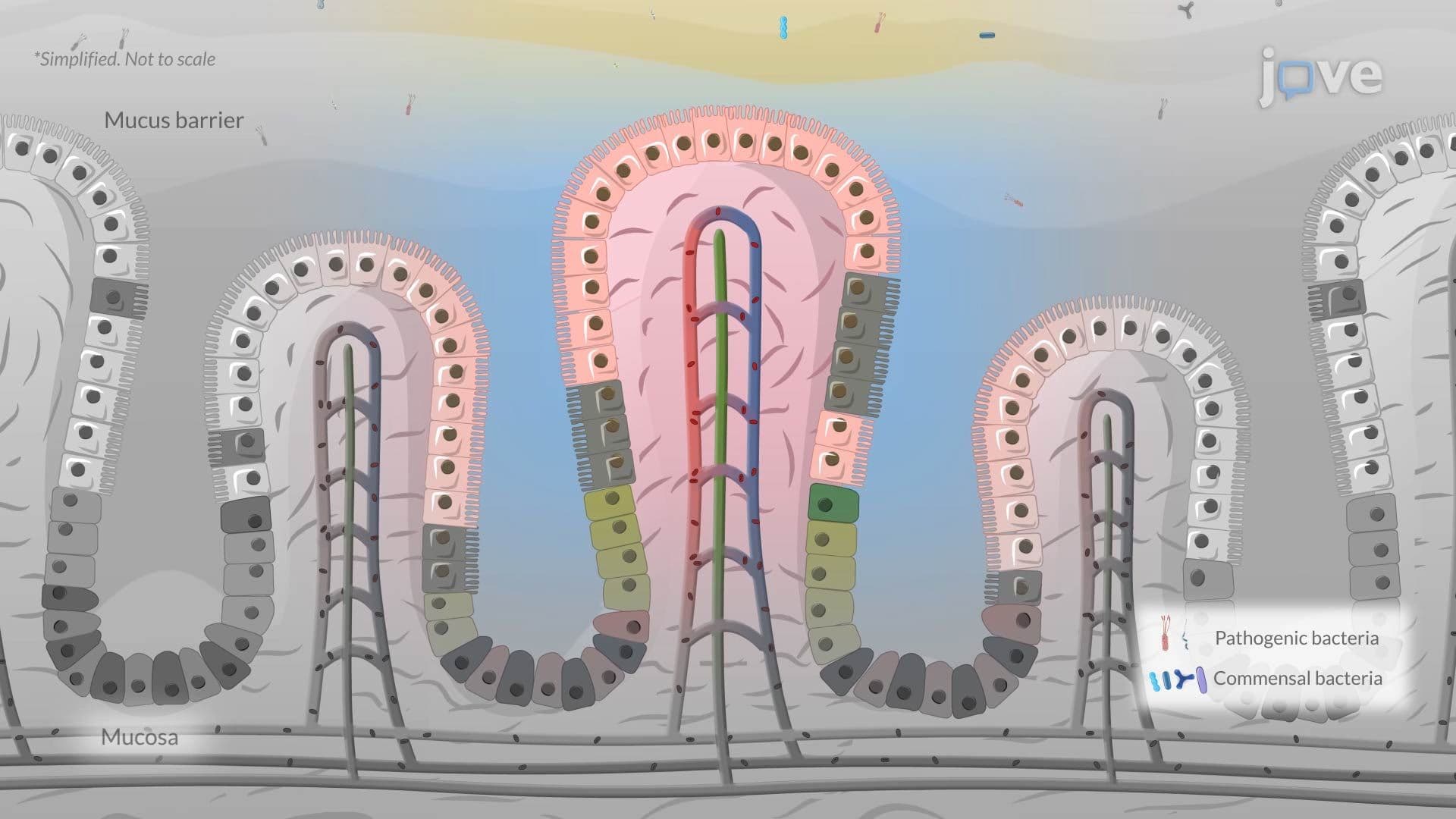
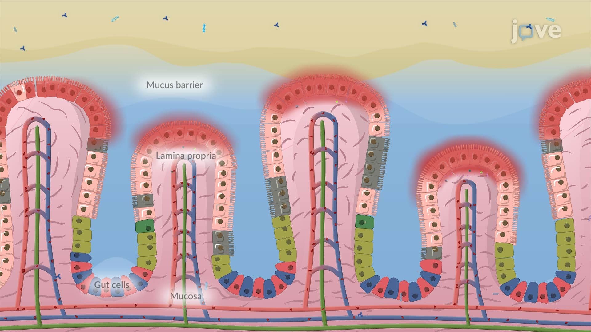
5. Inflammatory Bowel Disease (IBD): Ulcerative Colitis and Crohn's Disease IBD encompasses two major conditions driven by abnormal immune responses to gut flora. Ulcerative colitis is confined to the mucosa and submucosa of the colon and rectum, spreading continuously from the rectum proximally. It involves neutrophil infiltration into the crypts of Lieberkühn, forming crypt abscesses that impair water and electrolyte absorption and cause rectal bleeding. Crohn's disease causes transmural inflammation and can affect any GI segment from mouth to anus. Driven by Th1/Th17 immune responses and cytokines such as TNF-alpha and IL-12, it leads to complications like fistulas, strictures, and malabsorption, including vitamin B12 deficiency when the ileum is involved.
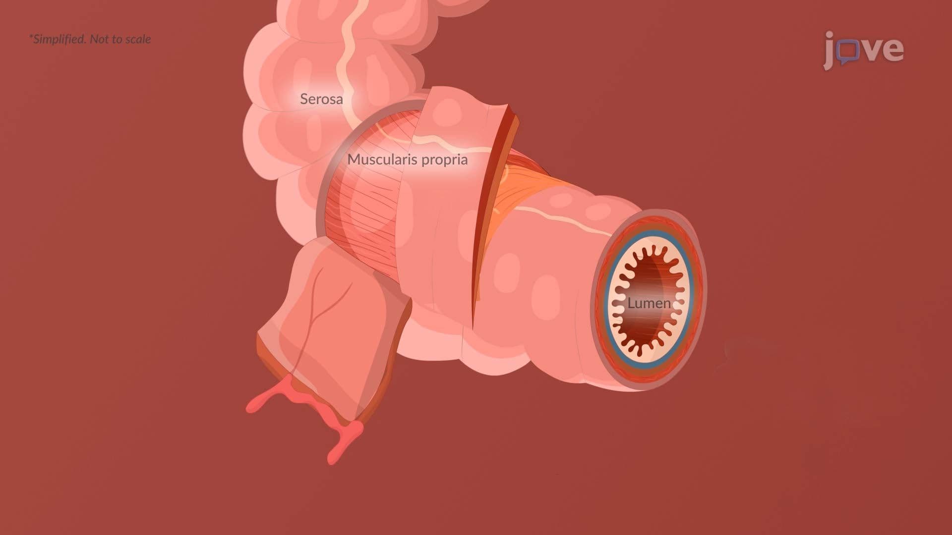
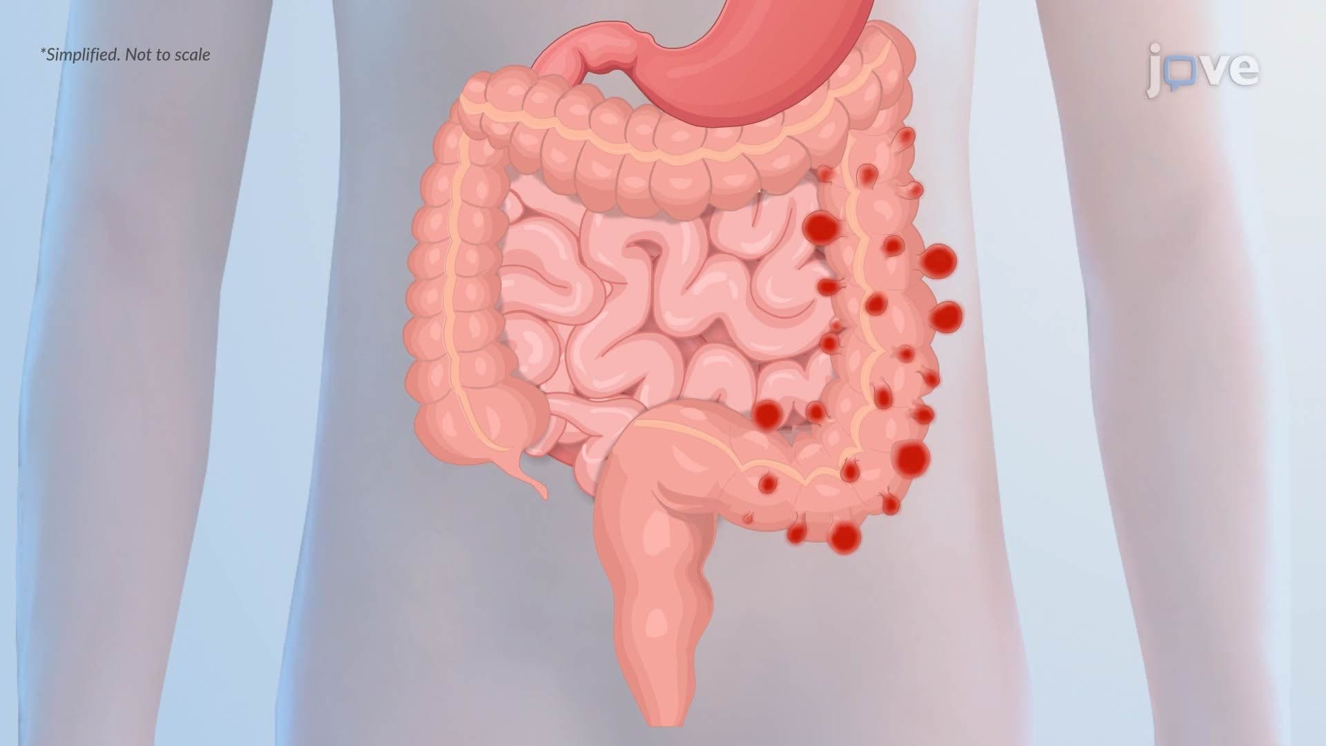
6. Irritable Bowel Syndrome (IBS) and Diverticular Disease IBS is a functional GI disorder characterized by altered motility, visceral hypersensitivity, gut-brain axis dysregulation, dysbiosis, and increased intestinal permeability. Symptoms vary by subtype: IBS-D features rapid transit and loose stools, IBS-C involves delayed transit and hard stools, and IBS-M alternates between both. Diverticular disease involves outpouching of the colonic mucosa and submucosa through weak points in the muscularis propria, most commonly in the sigmoid colon. According to the Law of Laplace, the sigmoid's smaller diameter generates higher wall tension. Low-fiber diets, chronic constipation, and muscle thickening increase intraluminal pressure, promoting diverticula formation and, when inflamed, diverticulitis.
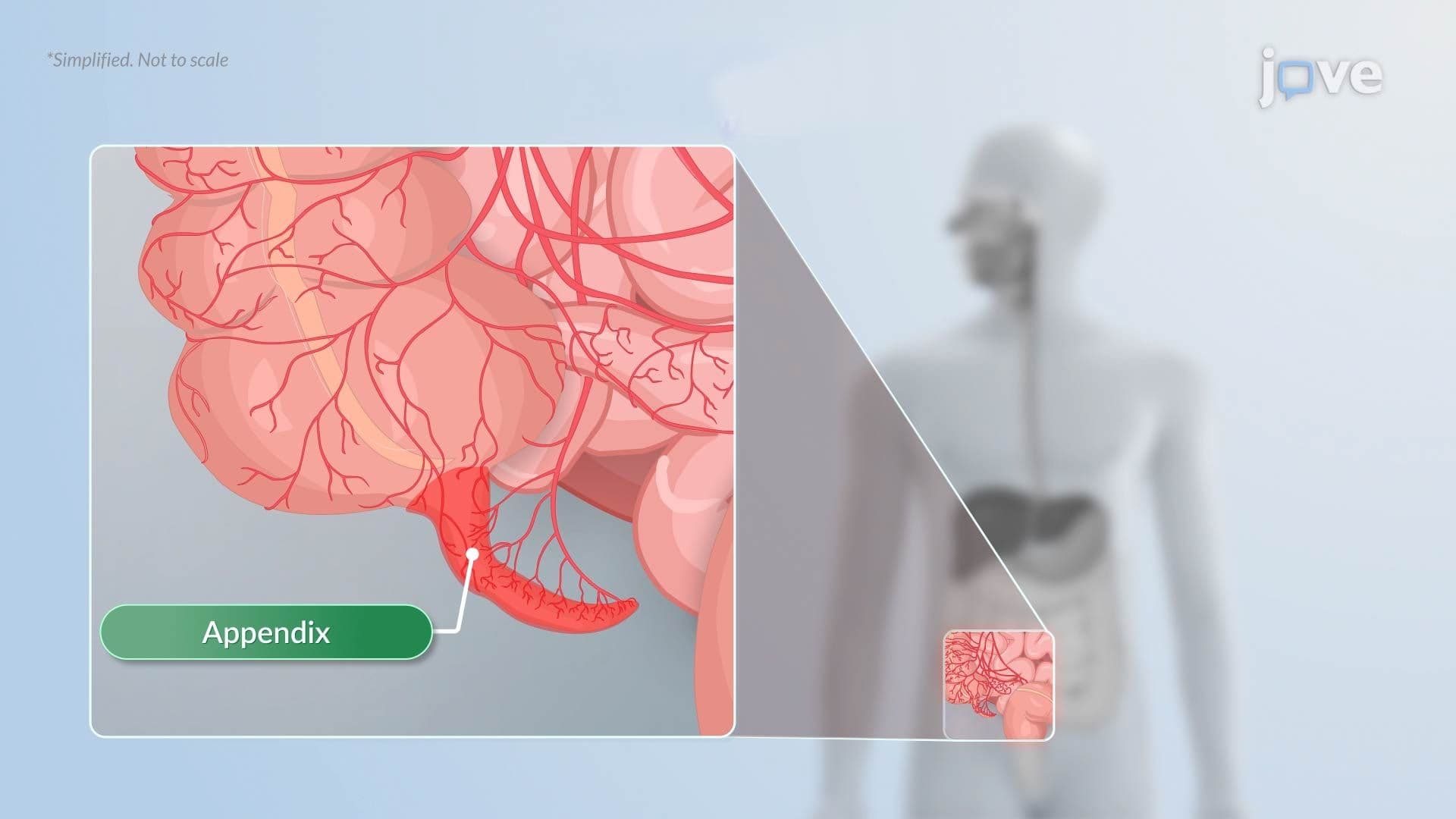
7. Appendicitis Appendicitis begins with obstruction of the appendiceal lumen, typically by a fecalith, lymphoid hyperplasia, or foreign body, which prevents mucosal drainage and causes pressure to build. Vascular compromise follows, producing ischemia and mucosal ulceration. Gut bacteria such as E. coli and Bacteroides invade the weakened wall, triggering acute inflammation. If left untreated, the appendix can progress to gangrene and perforation, releasing infected contents into the peritoneal cavity. Clinically, appendicitis is one of the most common causes of acute abdominal pain requiring emergency surgery in the United States.
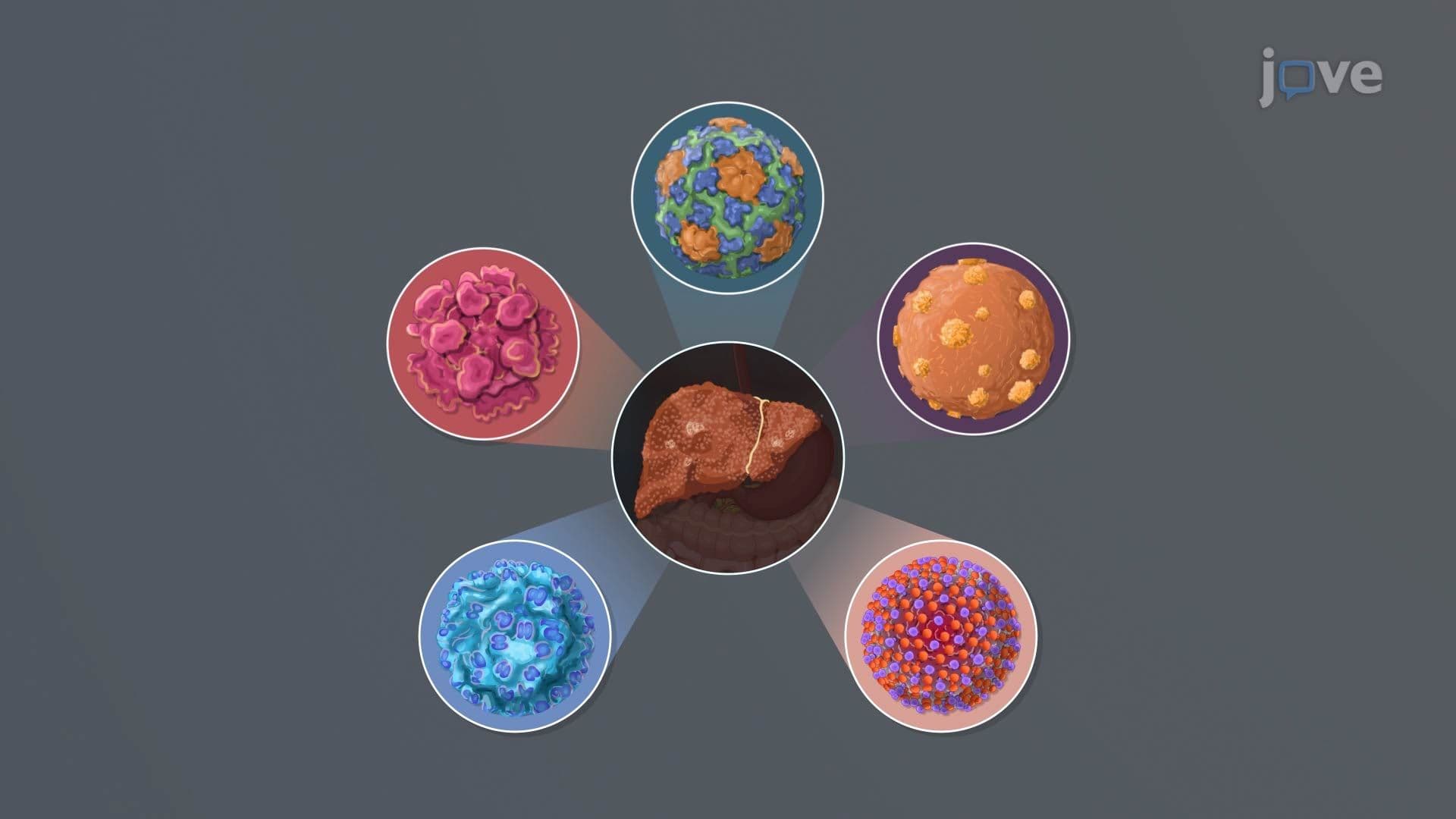
8. Viral Hepatitis Viral hepatitis involves liver inflammation caused by the hepatitis A-E viruses, each with a distinct transmission route and clinical course. Hepatitis A and E spread via the fecal-oral route through contaminated food or water. Hepatitis B is a DNA virus transmitted through blood, sexual contact, and perinatal exposure, and can progress to chronic liver disease. Hepatitis C, an RNA virus spread primarily through blood contact such as shared needles, carries a high risk of chronicity and cirrhosis. Hepatitis D requires co-existing hepatitis B infection. Other viruses, including Epstein-Barr virus and cytomegalovirus, can also cause hepatitis in certain clinical settings.
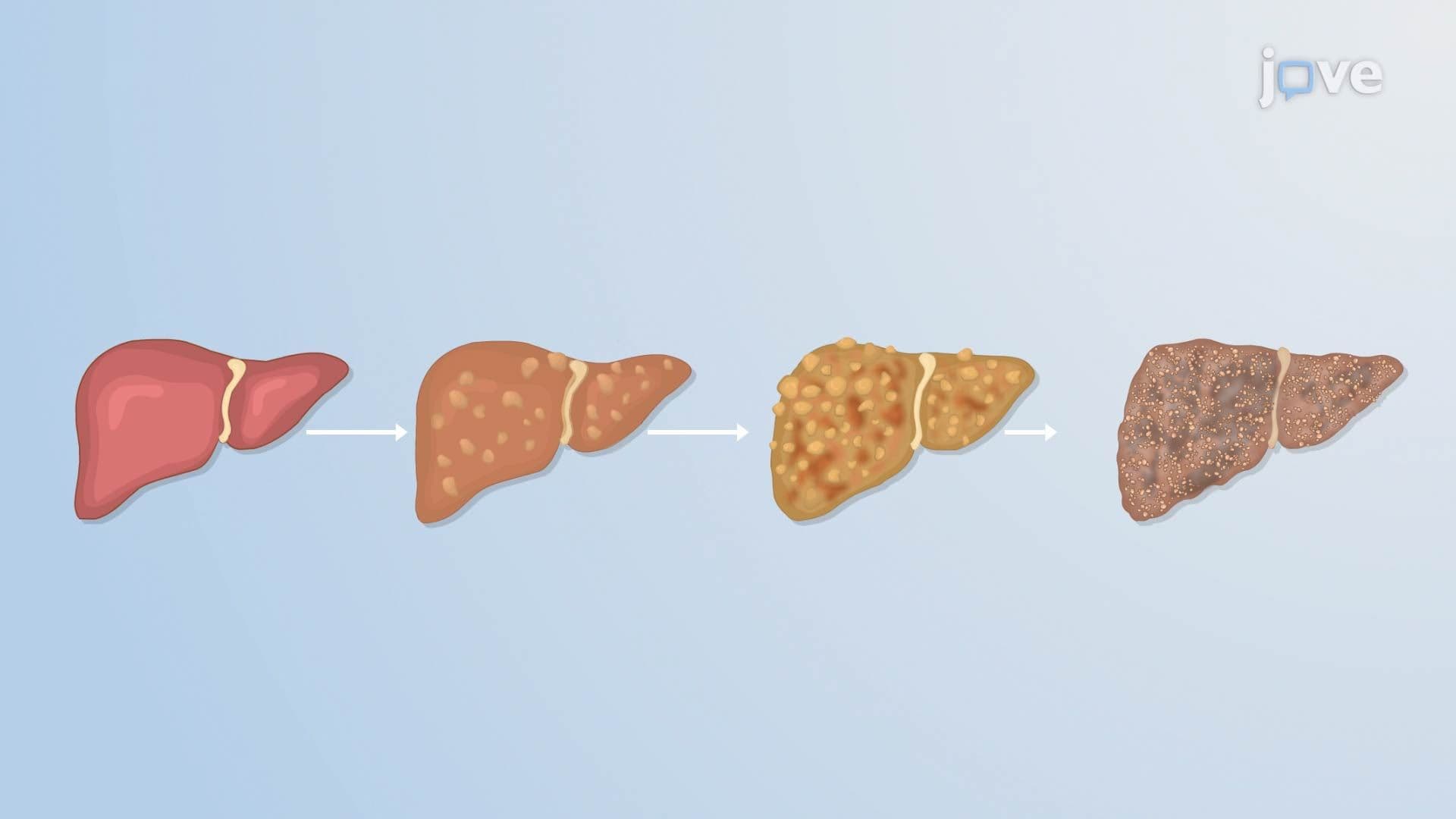
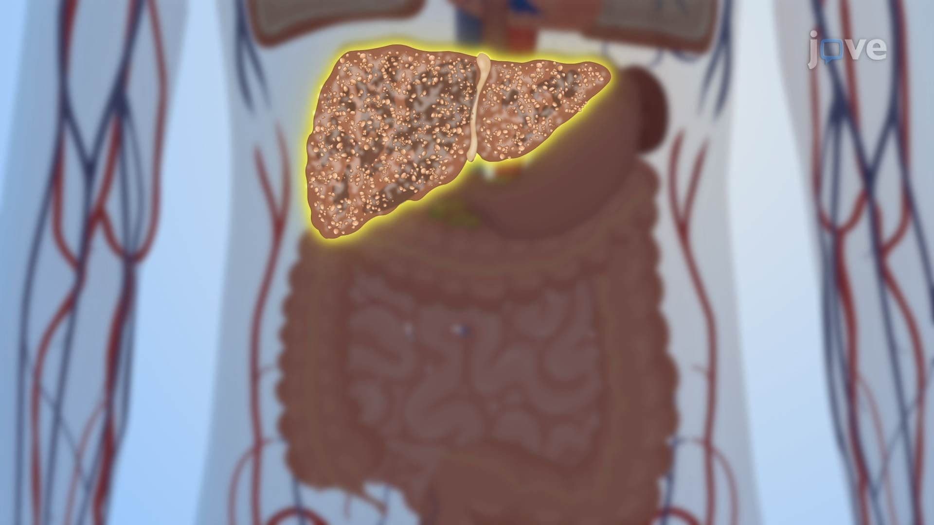
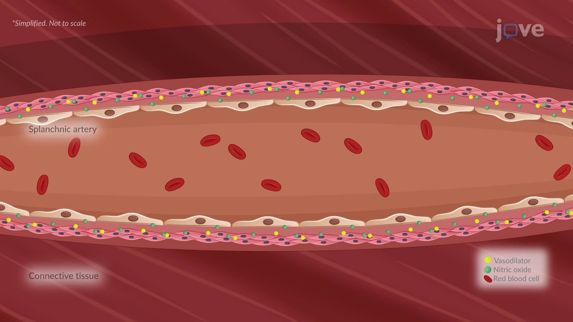
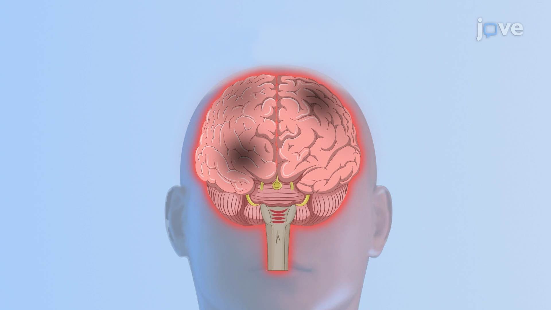
9. Cirrhosis, Portal Hypertension, and Systemic Complications Cirrhosis results from chronic liver injury, most commonly from alcohol use disorder, hepatitis B or C, or nonalcoholic fatty liver disease, leading to fibrosis and regenerative nodule formation. Activated hepatic stellate cells overproduce collagen, distorting liver architecture. This increases resistance to portal blood flow, causing portal hypertension. Blood is rerouted through collateral vessels such as esophageal varices, which may rupture and cause severe gastrointestinal bleeding. Elevated portal pressure, combined with splanchnic vasodilation and activation of the renin-angiotensin-aldosterone system, leads to ascites. Failure to clear ammonia results in hepatic encephalopathy, manifesting as confusion, lethargy, and disorientation.
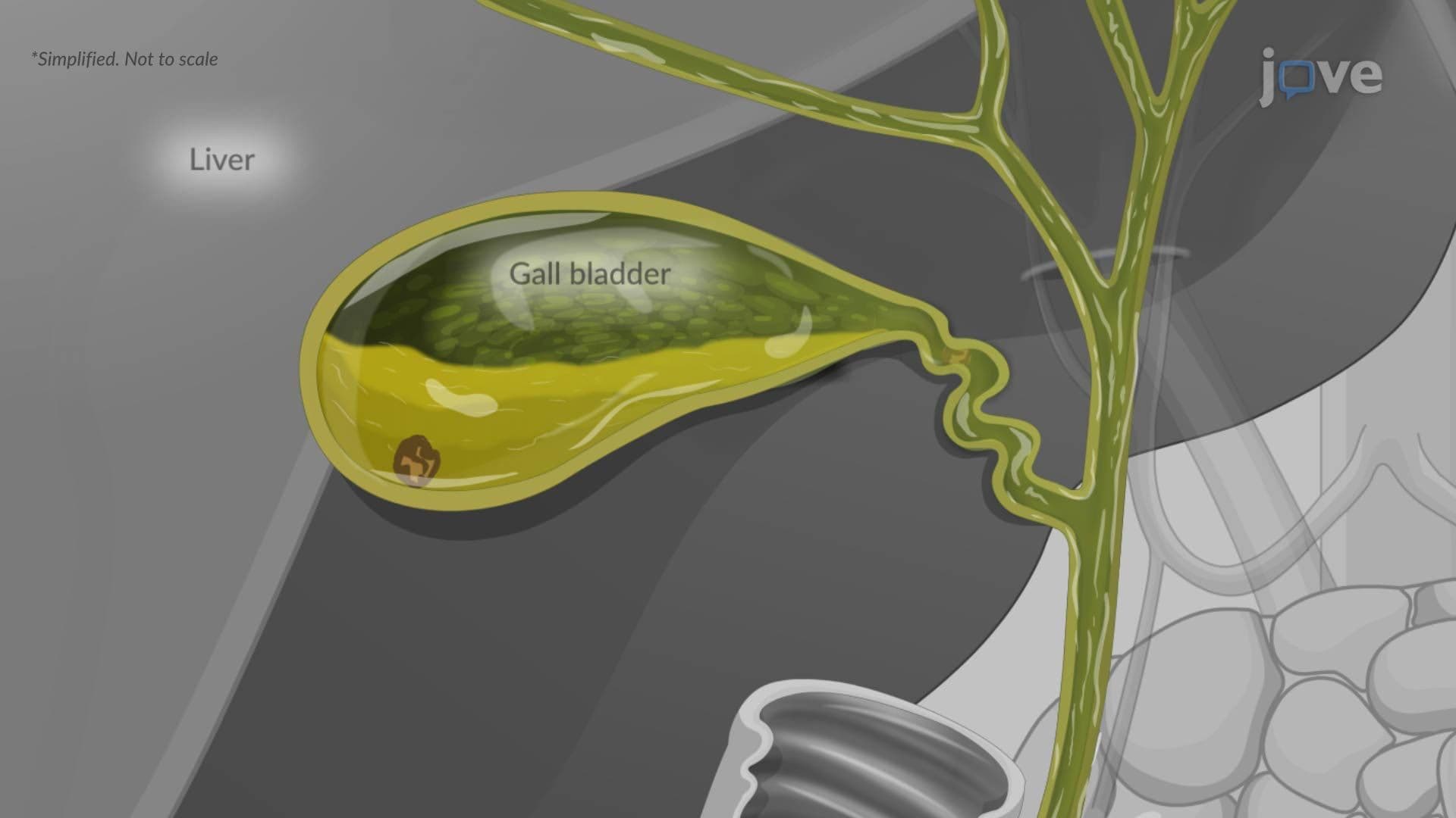
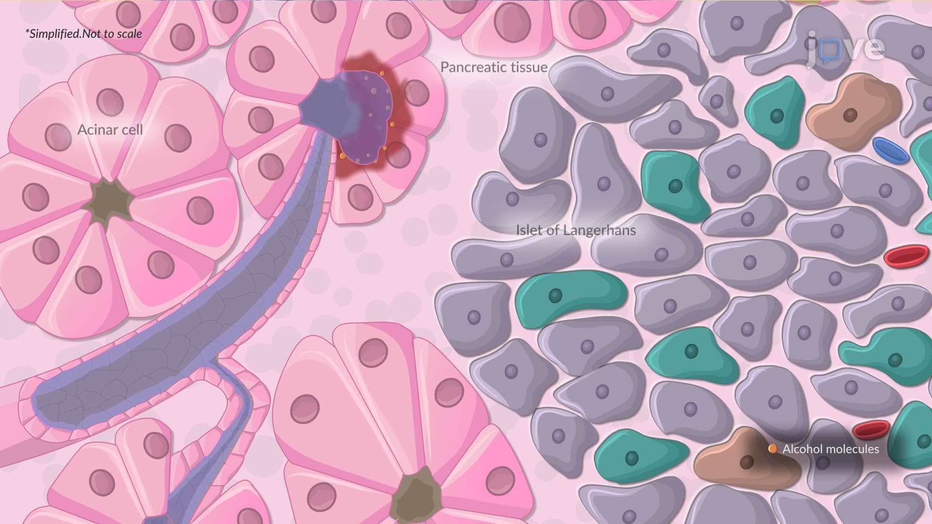
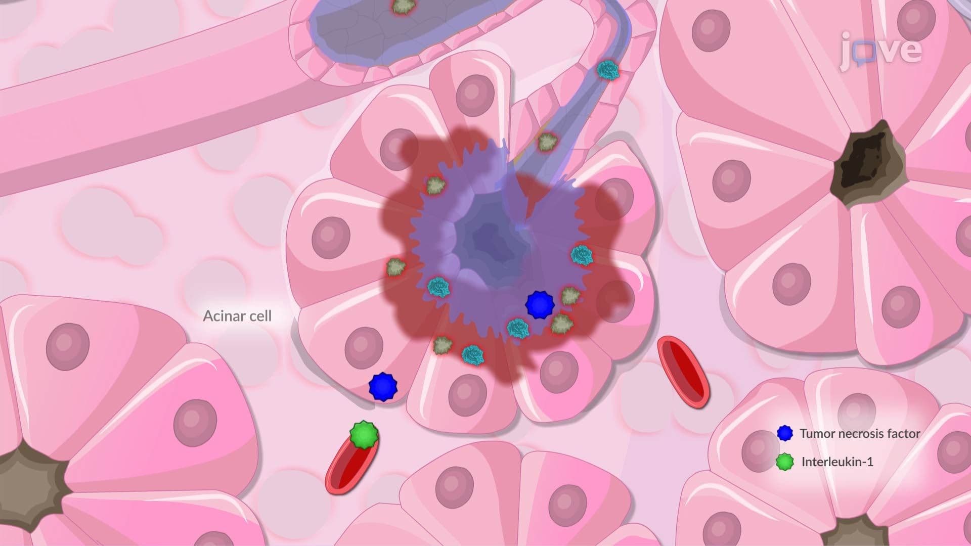
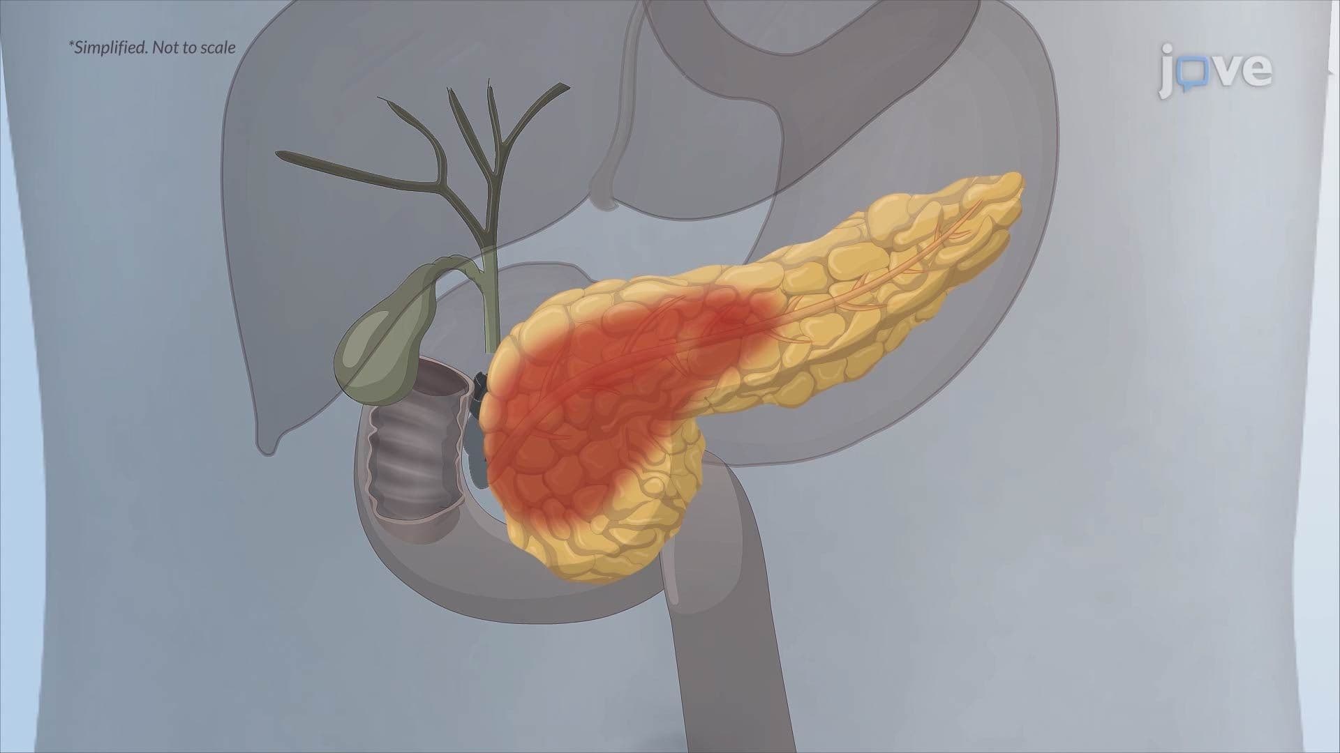
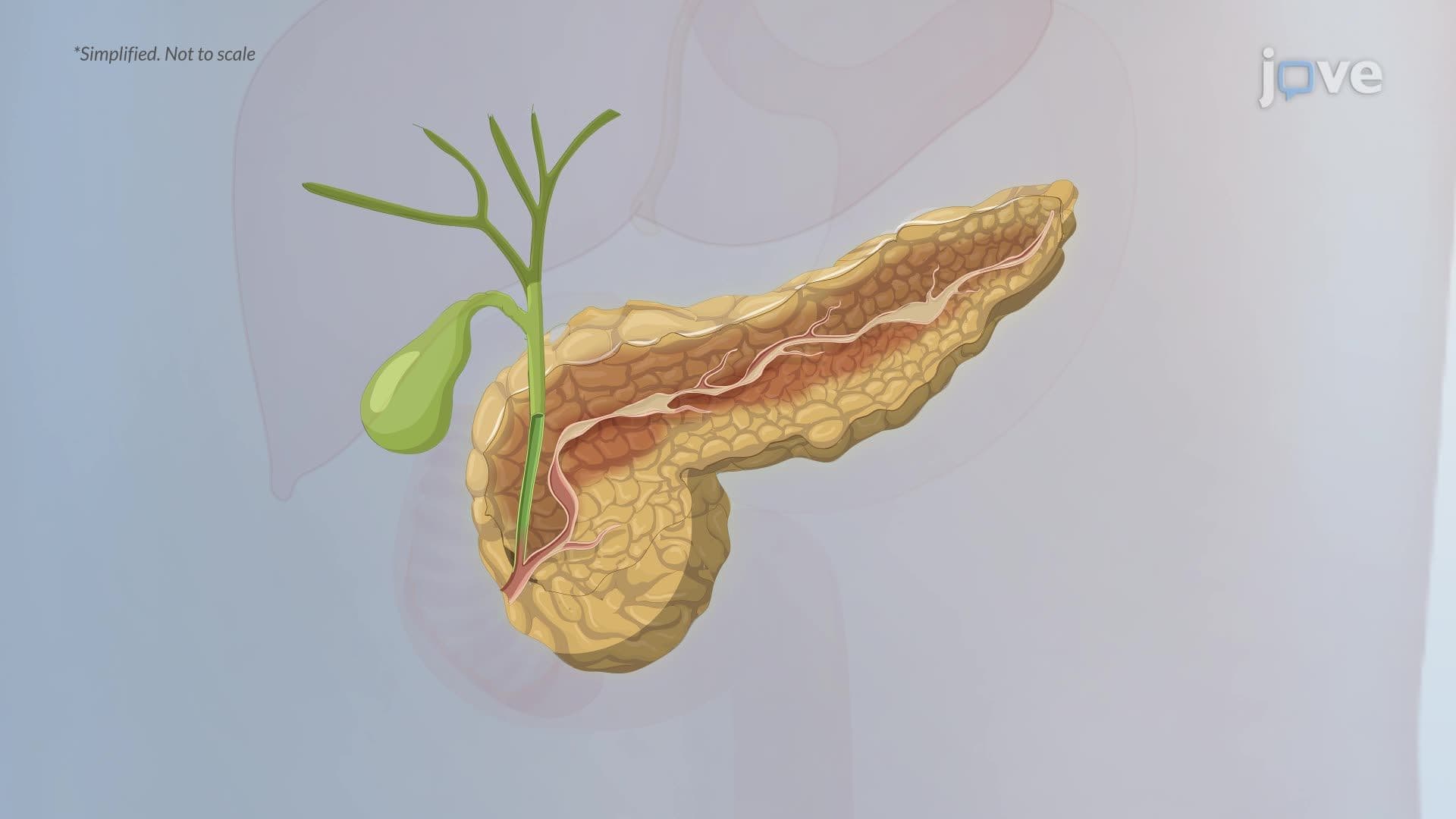
10. Jaundice, Cholecystitis, and Pancreatitis Jaundice develops when plasma bilirubin exceeds approximately 2.5-3 mg/dL, causing yellow discoloration of the skin and sclerae. It can be classified as prehepatic (excess bilirubin production), hepatic (impaired processing), or posthepatic (biliary obstruction). Cholecystitis involves gallbladder inflammation, most often from gallstone obstruction. Acute pancreatitis, triggered by gallstones or alcohol, causes autodigestion of pancreatic tissue by prematurely activated digestive enzymes, leading to inflammation, necrosis, and systemic complications. Chronic pancreatitis involves progressive fibrosis and loss of exocrine and endocrine function, ultimately resulting in malabsorption, steatorrhea, and diabetes mellitus.