Loading video…


 29 Concepts
 4.5K views

Video Summary: Pathophysiology in Intestinal Obstruction Ii
Did you know a simple intestinal blockage can cascade into a life-threatening infection within hours? The pathophysiology in intestinal obstruction II explores exactly how this dangerous chain reaction unfolds inside the body. In US emergency departments, bowel obstruction is among the top causes of acute abdominal surgery. Understanding pathophysiology in intestinal obstruction II basics helps explain how trapped fluid, pressure buildup, and tissue death ultimately lead to peritonitis. Watch the full video on JoVE Coach to master this concept with expert-led visuals and step-by-step explanations.
Intestinal obstruction is not a single event, it is a domino sequence of physiological failures. To truly understand pathophysiology in intestinal obstruction II, you need to trace how one blocked segment of bowel can escalate into a systemic emergency. This is a concept tested in college anatomy and physiology courses, nursing programs (NCLEX prep), and medical school entry pathways (MCAT and USMLE Step 1).
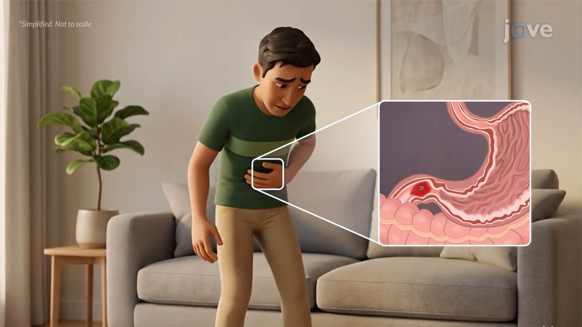
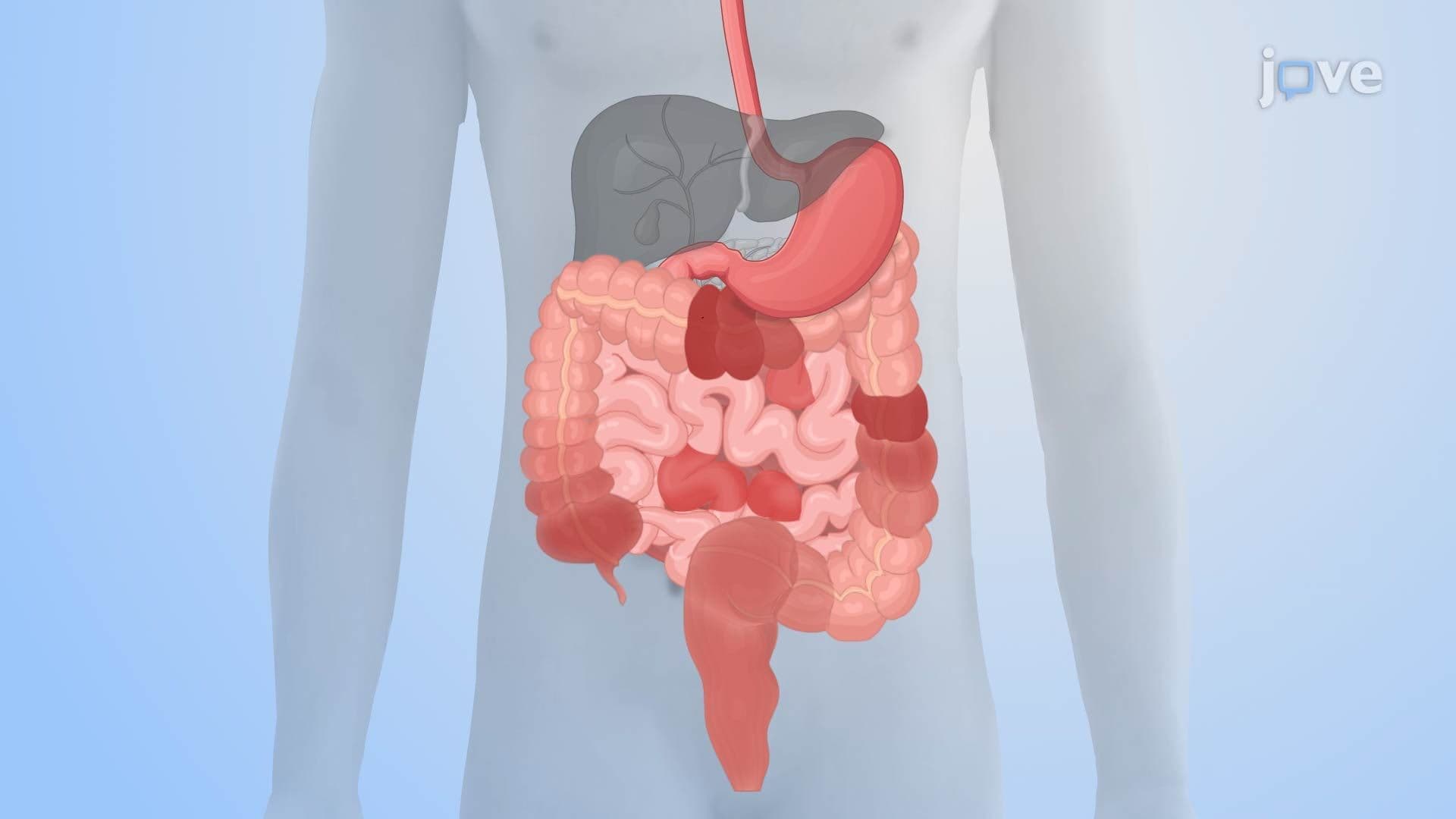
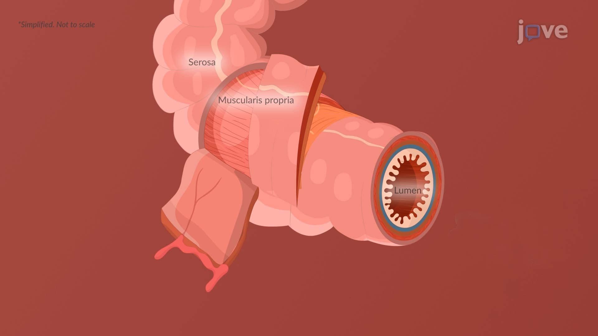
When the intestinal lumen is physically blocked, by adhesions, a hernia, or a tumor, contents can no longer move forward. Air and fluid accumulate proximal to the blockage, stretching the bowel wall. The intestine responds with forceful, rhythmic contractions trying to push through the obstruction. These contractions produce the classic colicky abdominal pain that patients describe as wave-like cramping, a hallmark symptom recognized in US emergency room assessments nationwide.
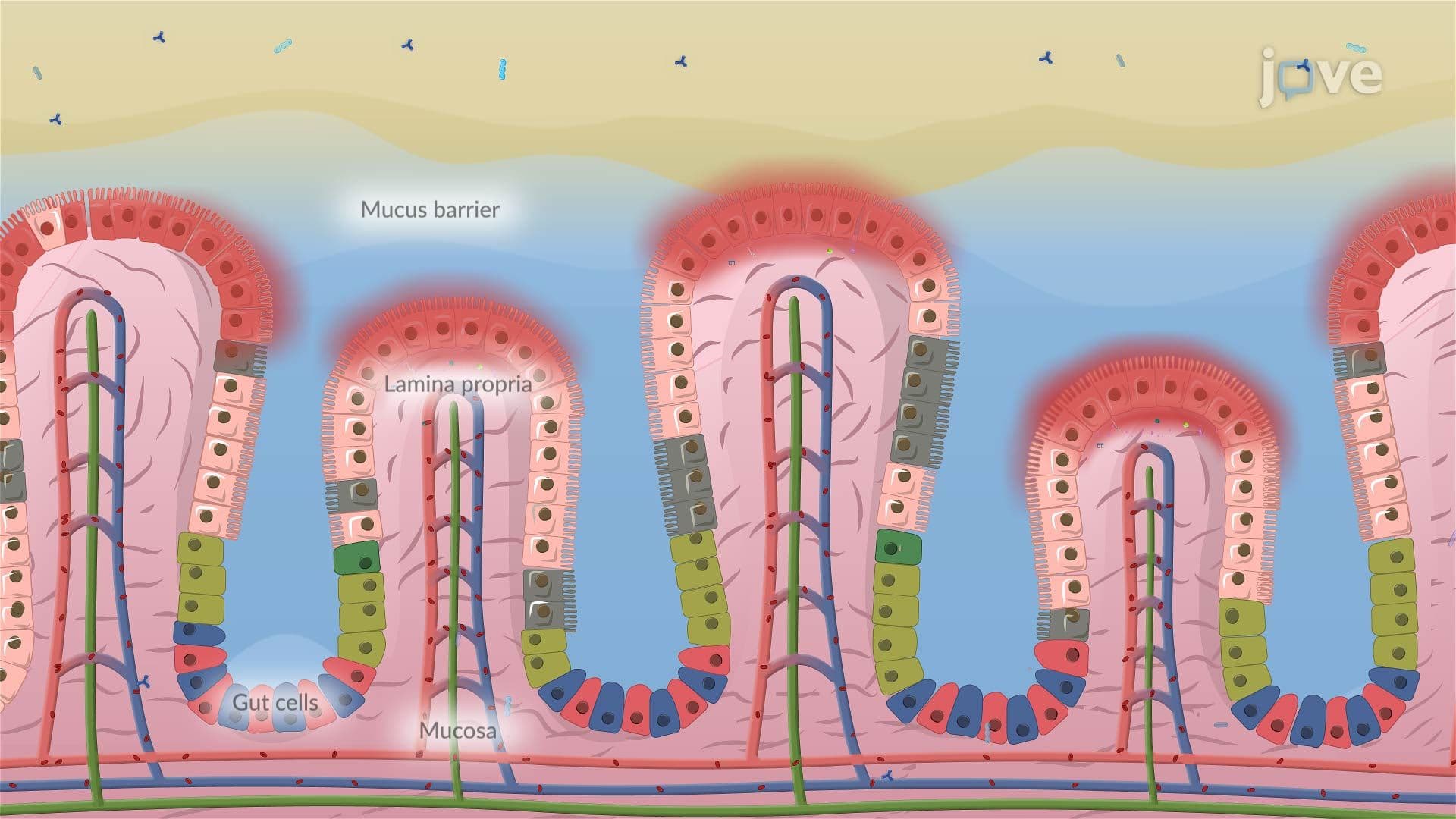
As the bowel stretches, inflamed capillaries begin leaking protein-rich fluid (exudate) directly into the bowel lumen. This is a critical turning point: the fluid that should stay in circulation is now trapped inside the gut. Intraluminal pressure rises sharply. This fluid sequestration, sometimes called "third-spacing", can rapidly lead to dehydration and electrolyte imbalances. In a clinical US setting, this is why IV fluid resuscitation is started immediately when a bowel obstruction is diagnosed.
Sustained high intraluminal pressure first compresses the thin-walled veins in the bowel wall, blocking venous return. Blood backs up, causing bowel wall edema. As pressure climbs further, even the arterial supply is compromised. Without oxygen delivery, intestinal cells shift to anaerobic metabolism and begin dying, a state called ischemia. Students studying for AP Biology or college physiology exams should recognize this as the same oxygen-deprivation mechanism that causes myocardial infarction, just occurring in the intestinal tissue instead.
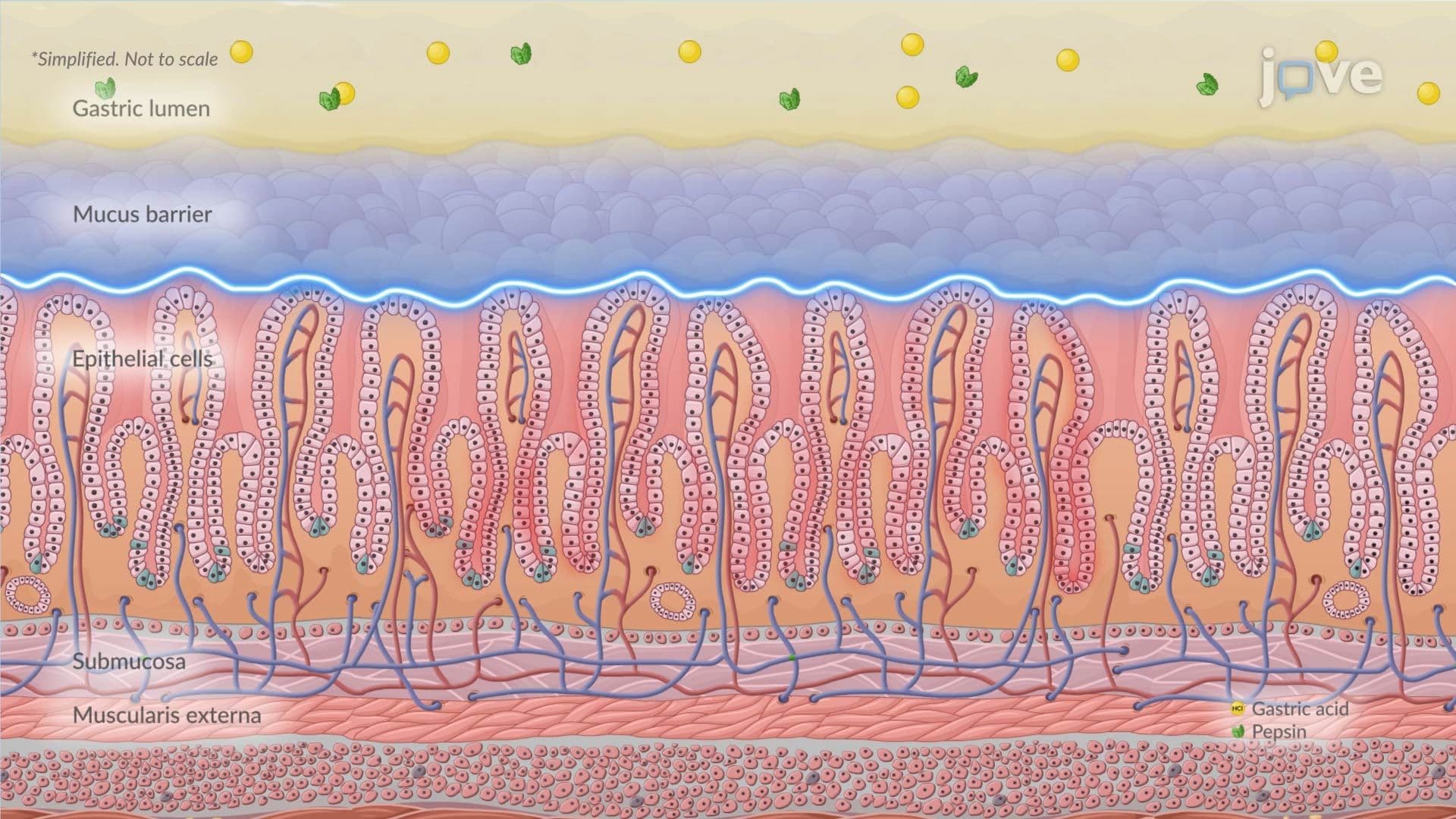
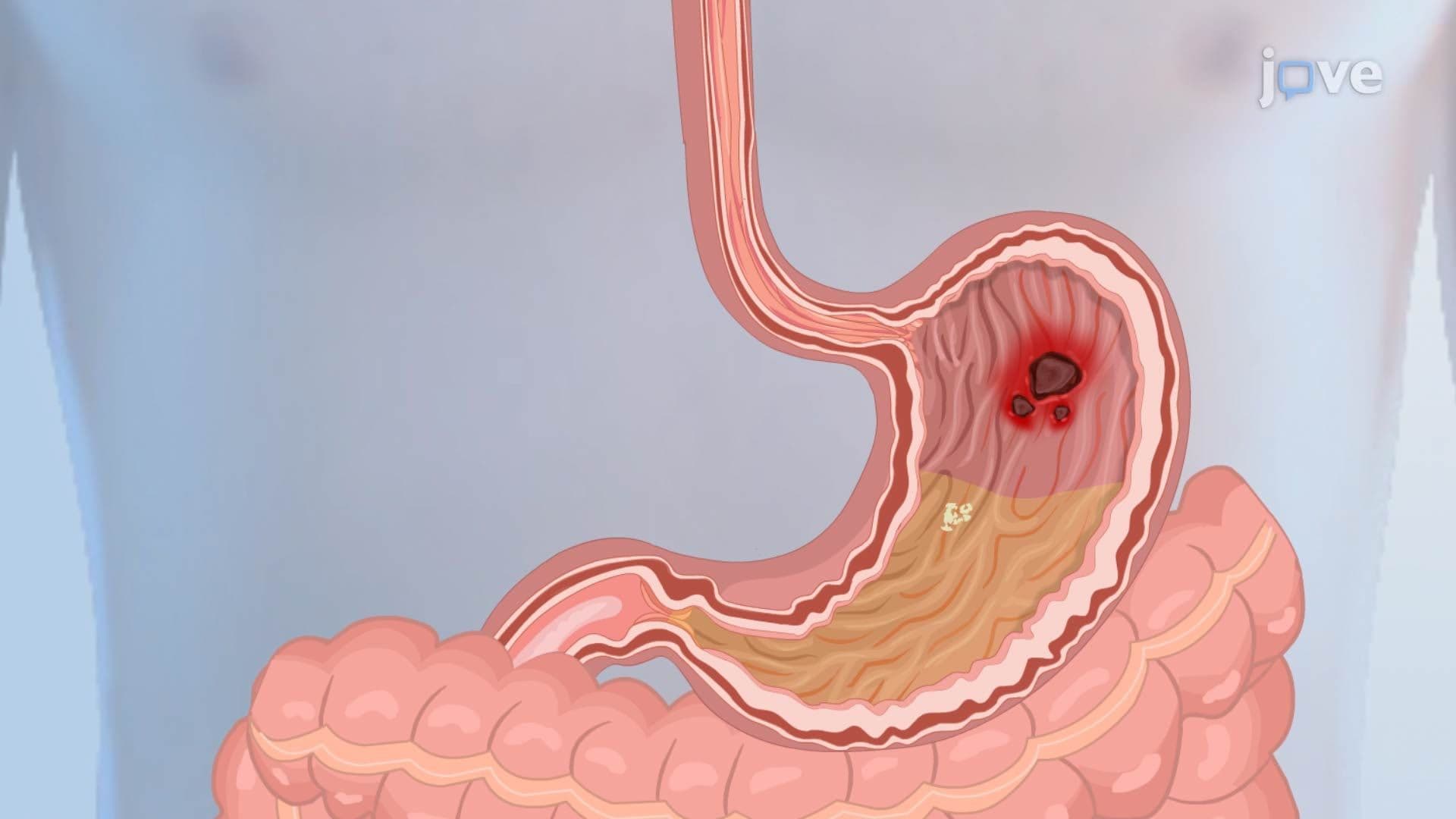
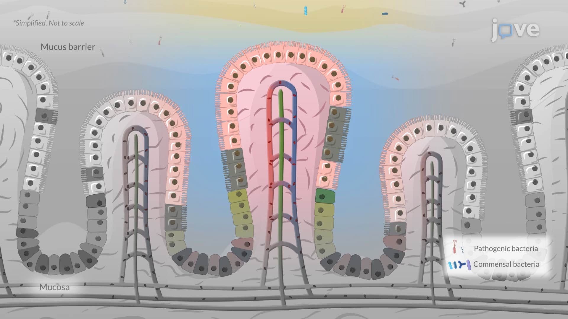
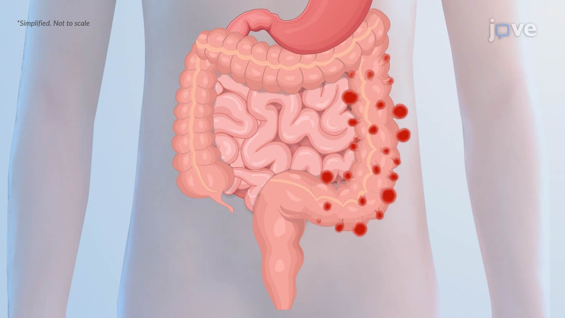
Prolonged ischemia leads to full-thickness tissue necrosis (death). The bowel wall becomes fragile and eventually ruptures, perforation. Intestinal contents, loaded with bacteria like *E. coli* and *Bacteroides*, plus corrosive digestive enzymes, spill into the normally sterile peritoneal cavity. The result is peritonitis, a severe inflammatory response of the abdominal lining. In the US, bowel perforation is a surgical emergency with significant mortality risk if not treated within hours. This pathophysiological sequence also shares overlapping mechanisms with conditions like gastrointestinal bleeding, peptic ulcer disease, and severe inflammatory bowel disease (IBD), all of which can compromise the mucosal barrier and allow bacterial translocation.
Understanding this cascade from obstruction to peritonitis is not just academically important, it explains every clinical decision made in the ER, from pain management to emergency surgery.
Related Micro-courses