Loading video…

 29 Concepts
 4.5K views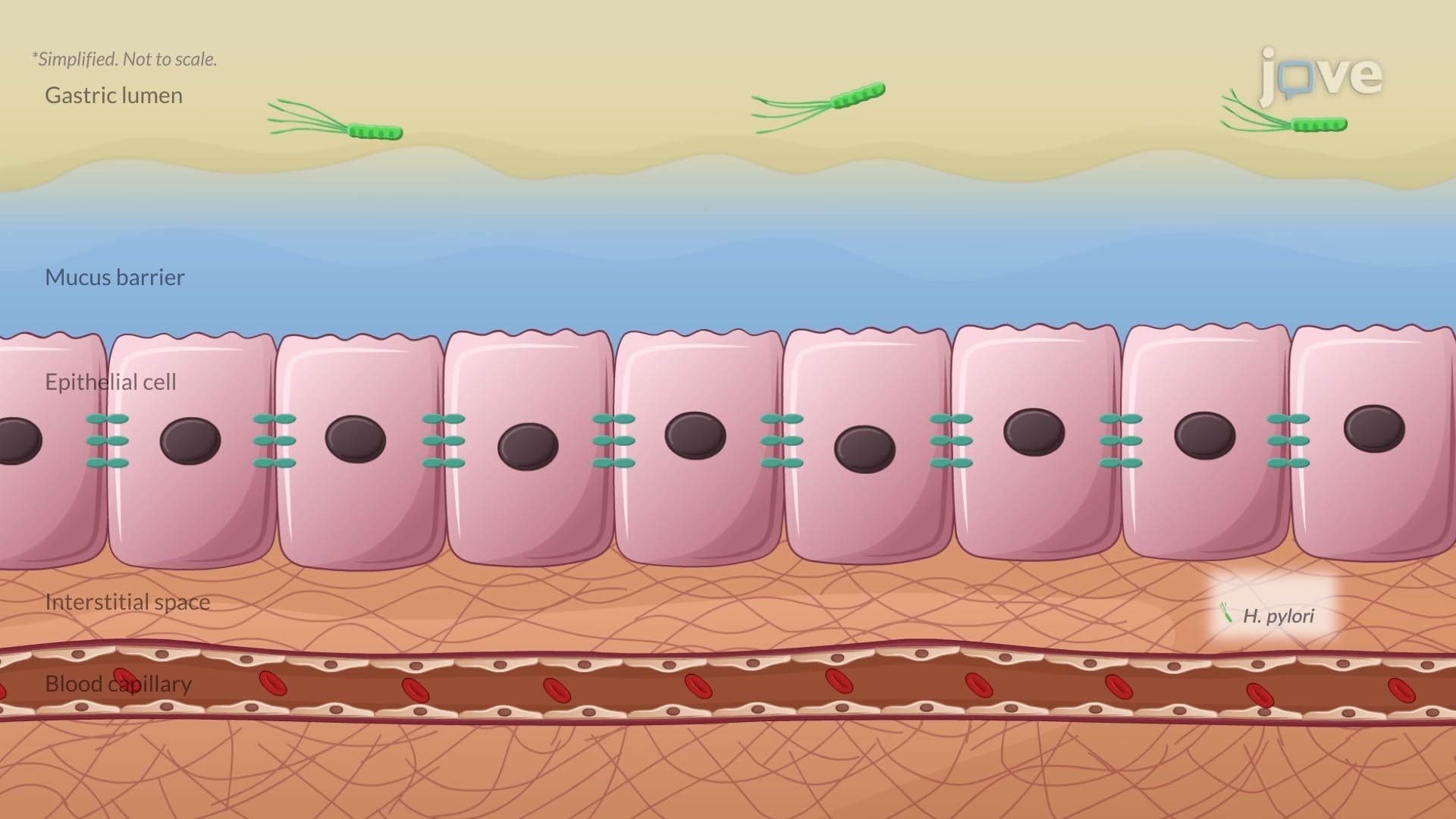

Video Summary: Pathophysiology in Gastritis Ii
Did you know a single bacterium can outsmart your immune system and quietly destroy your stomach lining? Pathophysiology in Gastritis II breaks down exactly how *H. pylori* hijacks host defenses, evades T-cell attacks using PD-L1, and triggers chronic inflammation, the same mechanism studied in gastroenterology programs at institutions like Johns Hopkins Medicine. Watch the full video on JoVE Coach to master this concept with expert-led visuals and step-by-step explanations.
Gastritis is not simply an upset stomach, it is a clinically significant inflammatory condition of the gastric mucosa with well-defined molecular mechanisms. Pathophysiology in Gastritis II focuses specifically on *Helicobacter pylori*, the gram-negative bacterium estimated by the CDC to infect roughly 35-40% of the U.S. population. Understanding *how* this organism causes disease is essential for biology, microbiology, and pre-health students preparing for exams like the MCAT, AP Biology, or college physiology coursework.
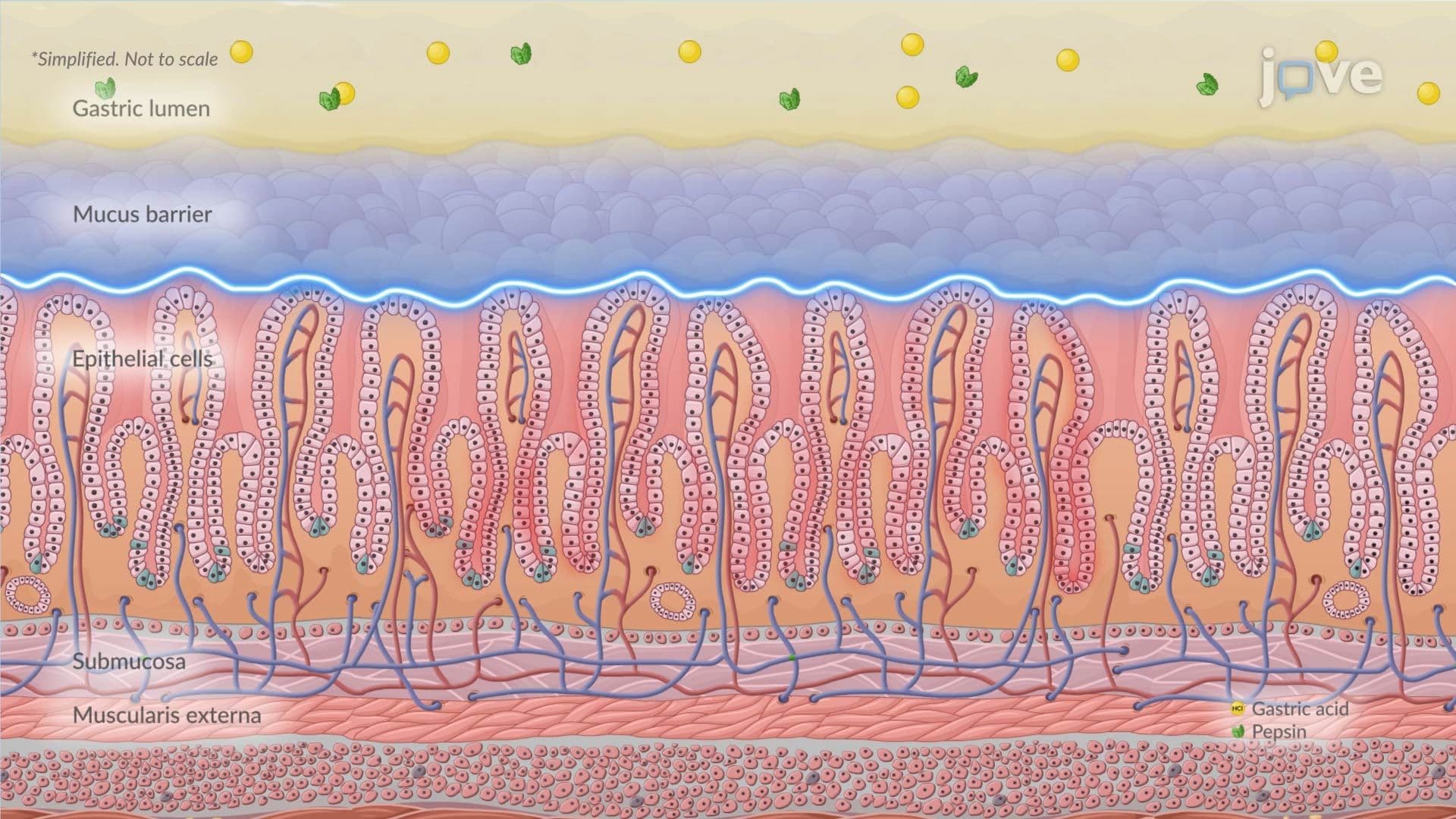
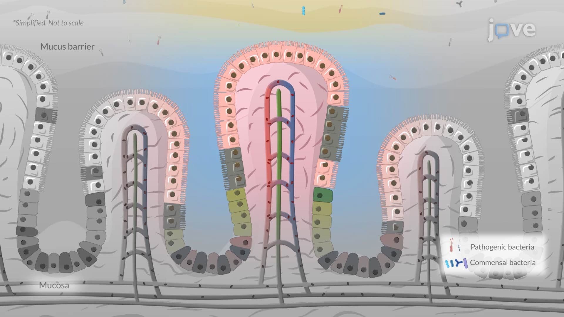
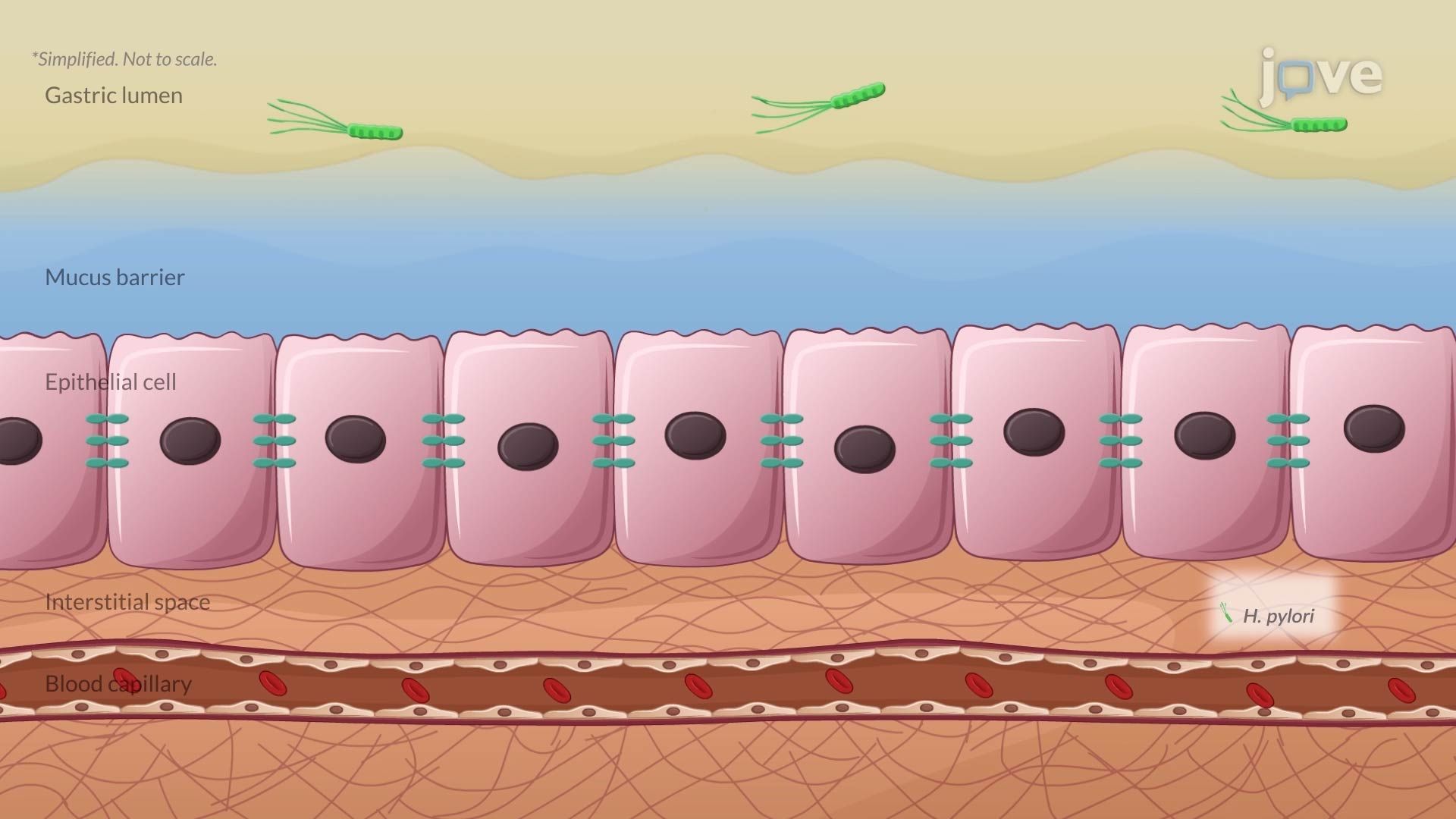
The stomach maintains a pH between 1.5 and 3.5, lethal to most microorganisms. *H. pylori* overcomes this by secreting urease, an enzyme that catalyzes the breakdown of urea into ammonia and carbon dioxide. The ammonia locally neutralizes gastric acid, creating a microenvironment with a survivable pH. This biochemical trick is also the basis of the urea breath test, a non-invasive diagnostic tool used in U.S. clinics to confirm active *H. pylori* infection. Once pH is stabilized, the bacterium uses spiral-shaped flagella and mucolytic enzymes to drill through the protective mucus layer and physically attach to gastric epithelial cells.
This is where the pathophysiology becomes especially sophisticated. Upon attachment, *H. pylori* induces gastric epithelial cells to upregulate PD-L1 (Programmed Death-Ligand 1). PD-L1 binds to the PD-1 receptor on cytotoxic T-cells, sending an inhibitory signal that effectively "turns off" those immune cells. This is the same checkpoint pathway that cancer cells exploit to evade immune destruction, and it is a major topic in immunology and pharmacology courses. The result is that macrophages and T-cells, despite being recruited to the site, cannot effectively eliminate the bacteria. This partial immune response actually worsens mucosal damage without clearing the infection.
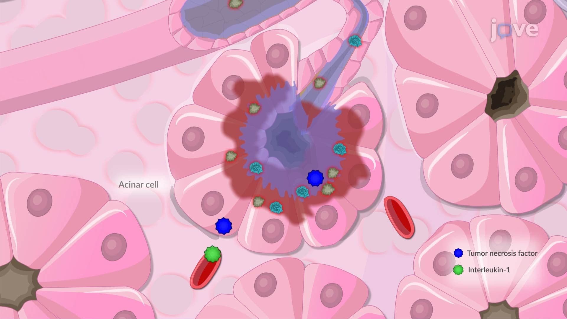
While immune evasion suppresses adaptive immunity, *H. pylori* simultaneously drives innate inflammation. It stimulates gastric epithelial cells to secrete interleukin-8 (IL-8), a powerful chemokine that recruits neutrophils to the site of infection. Neutrophil accumulation amplifies oxidative damage to the mucosal lining, a hallmark finding in active chronic gastritis seen on endoscopic biopsy. Additionally, strains carrying the Cytotoxin-associated gene A (CagA), a bacterial gene encoding an oncoprotein, use a molecular syringe-like apparatus called the Type IV secretion system to inject CagA directly into host epithelial cells. Inside the cell, CagA disrupts intracellular signaling pathways and degrades tight junctions between epithelial cells. Disrupted tight junctions compromise mucosal barrier integrity, accelerating tissue damage and increasing susceptibility to peptic ulcer disease and even gastric adenocarcinoma with chronic exposure.
For students in AP Biology or undergraduate pathophysiology courses, *H. pylori* is a model organism for understanding how pathogens manipulate host biology at the molecular level. On the MCAT, questions may connect *H. pylori* virulence to immune checkpoints, enzyme activity, or acid-base chemistry. Clinically, *H. pylori*-induced gastritis is a leading cause of peptic ulcer disease in the U.S., managed with "triple therapy", a combination of two antibiotics and a proton pump inhibitor, prescribed widely in American primary care settings. Recognizing the underlying mechanisms helps contextualize why treatment targets both the bacterium and gastric acid simultaneously.
Related Micro-courses