Loading video…

 29 Concepts
 4.5K views

Video Summary: What Is Jaundice
Ever wonder why a newborn's skin turns yellow in the hospital nursery? That's jaundice basics in action. Jaundice occurs when bilirubin, a yellow pigment from broken-down red blood cells, builds up in the blood above roughly 2.5 mg/dL, staining the skin, eyes, and mucous membranes. In US neonatal units, it's one of the most commonly treated newborn conditions. Watch the full video on JoVE Coach to master this concept with expert-led visuals and step-by-step explanations.
Jaundice, also called icterus, is the yellow discoloration of the skin, whites of the eyes (sclerae), and mucous membranes caused by excess bilirubin circulating in the blood. It becomes clinically visible when plasma bilirubin exceeds approximately 2.5 to 3 milligrams per deciliter (mg/dL). Importantly, jaundice is not a disease itself, it is a symptom pointing to an underlying disruption somewhere along the bilirubin metabolic pathway. Understanding that pathway in detail is essential for students in AP Biology, college anatomy and physiology, and anyone preparing for exams like the MCAT, USMLE, NCLEX, or HESI A2.
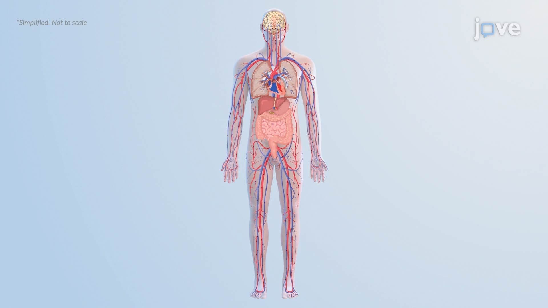
Every red blood cell has a lifespan of roughly 120 days. When these cells age or are damaged, macrophages in the spleen and liver break down hemoglobin, the oxygen-carrying protein inside, into two components: globin (the protein portion) and heme (the iron-containing ring structure). Globin is recycled into amino acids, while heme undergoes a two-step enzymatic conversion. First, heme oxygenase converts heme into biliverdin, a green pigment. Then biliverdin reductase reduces biliverdin into unconjugated bilirubin, which is yellow and lipid-soluble. Because it cannot dissolve in water, unconjugated bilirubin must bind to albumin, a transport protein in the bloodstream, to travel safely to the liver without causing cellular damage.
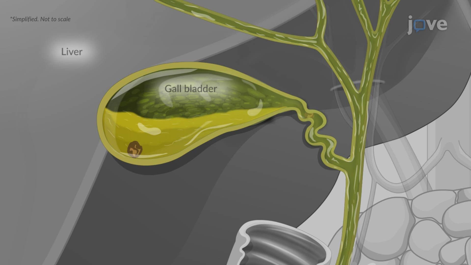
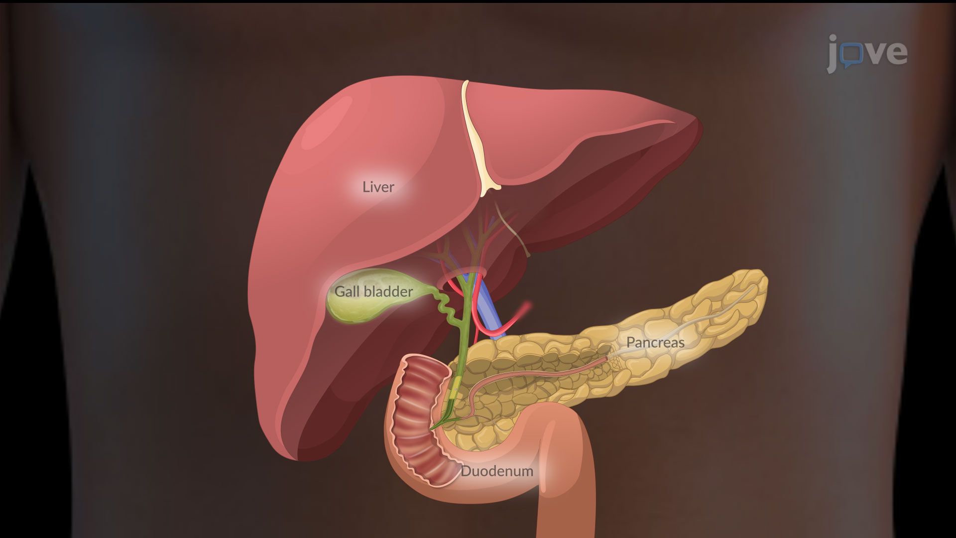
Once unconjugated bilirubin reaches the liver, hepatocytes (liver cells) take it up and attach it to glucuronic acid through an enzyme called UDP-glucuronosyltransferase (UGT). This process, called conjugation, converts bilirubin into its water-soluble form, conjugated bilirubin. The liver then secretes conjugated bilirubin into bile, which travels through the bile ducts into the small intestine. There, intestinal bacteria convert it into urobilinogen, which gives stool its brown color. Some urobilinogen is reabsorbed into the bloodstream and excreted by the kidneys, which is why urine turns dark in certain liver diseases. This entire pathway is a high-yield topic in college biochemistry and physiology courses across the US.
Physicians and nurses in US hospitals classify jaundice by where the breakdown occurs:
On the MCAT and USMLE, distinguishing these three types using lab values (elevated direct vs. indirect bilirubin) is a commonly tested skill. AP Biology and college-level physiology courses frequently use jaundice as a case study to illustrate enzyme function, membrane transport, and organ system integration. Recognizing which enzyme or transporter is deficient, and predicting the downstream effects, builds exactly the systems-level thinking these exams reward.
Related Micro-courses