Loading video…

 29 Concepts
 4.5K views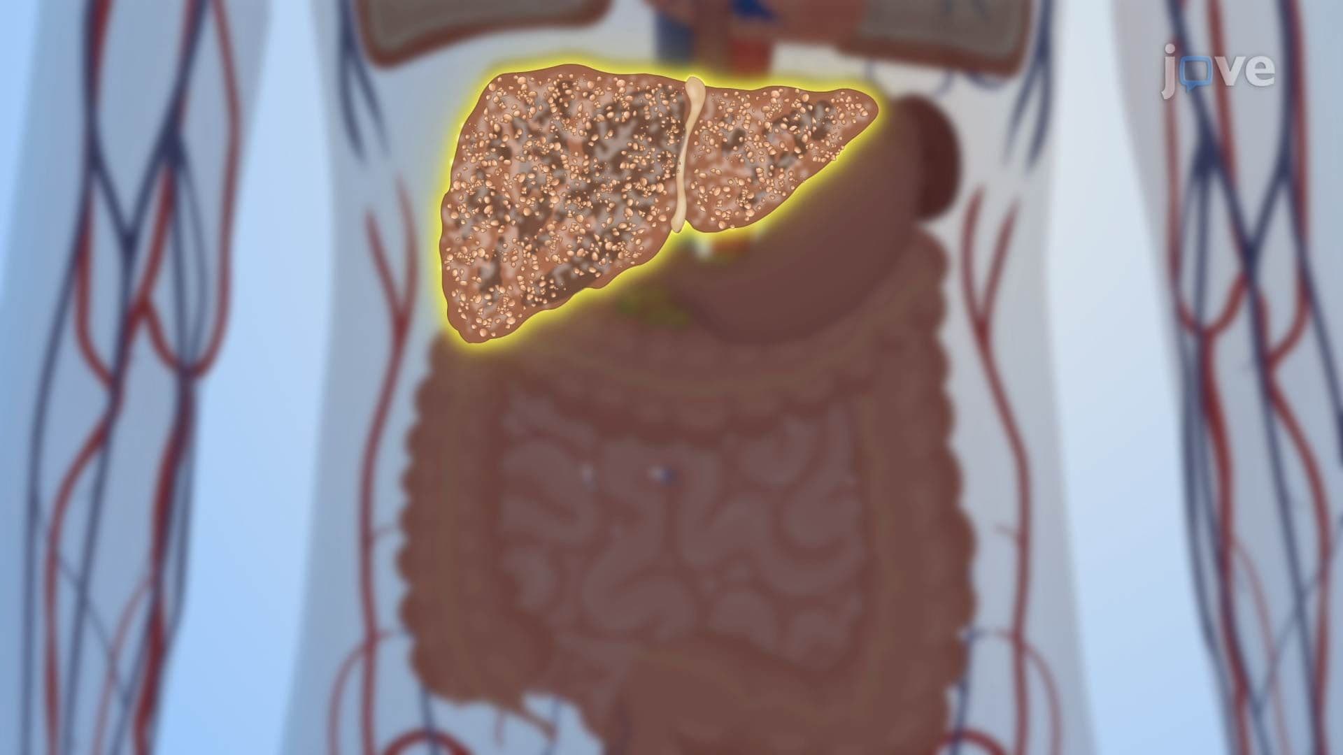

Video Summary: Pathophysiology in Cirrhosis Ii
Did you know that your liver can silently scar itself for years before symptoms ever appear? The pathophysiology in cirrhosis II reveals how repeated liver injury triggers a cascade of inflammation, immune activation, and cell death, ultimately reshaping liver architecture. American gastroenterology clinics see thousands of cirrhosis cases annually linked to alcohol use and hepatitis C. Watch the full video on JoVE Coach to master this concept with expert-led visuals and step-by-step explanations.
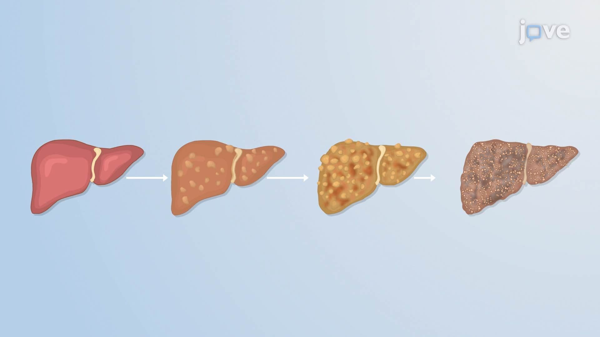
Cirrhosis is not a single event, it is the final outcome of years of progressive, self-reinforcing liver damage. Understanding the pathophysiology in cirrhosis II means tracing an interconnected chain of cellular and molecular events that transform a healthy, functional organ into a scarred, dysfunctional one. This process is highly relevant to courses in anatomy and physiology, pathophysiology, and pre-health programs across US colleges and universities.
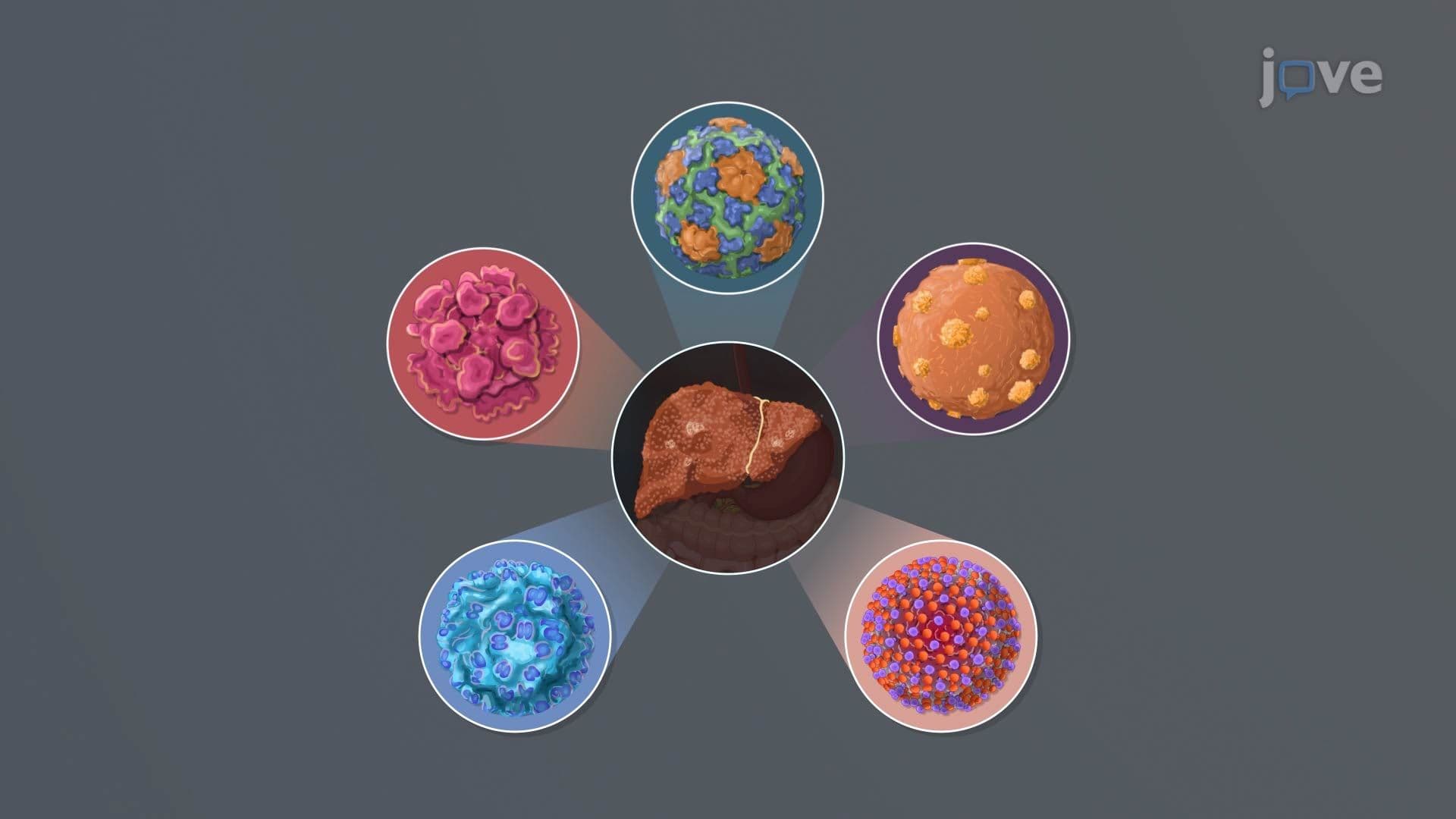
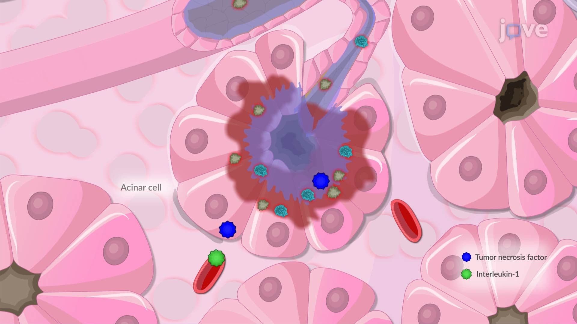
Every episode of liver injury, whether from chronic alcohol use, hepatitis B or C infection, or non-alcoholic fatty liver disease (NAFLD), one of the fastest-growing liver conditions in the United States, triggers an inflammatory response. Immune cells flood the liver tissue and begin releasing pro-inflammatory signaling proteins called cytokines. Two of the most important are tumor necrosis factor-alpha (TNF-α) and interleukin-1 (IL-1). These cytokines do not simply cause local inflammation; they also act as upstream signals that set the entire fibrotic process in motion. In US pathophysiology courses, students are frequently tested on the cytokine signaling sequence because it appears in MCAT biological sciences passages and USMLE Step 1 questions.
The cytokines released during inflammation stimulate the production of transforming growth factor-beta (TGF-β), widely considered the master regulator of fibrosis. TGF-β acts on hepatic stellate cells, specialized cells normally responsible for storing vitamin A in the healthy liver. When activated, these cells undergo a dramatic transformation: they stop storing vitamin A and start producing excessive amounts of collagen and other extracellular matrix proteins. Over time, this continuous collagen deposition forms dense fibrous bands throughout the liver. Think of it as scar tissue replacing the liver's functional scaffolding. This mechanism is a high-yield concept in US nursing programs, where NCLEX preparation frequently includes questions on liver disease progression.
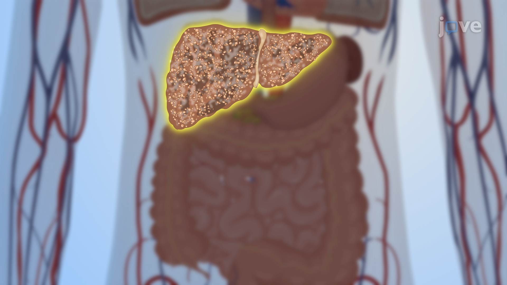
Alongside fibrosis, hepatocytes, the primary functional cells of the liver, undergo both apoptosis (programmed cell death) and necrosis (uncontrolled cell death). In a healthy liver, cell regeneration is orderly and precise. But in an environment saturated with inflammatory signals and fibrous tissue, regeneration becomes chaotic. New clusters of hepatocytes form but are encased in fibrous tissue rather than integrated into normal liver architecture. These isolated clusters are called regenerative nodules, and their presence, surrounded by fibrous bands, is actually the defining histological feature of cirrhosis. US pathology courses and AP Biology students studying cell cycle regulation will recognize this as a breakdown in normal tissue homeostasis.
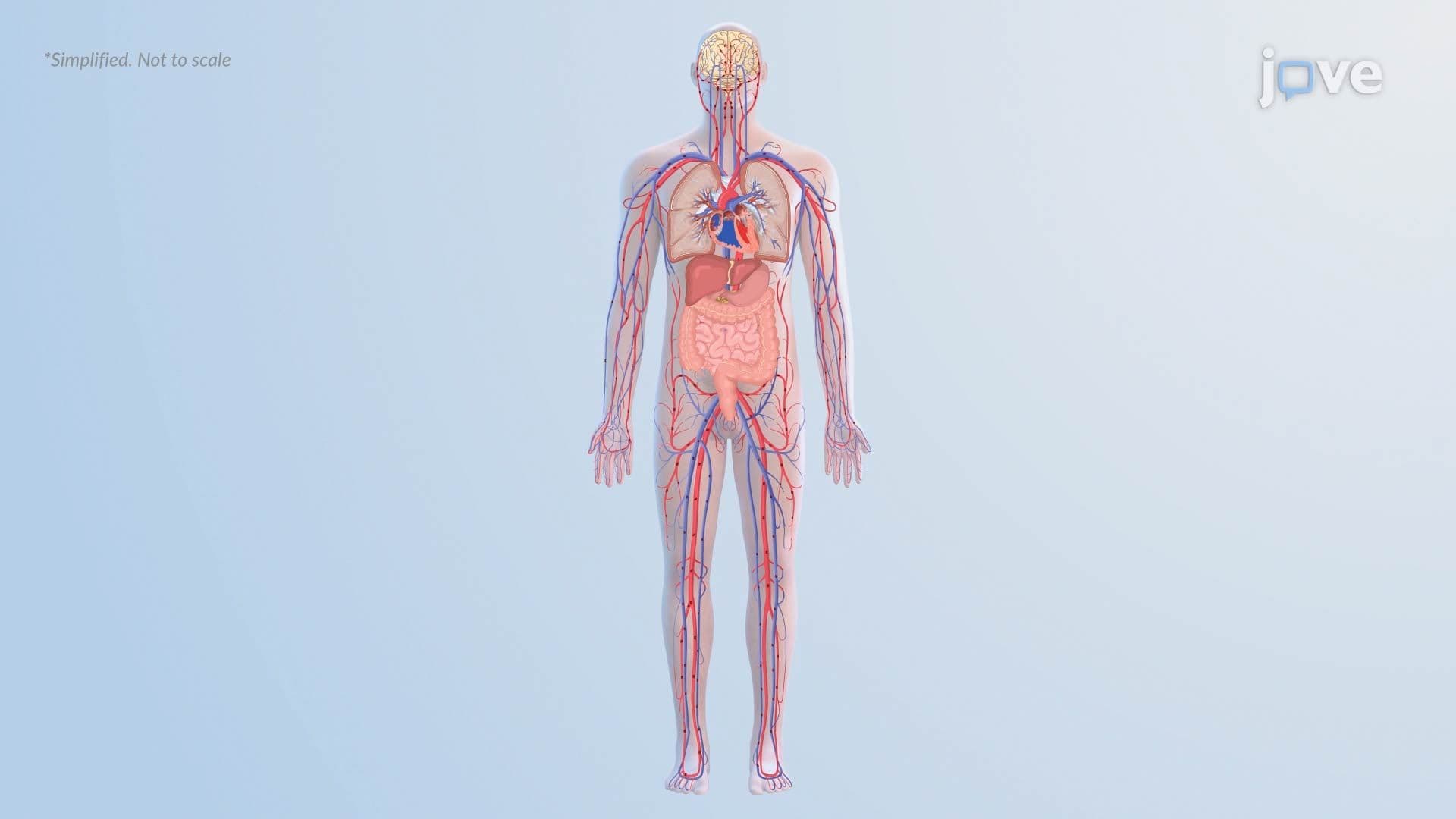
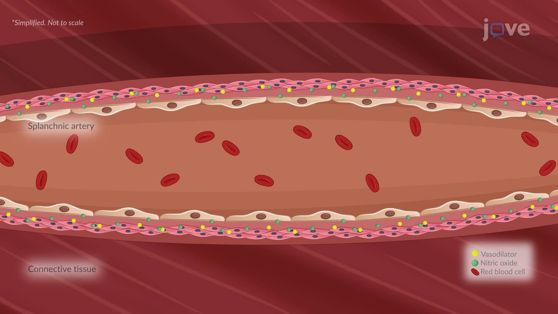
The physical distortion caused by fibrosis and regenerative nodules dramatically increases resistance to blood flowing through the portal vein, the major vessel delivering nutrient-rich blood from the intestines to the liver. This increased resistance forces blood pressure within the portal system to rise, a condition called portal hypertension. Portal hypertension has serious downstream consequences, including the development of esophageal varices (enlarged, fragile veins that can rupture and cause gastrointestinal bleeding), ascites (fluid accumulation in the abdomen), and hepatic encephalopathy. In US emergency medicine and internal medicine training, recognizing the clinical signs of portal hypertension is an essential competency, and understanding its root cause begins with the cellular mechanisms explored here.
Related Micro-courses