Loading concepts…

 27 Concepts
 4.7K Views
 Popular topics



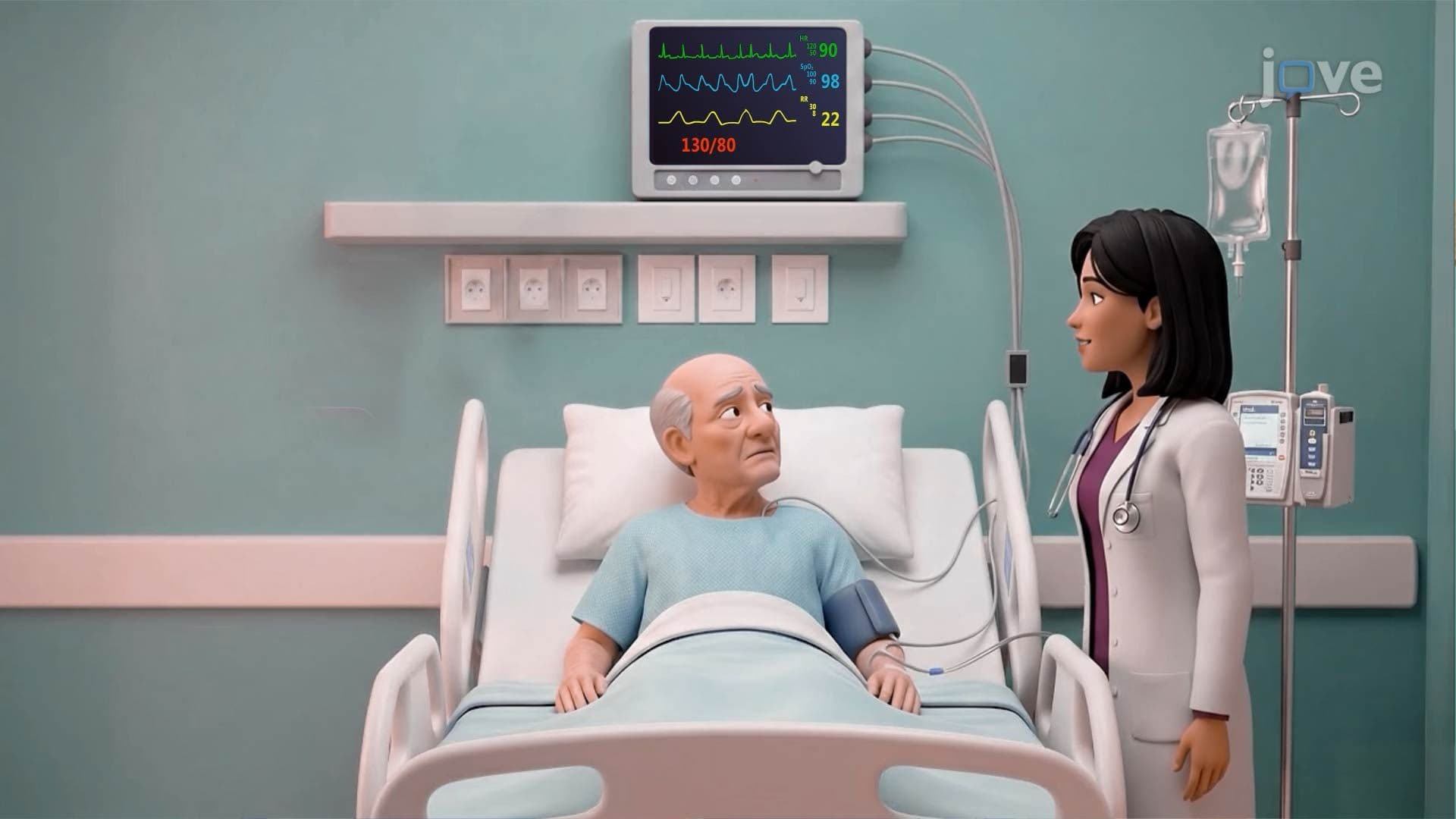






Endocrine disorders arise when hormone production, regulation, or tissue response breaks down, triggering widespread metabolic consequences. This micro-course covers the full spectrum of common endocrine system diseases, from diabetes mellitus and hormonal imbalance disorders like hypothyroidism and Cushing's syndrome to acute crises such as diabetic ketoacidosis. Built with JoVE Coach, the content connects pathophysiology to real clinical presentations, supporting US exam readiness and deeper scientific understanding.
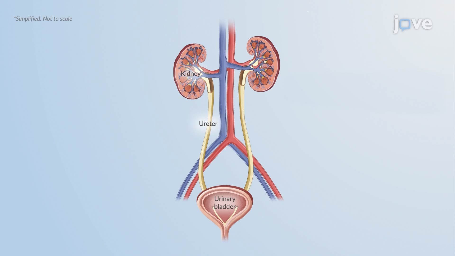
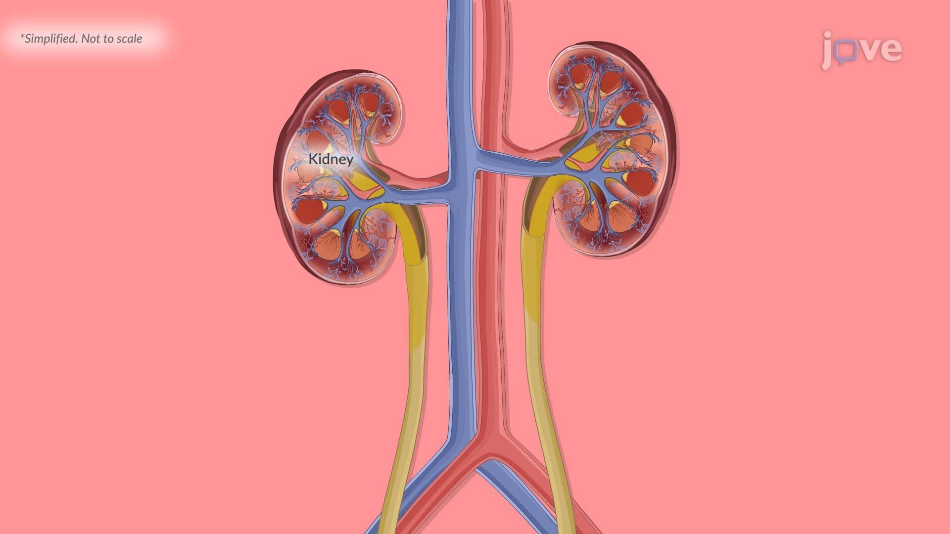
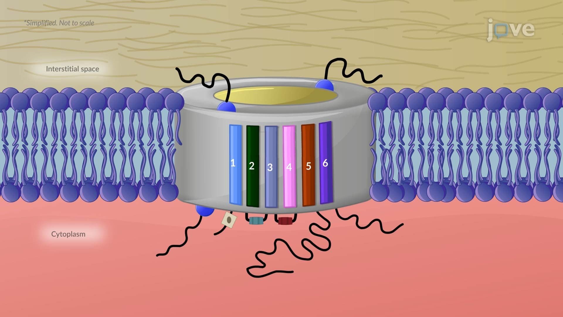
1. Diabetes Insipidus: Types and Mechanisms Diabetes insipidus (DI) disrupts water balance by impairing antidiuretic hormone (ADH) action, causing the kidneys to excrete up to 10-20 liters of dilute urine daily. Central DI results from insufficient ADH production, often following brain injury or intracranial tumors. Nephrogenic DI occurs when the kidneys cannot respond to ADH, a pattern seen with chronic kidney disease or lithium carbonate use. Gestational DI is triggered by placental vasopressinase breaking down ADH during pregnancy. In all forms, loss of aquaporin-2 channel insertion in collecting ducts prevents water reabsorption, raising plasma osmolality and driving intense thirst, dehydration, and polyuria.
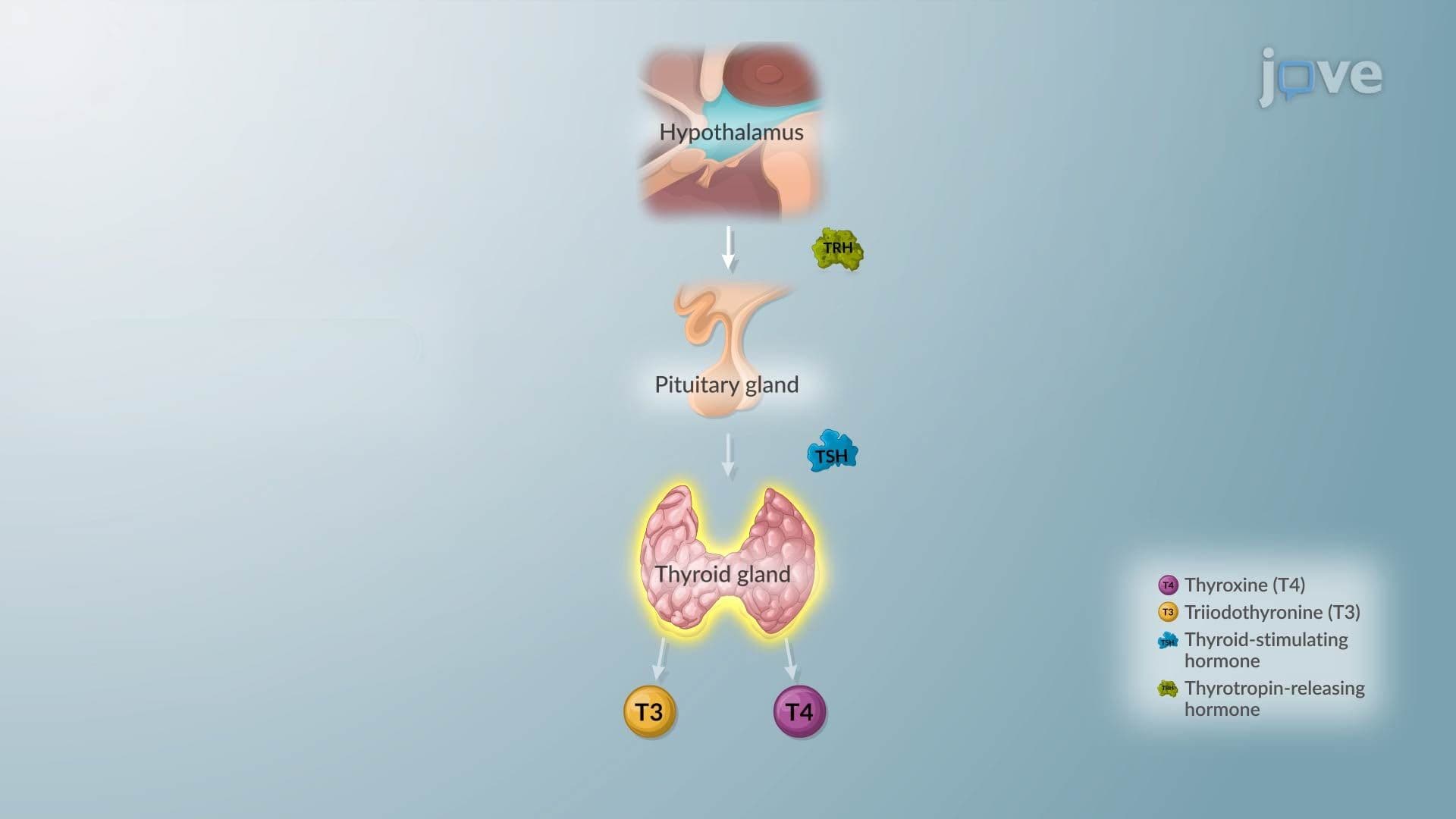
2. Hyperthyroidism and Graves' Disease Hyperthyroidism results from excessive thyroid hormone production, suppressing TSH below 0.4 mIU/L while elevating free T3 and T4. The most common cause is Graves' disease, an autoimmune condition in which thyroid-stimulating immunoglobulins (TSI) bind TSH receptors and continuously overstimulate the thyroid, independent of normal regulatory control. This elevates basal metabolic rate and increases beta-adrenergic sensitivity, producing palpitations, tremors, heat intolerance, and weight loss. Graves' disease also targets orbital fibroblasts, causing exophthalmos and periorbital edema. Genetic susceptibility through HLA-DR3 and HLA-B8 variants, combined with environmental triggers like smoking, increases disease risk significantly.
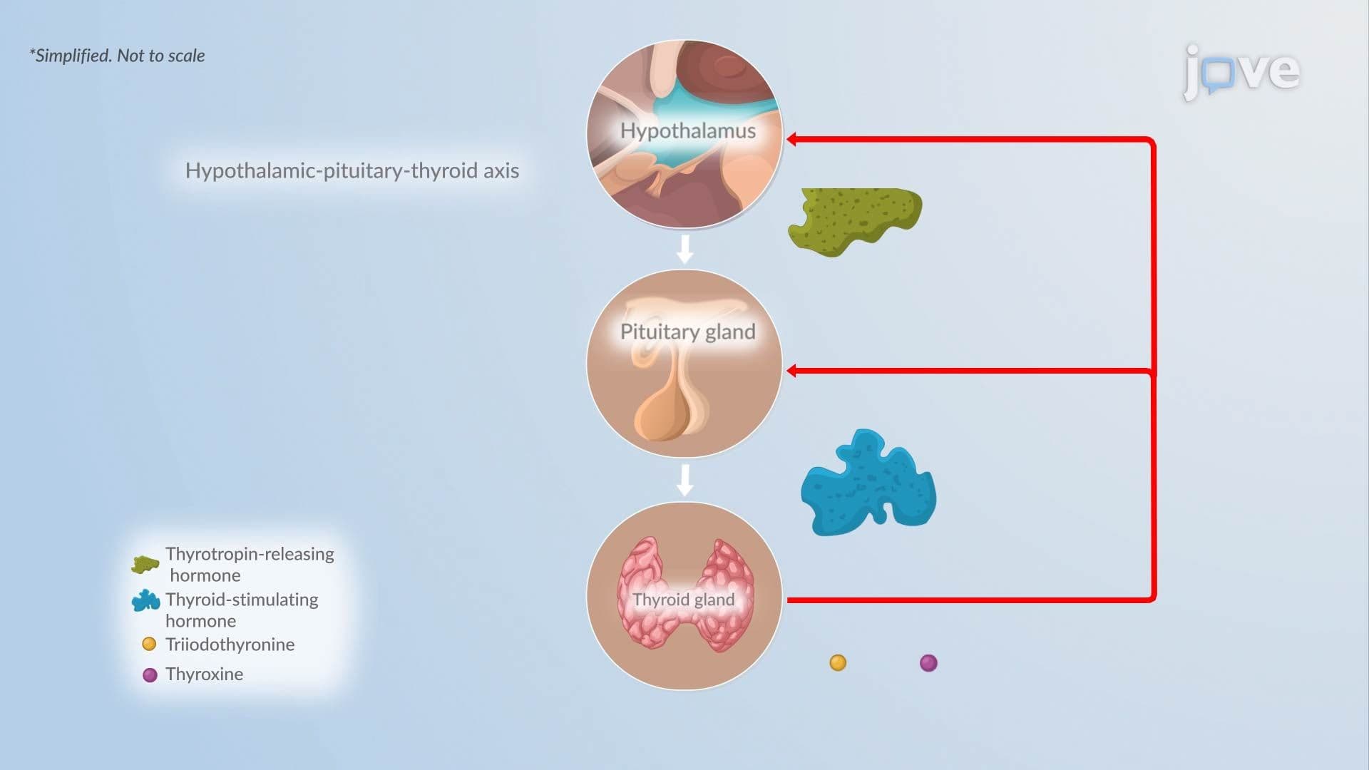
3. Goiter and Hypothyroidism A goiter, an abnormal thyroid enlargement, reflects chronic stimulation of thyroid tissue from elevated TSH, autoimmune activation (as in Graves' disease or Hashimoto's thyroiditis), or iodine deficiency. Over time, diffuse goiters may become nodular and develop autonomous hormone production, resulting in toxic multinodular goiter. Hypothyroidism, by contrast, involves insufficient thyroid hormone output. Primary hypothyroidism elevates TSH due to lost negative feedback; secondary hypothyroidism results from pituitary TSH deficiency; and tertiary hypothyroidism stems from insufficient hypothalamic TRH. All three forms reduce circulating T3 and T4, slowing metabolism across organ systems.
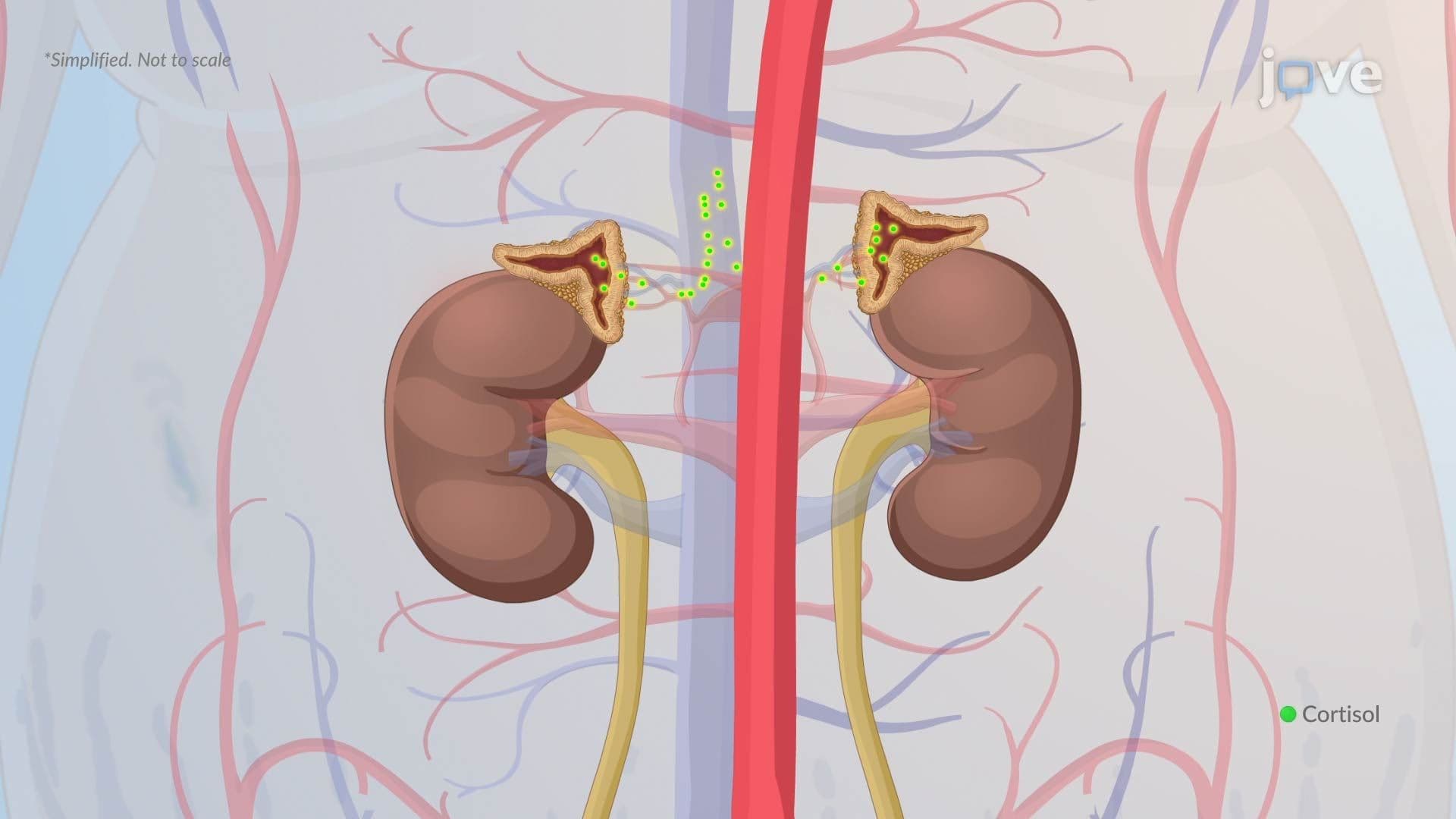
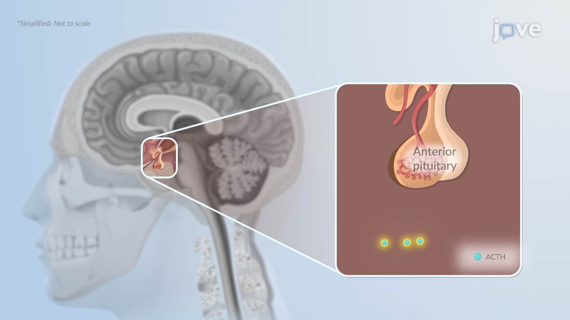
4. Cushing's Syndrome Cushing's syndrome develops from prolonged cortisol excess. The most common cause in the US is iatrogenic, long-term corticosteroid use (e.g., prednisone for autoimmune conditions). Endogenous causes include Cushing's disease, where a pituitary adenoma secretes excess ACTH, driving adrenal cortisol overproduction despite negative feedback. Ectopic ACTH syndrome, often from small-cell lung carcinoma or pancreatic tumors, independently elevates ACTH and cortisol. ACTH-independent causes include autonomous adrenal adenomas. Disruption of the HPA axis leads to classic features: central obesity, moon facies, buffalo hump, hypertension, hyperglycemia, and immunosuppression, all reflecting sustained glucocorticoid excess.
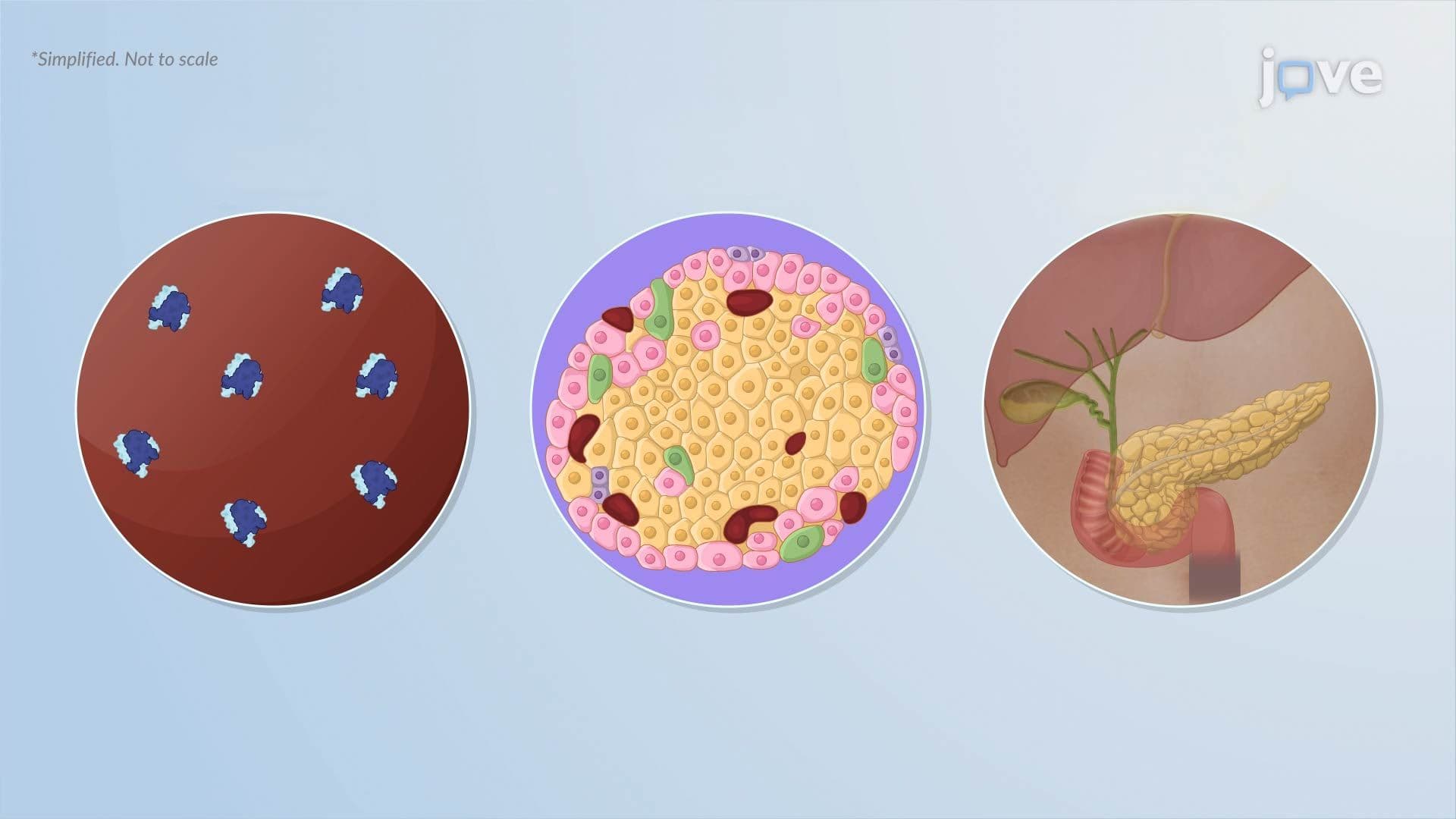
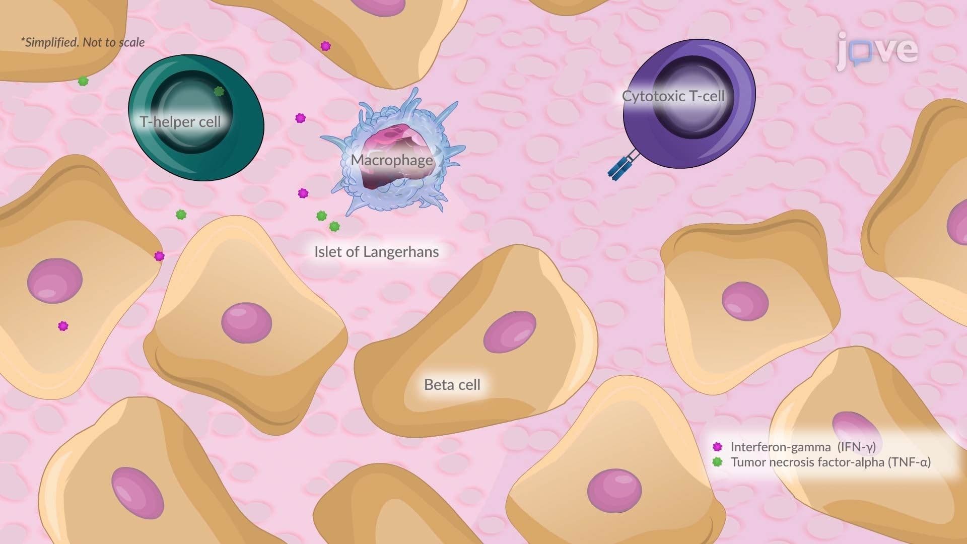
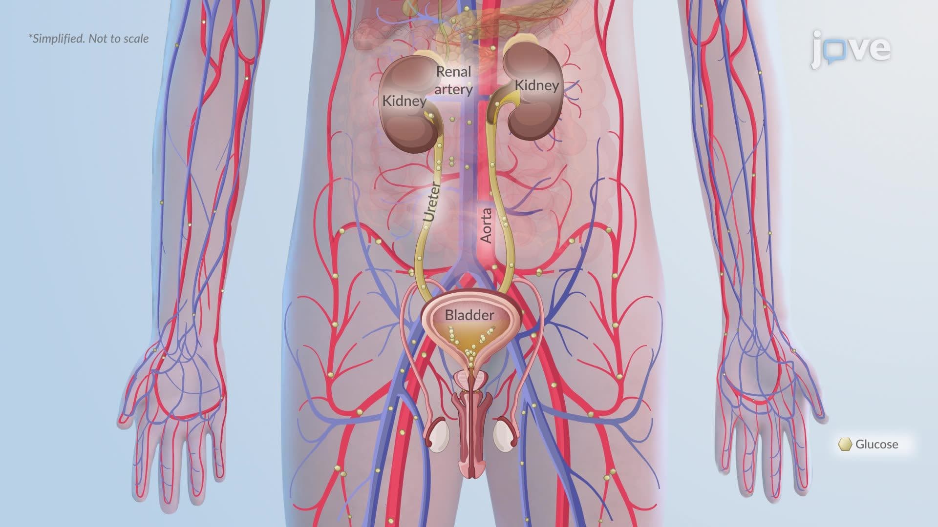
5. Type 1 Diabetes Mellitus: Pathophysiology and Clinical Features Type 1 diabetes mellitus (T1DM) is an autoimmune disease in which T helper 1 cells release inflammatory cytokines, including IFN-γ and TNF-α, activating cytotoxic T cells that destroy pancreatic beta cells in the islets of Langerhans. This process, called insulitis, results in absolute insulin deficiency. Without insulin, glucose cannot enter insulin-dependent tissues, causing hyperglycemia. When blood glucose exceeds the renal threshold (~180 mg/dL), osmotic diuresis produces polyuria, polydipsia, and dehydration. Patients also experience polyphagia, unexplained weight loss, and fatigue. HLA-DR3 and HLA-DR4 gene variants significantly increase genetic susceptibility.
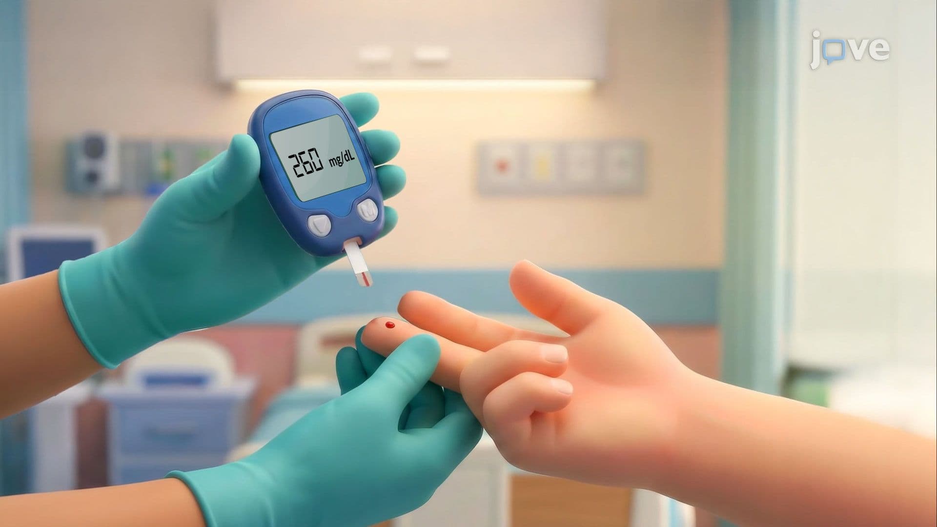
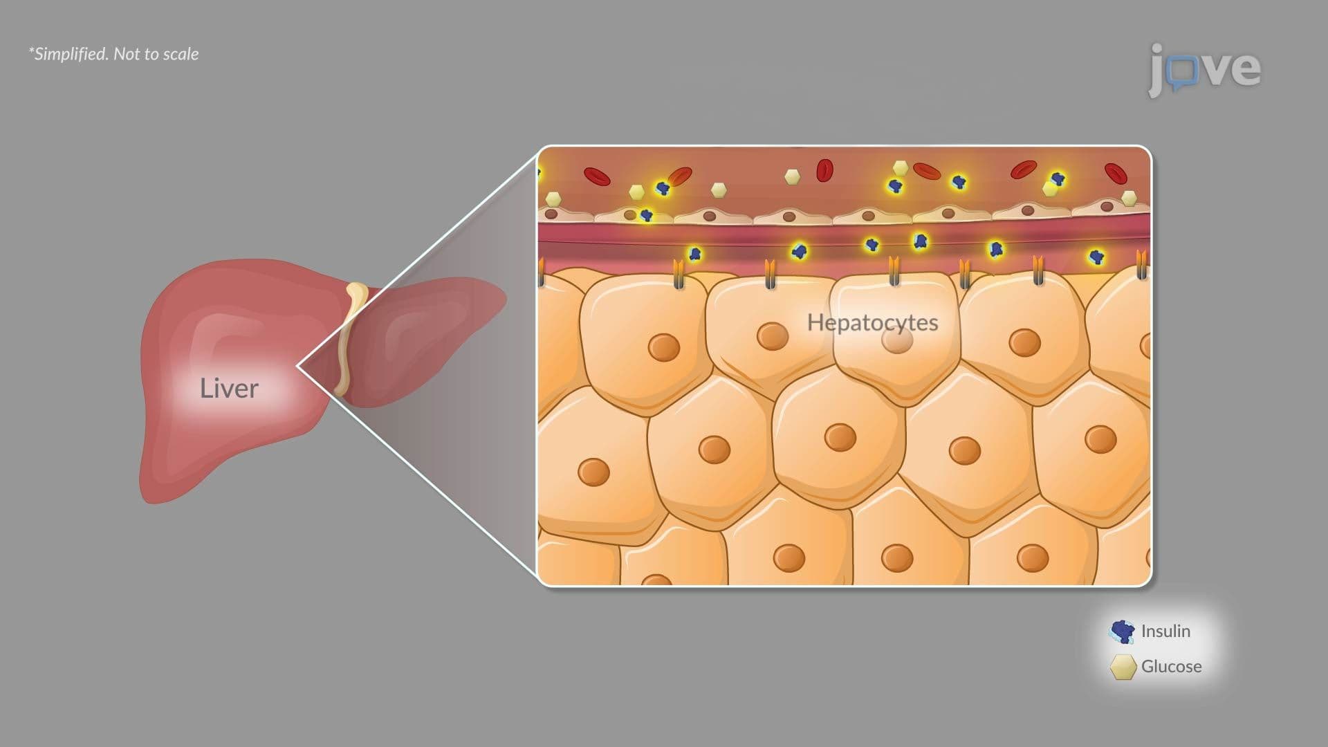
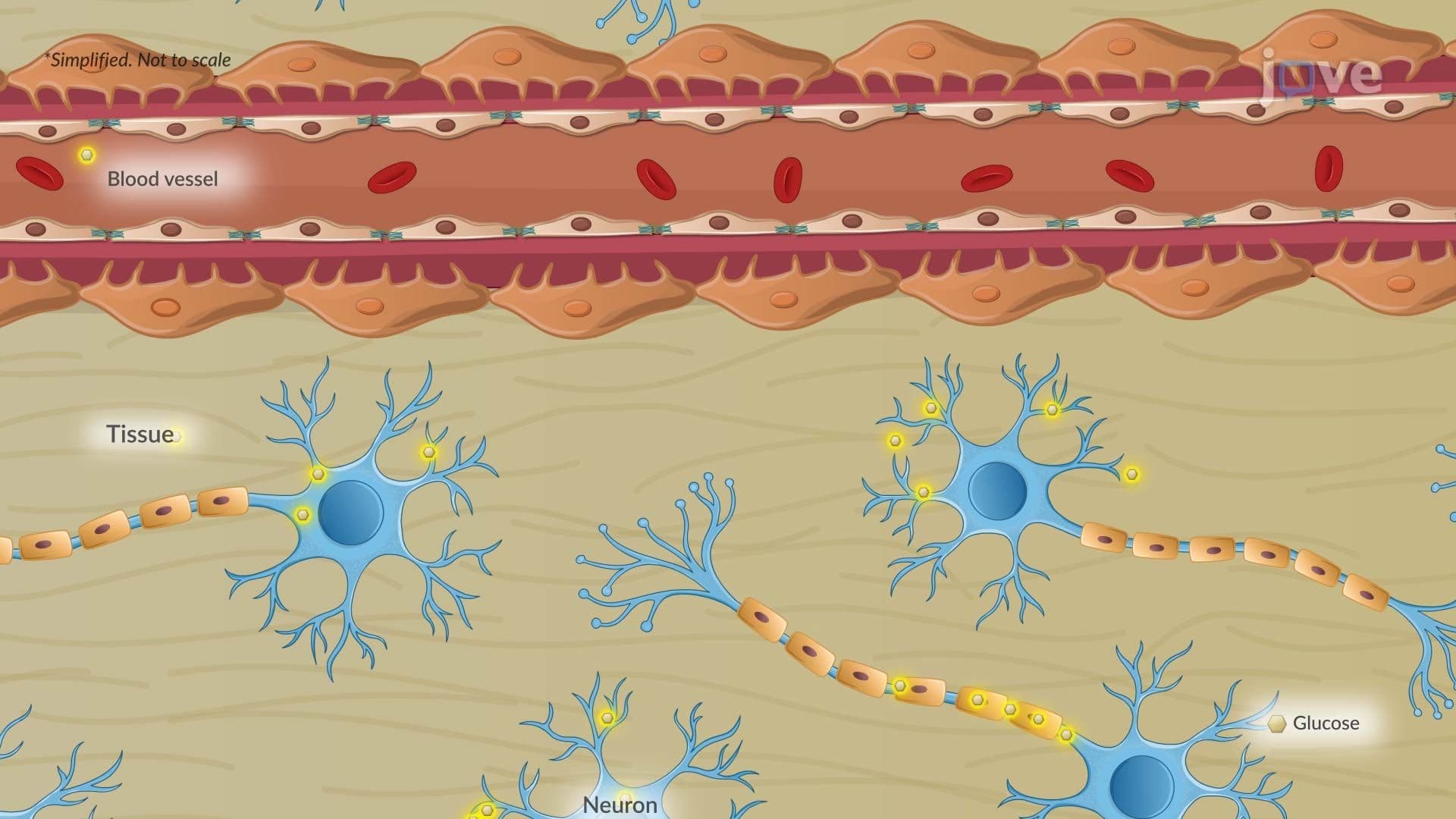
6. Type 2 Diabetes Mellitus: Pathophysiology and Diagnosis Type 2 diabetes mellitus (T2DM) develops through progressive insulin resistance in skeletal muscle, liver, and adipose tissue, combined with beta-cell exhaustion. In muscle, reduced insulin sensitivity decreases postprandial glucose uptake. In the liver, continued glucose production despite adequate insulin, worsened by dysregulated glucagon, raises fasting glucose. Adipose insulin resistance elevates free fatty acids, compounding lipotoxic damage to beta cells. Diagnosis is confirmed by fasting plasma glucose ≥126 mg/dL, 2-hour glucose ≥200 mg/dL on an oral glucose tolerance test, HbA1c ≥6.5%, or a random glucose ≥200 mg/dL with classic symptoms. Risk factors include obesity, physical inactivity, family history, and advancing age.
7. Acute Diabetic Emergencies: DKA, HHS, Hypoglycemia, and Hyperglycemia Diabetic ketoacidosis (DKA) occurs when insulin deficiency, most commonly in T1DM, triggers counterregulatory hormone release. Increased hepatic fatty acid oxidation produces ketone bodies, acetoacetate, β-hydroxybutyrate, and acetone, causing metabolic acidosis. DKA is classified by blood pH and bicarbonate levels. Hyperosmolar hyperglycemic state (HHS) primarily affects older adults with T2DM: blood glucose exceeds 600 mg/dL and serum osmolality surpasses 320 mOsm/kg, but enough residual insulin prevents ketosis. Hypoglycemia, glucose below 70 mg/dL, results from excess insulin or missed meals. Acute hyperglycemia from missed insulin doses or illness triggers osmotic diuresis, worsening dehydration.
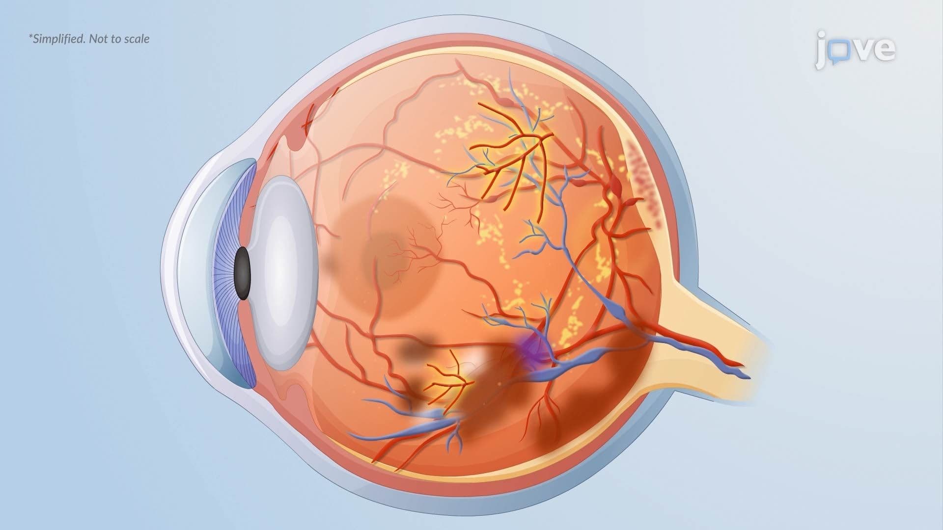
8. Chronic Complications of Diabetes: Microvascular and Macrovascular Disease Sustained hyperglycemia damages both large and small blood vessels. Microvascular complications include diabetic retinopathy, progressing from pericyte loss and microaneurysms (nonproliferative) to neovascularization (proliferative), diabetic nephropathy from glomerular injury, and peripheral and autonomic neuropathy. Diabetic foot ulcers arise from the combined effects of sensory neuropathy (loss of protective sensation), peripheral artery disease (hypoperfusion), impaired immune response, and poor wound healing due to hyperglycemia-driven dysfunction of neutrophils, fibroblasts, and keratinocytes. Macrovascular complications elevate risk for coronary artery disease, stroke, and peripheral artery disease, leading causes of diabetes-related mortality in the United States.