Loading video…

 27 Concepts
 4.7K views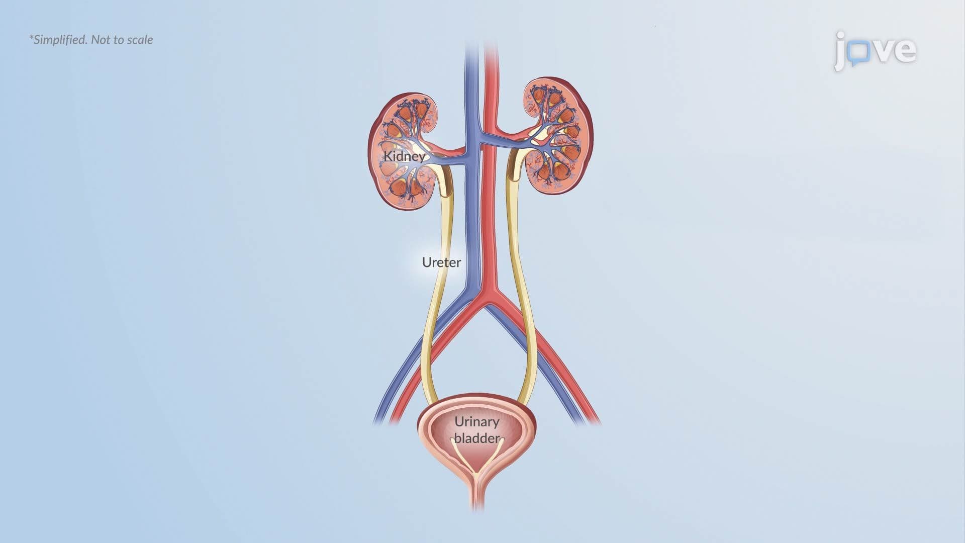

Video Summary: Pathophysiology in Diabetes Insipidus Ii
Did you know your kidneys can waste over 20 liters of water daily when a single hormone fails? Understanding Pathophysiology in Diabetes Insipidus II reveals how a breakdown in ADH signaling, whether in the brain or kidneys, triggers dangerous fluid loss. At US hospitals, this condition is commonly confused with uncontrolled diabetes mellitus, yet the mechanisms differ entirely. Watch the full video on JoVE Coach to master this concept with expert-led visuals and step-by-step explanations.
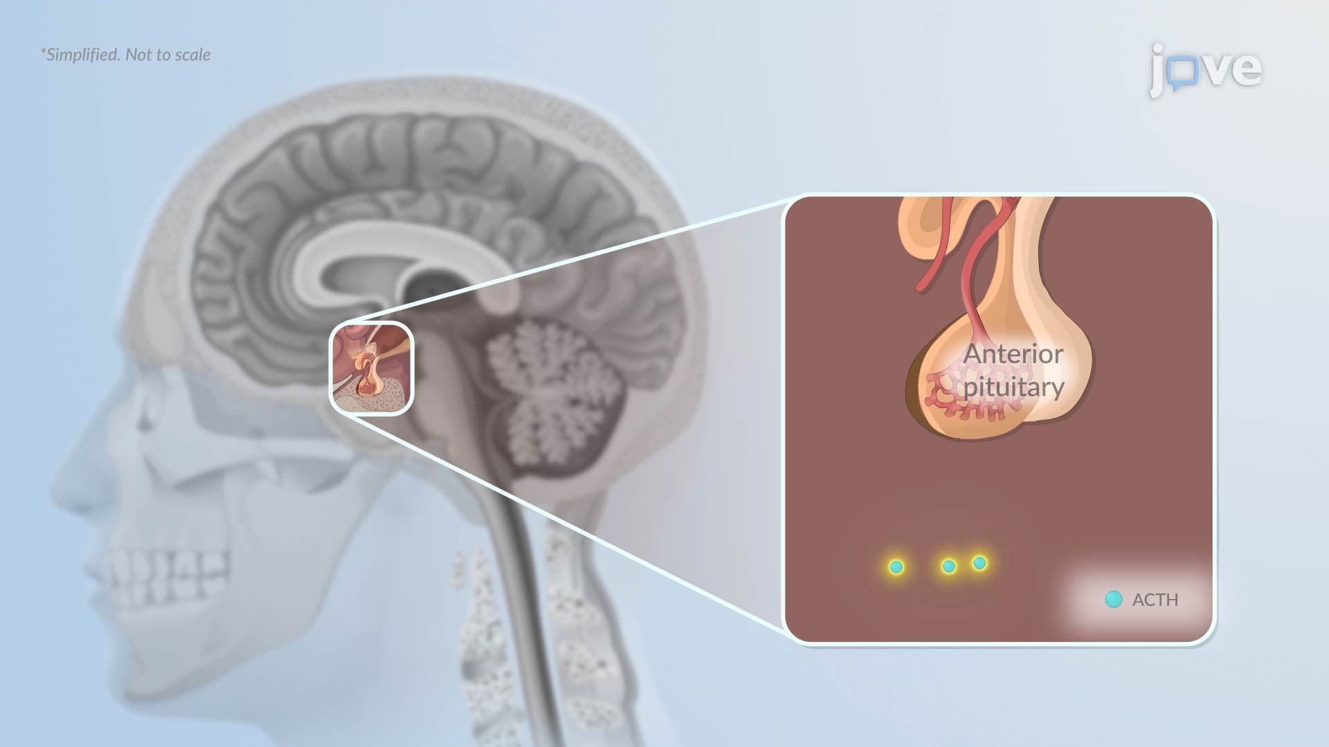
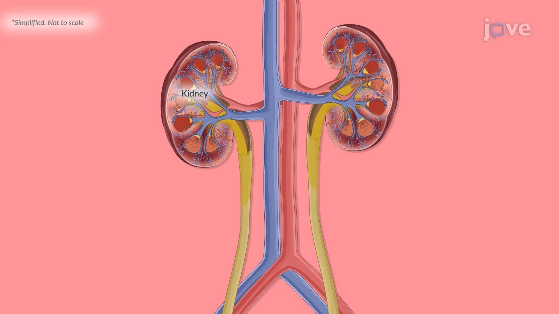
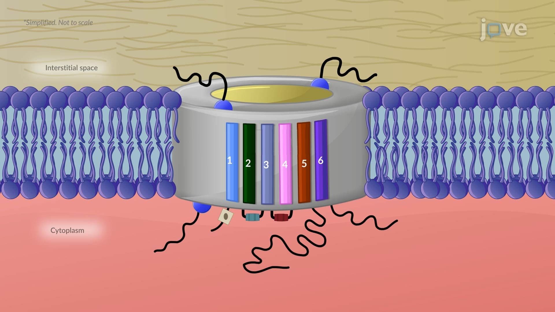
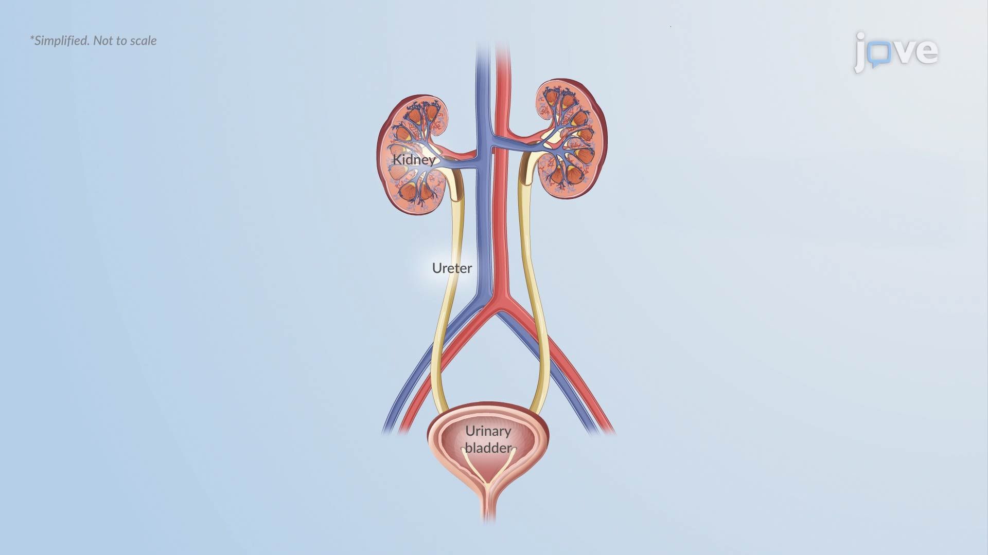
To understand Pathophysiology in Diabetes Insipidus II, you first need to appreciate how elegantly the body controls water. Under normal conditions, the hypothalamus monitors blood concentration and signals the posterior pituitary gland to release antidiuretic hormone (ADH), also called vasopressin. ADH then travels through the bloodstream to the kidneys, where it triggers the insertion of aquaporin-2 water channels into the collecting duct cells. These channels act like tiny doors, allowing water to flow back into the body rather than being lost as urine. The result is concentrated urine and stable plasma osmolality, a precise chemical balance your body works hard to maintain.
Diabetes insipidus (DI) disrupts this pathway at one of two points. In central DI, the hypothalamus or posterior pituitary fails to produce or release adequate ADH. This can occur after traumatic brain injury, a brain tumor, or surgery, conditions seen regularly in US neurosurgical and neurology units. In nephrogenic DI, ADH is produced normally, but the kidney tubules do not respond to it. This unresponsiveness may stem from genetic mutations, chronic lithium use (a common psychiatric medication in the US), or hypercalcemia. In both types, the collecting ducts remain impermeable to water, and free water pours out in the urine.
When aquaporin-2 channels are absent or non-functional, urine output skyrockets, exceeding three liters per day in most clinical cases and sometimes reaching 15-20 liters. This massive fluid loss raises plasma osmolality above the normal threshold of 285-295 milliosmoles per kilogram, which the hypothalamus detects and interprets as a dehydration signal. The thirst center activates intensely, driving polydipsia. In children, this fluid imbalance often manifests as nocturnal enuresis (bedwetting), a symptom that can be mistaken for a behavioral issue rather than a hormonal one. If fluid intake cannot keep pace with losses, common during illness or sleep, severe dehydration and hypernatremia can develop rapidly.
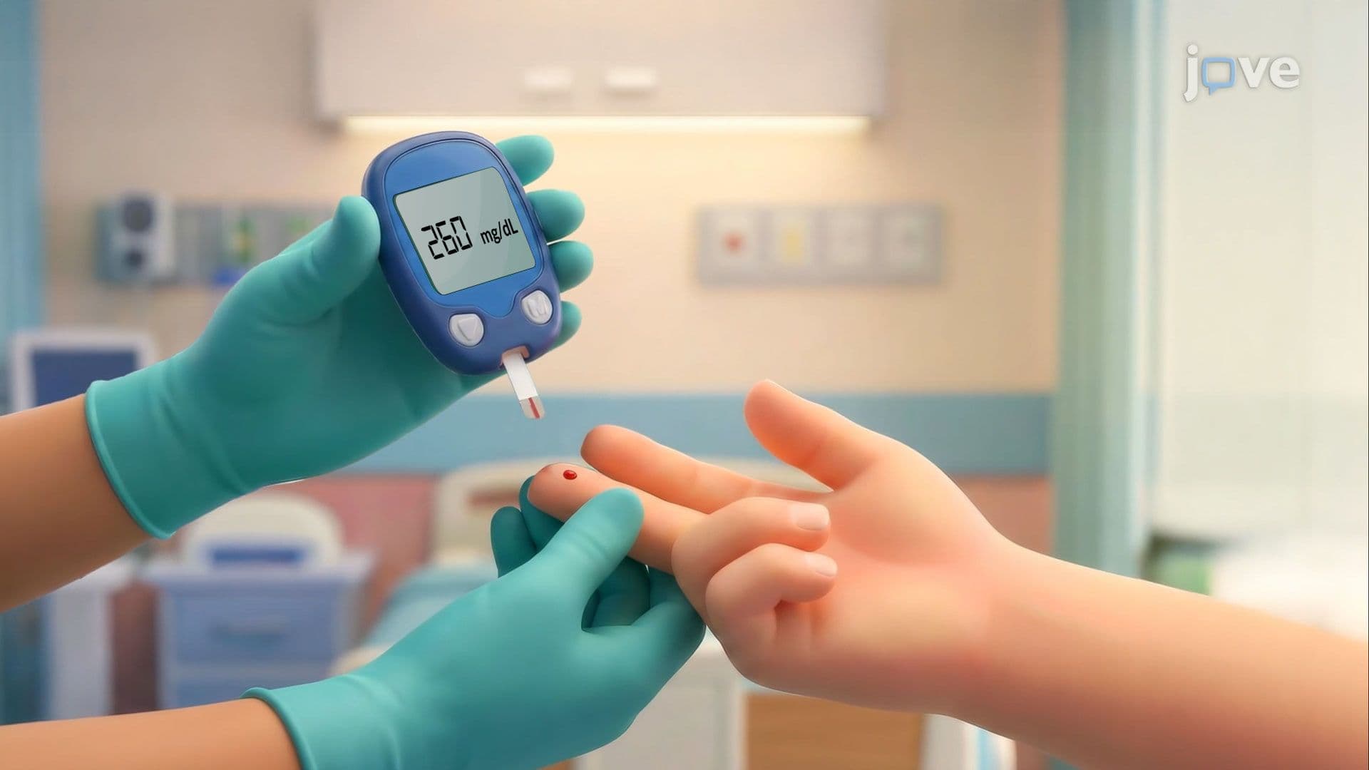
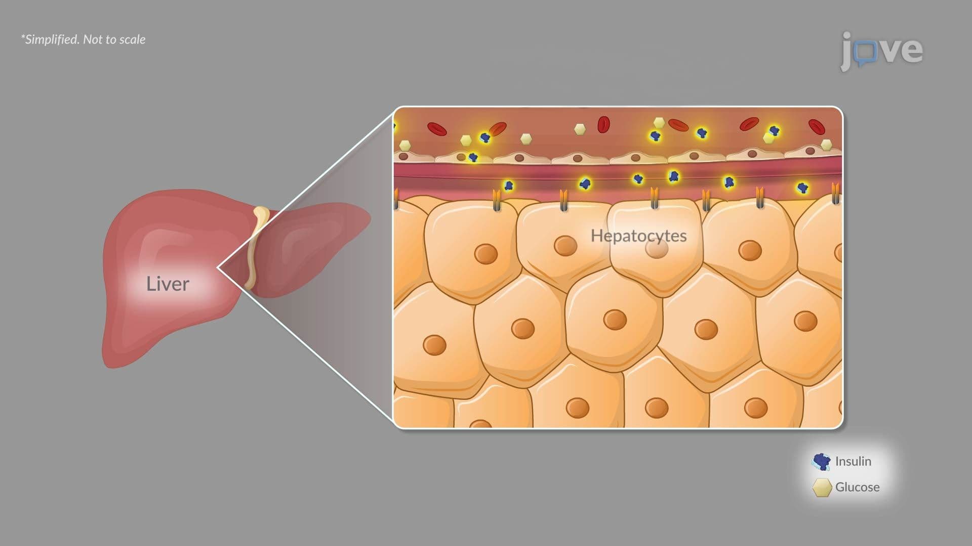
For students in AP Biology, Human Anatomy and Physiology, or pre-med college courses, DI is a gateway concept for understanding how endocrine disorders ripple across organ systems. Questions on exams like the MCAT frequently ask students to compare DI with diabetes mellitus (a glucose regulation disorder, not a water regulation disorder), or to distinguish central from nephrogenic causes based on lab findings and patient history. The concept also connects to Cushing's syndrome, adrenal insufficiency, and hypothyroidism, all conditions where a hormonal signaling failure creates systemic consequences. Understanding how plasma osmolality is measured and interpreted is also a common topic in college physiology midterms. Knowing that a water deprivation test helps diagnose DI, and that central DI responds to synthetic ADH (desmopressin) while nephrogenic DI does not, gives you a clinically grounded framework that translates directly to both exam success and real healthcare settings.
Related Micro-courses