Loading video…

 27 Concepts
 4.7K views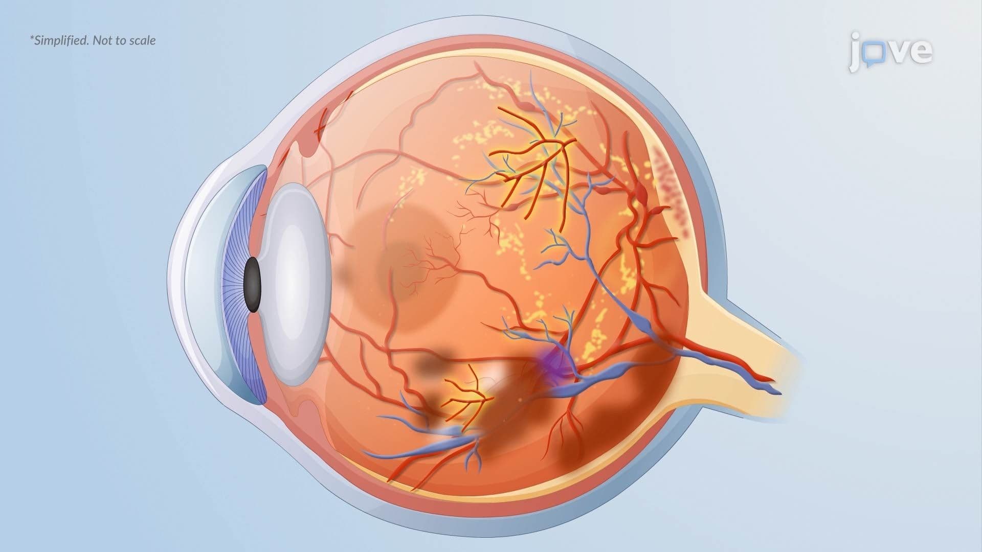

Video Summary: What Is Diabetic Retinopathy
Did you know that diabetes is the leading cause of new blindness among working-age adults in the United States? Diabetic retinopathy is the silent eye disease behind those staggering statistics, and understanding the basics of diabetic retinopathy could be the difference between preserving and losing your sight. From weakened blood vessels to dangerous new vessel growth, this condition tells the story of how chronically high blood sugar quietly destroys the retina. Watch the full video on JoVE Coach to master this concept with expert-led visuals and step-by-step explanations.
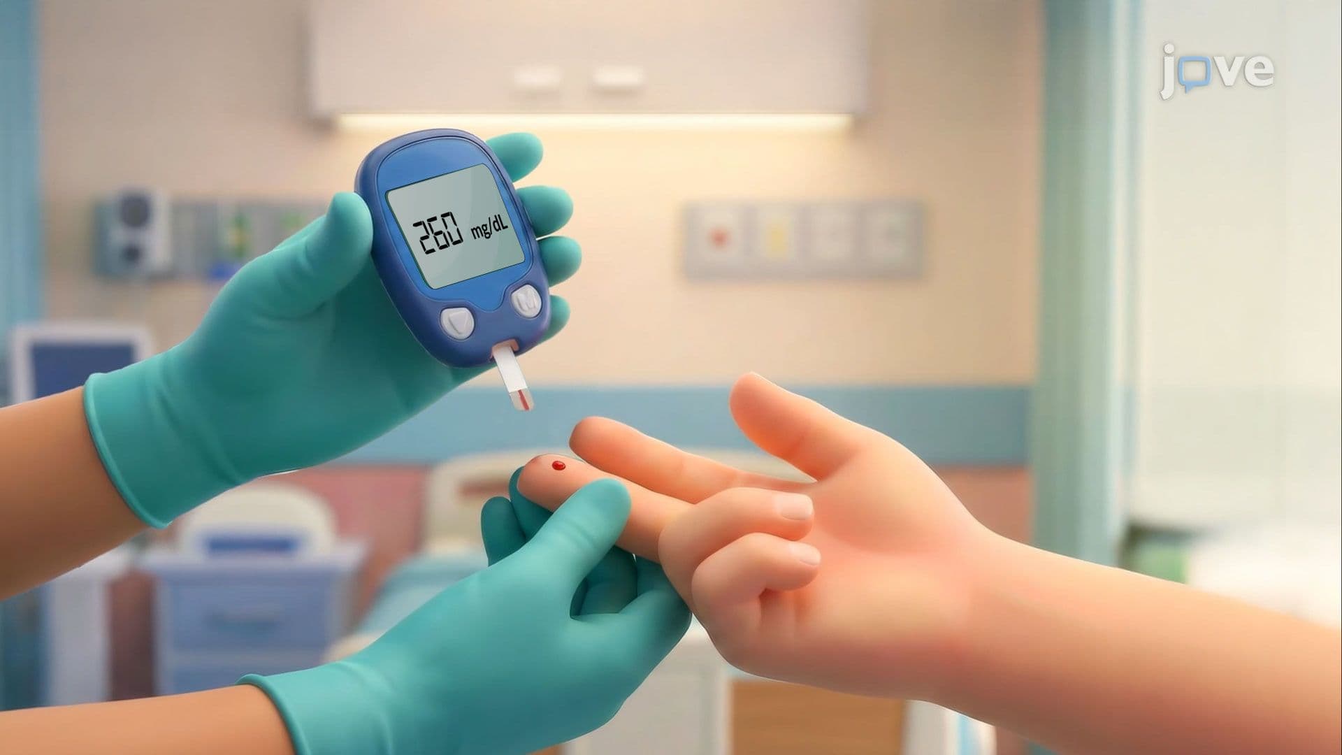
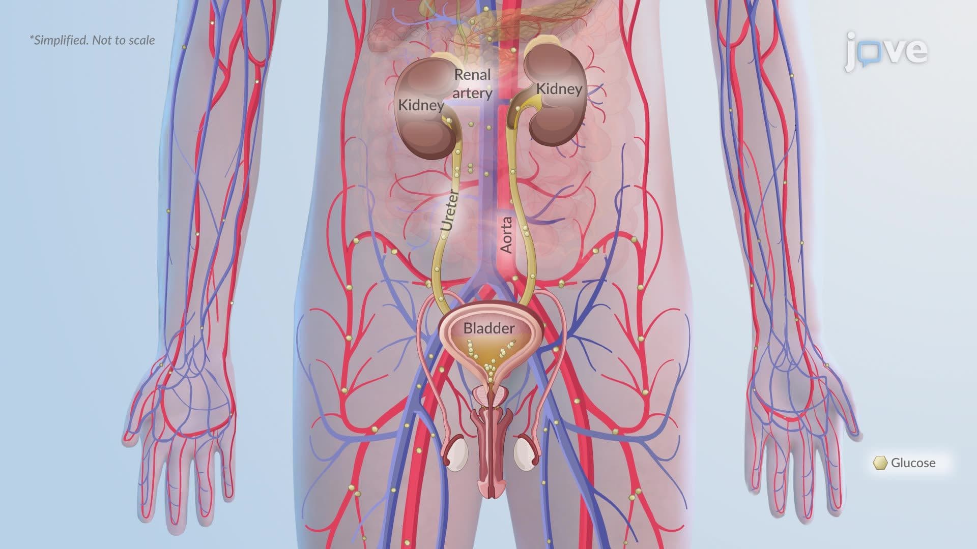
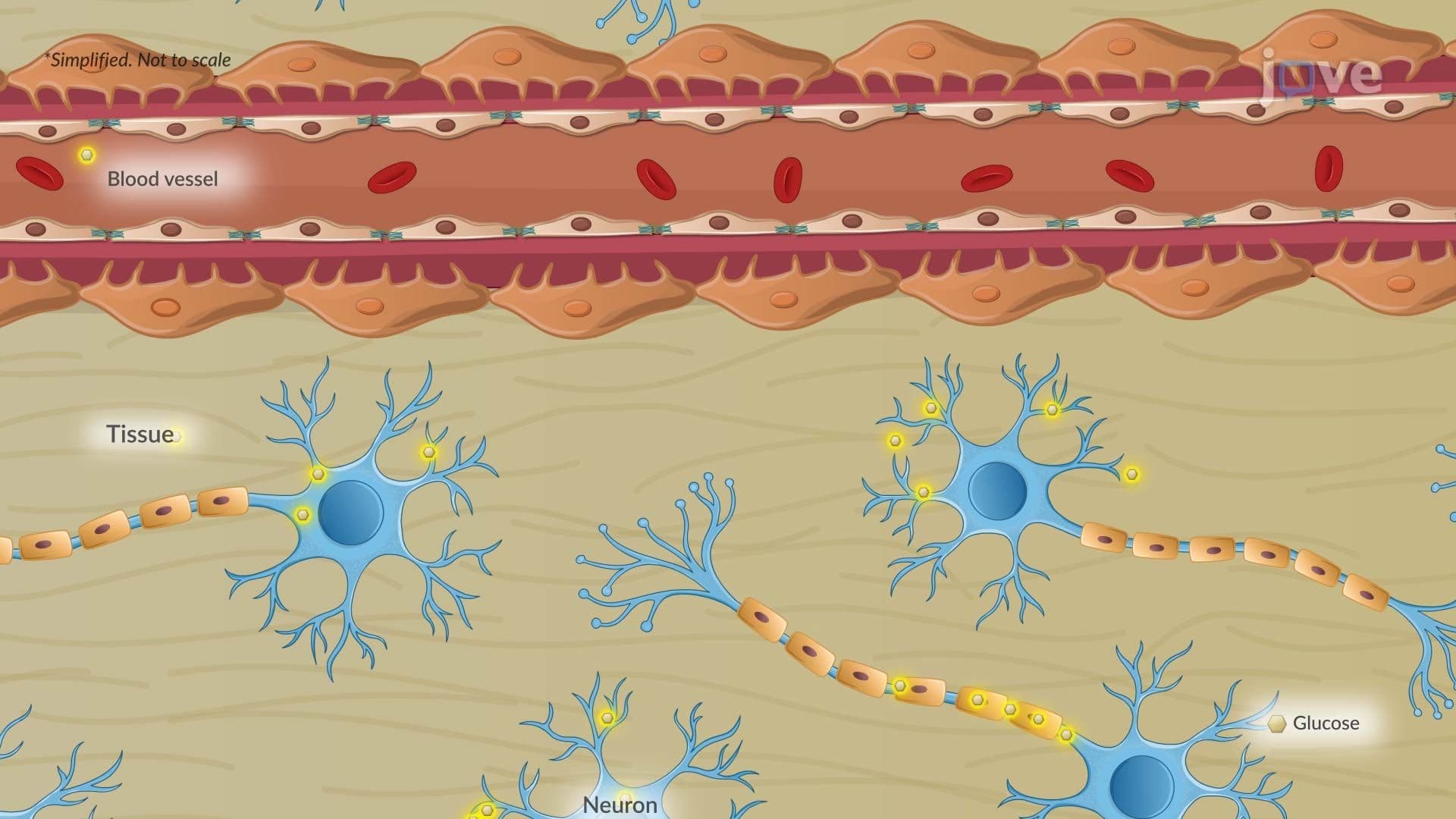
Diabetic retinopathy is a progressive, diabetes-related eye disease in which chronically elevated blood glucose levels, a state called hyperglycemia, gradually destroy the tiny blood vessels that supply the retina. The retina is the light-sensitive tissue lining the back of the eye, and it depends entirely on a healthy network of capillaries to function. When those vessels fail, so does vision. According to the Centers for Disease Control and Prevention (CDC), roughly 1 in 3 Americans with diabetes over age 40 shows signs of diabetic retinopathy, making it one of the most common and preventable causes of blindness in the country.
Understanding this disease is not just clinically important, it is also a core concept in high school AP Biology, college-level anatomy and physiology courses, and health science curricula across the US.
While diabetes mellitus is the root cause, several additional factors accelerate the progression of diabetic retinopathy. Hypertension compounds vascular stress, while dyslipidemia contributes to the lipid deposits seen in the retina. Smoking impairs circulation and increases oxidative damage to vessel walls. Perhaps more surprisingly, physiological states like pregnancy and puberty can also worsen the disease, hormonal shifts during these periods affect vascular permeability and metabolic demand. This overlap with hormonal imbalances and the broader endocrine system is why endocrinologists and ophthalmologists often co-manage diabetic patients. Understanding how these risk factors interact helps explain why tight glycemic and blood pressure control are the primary treatment strategies for slowing disease progression.
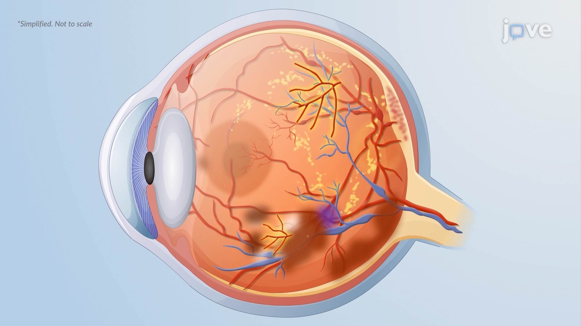
In the nonproliferative stage, structural breakdown begins at the cellular level. Pericytes, specialized cells that support and stabilize capillary walls, are lost early in the disease. Without this support, retinal capillaries weaken and balloon outward, forming microaneurysms, which appear as tiny red dots when viewed through a fundoscope. Increased vascular permeability allows lipids to escape into retinal tissue, creating hard exudates, yellow deposits with sharp borders that are a classic finding on eye examination.
As ischemia worsens, blocked capillaries and arterioles leave patches of retinal tissue severely oxygen-deprived. This produces cotton wool spots (areas of nerve fiber infarction), intraretinal hemorrhages, venous beading, and intraretinal microvascular abnormalities (IRMA). Critically, macular edema, swelling at the center of the retina, can develop at any stage and represents the leading cause of blindness in diabetic patients. On exams like the MCAT and USMLE Step 1, students are frequently tested on these clinical findings and what they reveal about underlying pathology.
In the proliferative stage, the retina's response to ischemia becomes the problem itself. Oxygen-starved tissue releases vascular endothelial growth factor (VEGF), a signaling protein that stimulates the growth of new blood vessels. However, these new vessels, a process called neovascularization, are structurally fragile and grow in abnormal locations, including along the surface of the retina and into the vitreous gel of the eye.
The consequences are severe. Fragile neovascular vessels can rupture, flooding the vitreous cavity with blood (vitreous hemorrhage) and causing sudden, dramatic vision loss. Additionally, fibrous tissue that forms alongside new vessels can pull on the retina, causing tractional retinal detachment, a true ocular emergency. In US clinical practice, anti-VEGF injections (such as ranibizumab or bevacizumab) and laser photocoagulation are frontline treatments to slow or stop these complications. Students studying pathophysiology for college midterms or health profession entrance exams should understand VEGF's role not just here, but across cancer biology, wound healing, and other disease contexts.
Related Micro-courses