Loading video…

 27 Concepts
 4.7K views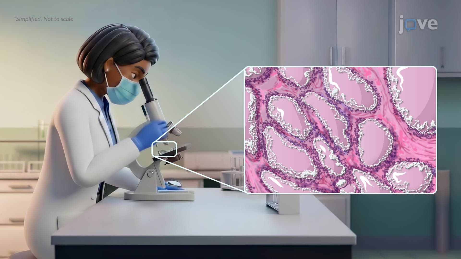

Video Summary: Pathophysiology in Graves Disease Ii
Your immune system is supposed to protect you, but what happens when it attacks your own thyroid? Understanding the pathophysiology in Graves disease II reveals how rogue antibodies hijack thyroid receptors, triggering uncontrolled hormone production. At major US teaching hospitals like Mayo Clinic, this autoimmune cascade explains symptoms from bulging eyes to racing hearts. Watch the full video on JoVE Coach to master this concept with expert-led visuals and step-by-step explanations.
Graves' disease is the most common cause of hyperthyroidism in the United States, affecting approximately 1 in 200 Americans, with a strong prevalence among women of reproductive age. But this isn't simply a thyroid problem, it's an autoimmune breakdown that rewires how the entire endocrine system functions. Grasping the pathophysiology in Graves disease II is essential for anyone studying biology, anatomy, or pre-health sciences, and it appears regularly on exams ranging from AP Biology to the MCAT and USMLE Step 1.
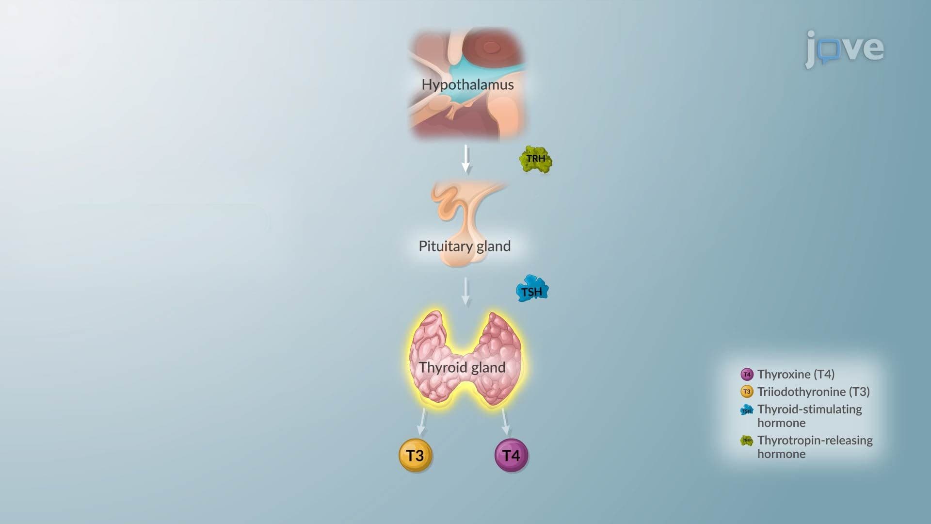
Under normal conditions, the immune system learns to ignore the body's own tissues, a process called self-tolerance. In Graves' disease, this system fails. Plasma cells begin producing thyroid-stimulating immunoglobulins (TSI), a class of autoantibodies that lock onto TSH receptors (TSH-R) on thyroid follicular cells. Unlike real TSH, which is released in carefully regulated pulses by the pituitary gland, TSI binds continuously and cannot be switched off by normal feedback mechanisms. The result is relentless stimulation of the thyroid, like a gas pedal stuck to the floor with no brake available.
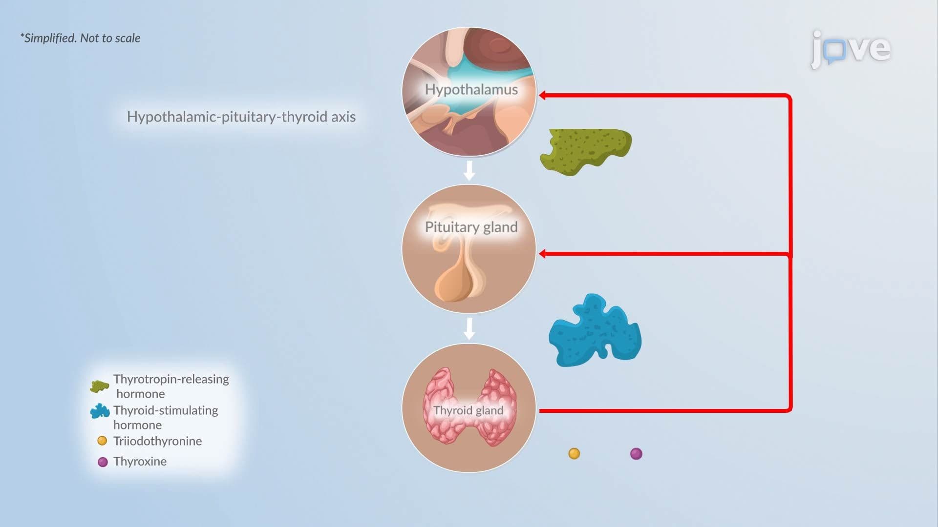
With TSH-R continuously activated, follicular cells ramp up synthesis and secretion of T4 (thyroxine) and T3 (triiodothyronine) far beyond physiological need. These hormones flood the bloodstream, raising the body's basal metabolic rate significantly. Patients, such as a college student at a US university noticing unexplained weight loss, tremors, and a heart rate consistently above 100 beats per minute, are experiencing the direct downstream effects of this hormonal surge. Elevated T3 and T4 also suppress TSH from the pituitary through negative feedback, which is why lab results in Graves' disease classically show low TSH alongside high free T4, a key distinction tested on NCLEX and MCAT exams.
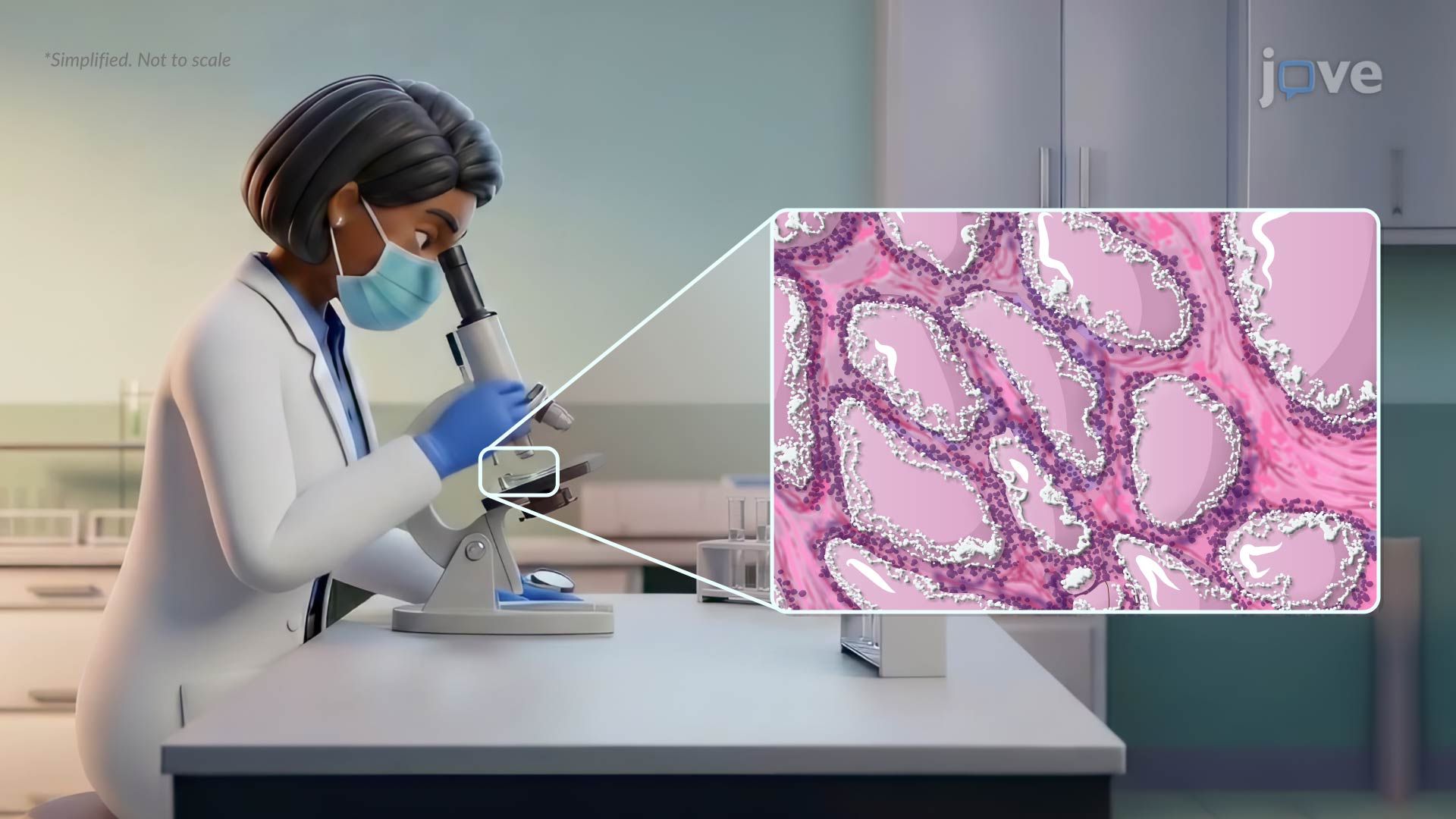
On histological examination, the thyroid tissue in Graves' disease shows dramatic changes. Follicular cells become tall and columnar rather than their usual flat or cuboidal shape, and they crowd together forming papillary infoldings. The gland becomes highly vascular, and the colloid, the stored gel-like substance inside follicles, is rapidly consumed and depleted, leaving characteristic scalloped or "wavy" edges on the follicle margins. These microscopic findings are frequently tested in college-level pathology and histology courses and may appear as image-based questions on the USMLE.
One of the most clinically striking features of Graves' disease is that it doesn't stop at the thyroid. Fibroblasts in the orbit (eye socket) and skin also express TSH receptors. The same immune attack that targets the thyroid causes these fibroblasts to accumulate glycosaminoglycans, leading to tissue swelling. In the eye socket, this produces periorbital edema and the hallmark exophthalmos, a protrusion of the eyeballs that can impair vision. On the shins, thickened, non-pitting skin plaques develop, called pretibial myxedema. Understanding these extrathyroidal effects helps students distinguish Graves' disease from other causes of hyperthyroidism, such as toxic multinodular goiter, and is a critical differentiator on AP, HESI A2, and TEAS assessments.
Related Micro-courses