Loading video…

 27 Concepts
 4.7K views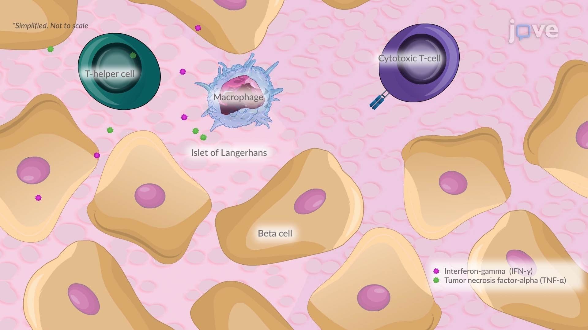

Video Summary: Pathophysiology in Type I Diabetes Ii
Did you know your own immune system can mistakenly destroy the cells that keep your blood sugar in balance? The pathophysiology in Type I Diabetes II reveals how a cascade of autoimmune attacks dismantles insulin-producing beta cells in the pancreas. In the US, nearly 1.6 million people live with Type 1 diabetes, managing daily glucose crises. Watch the full video on JoVE Coach to master this concept with expert-led visuals and step-by-step explanations.
Type 1 diabetes is far more than a sugar problem, it is a sophisticated autoimmune breakdown rooted in the body's own defense system turning against itself. The pathophysiology in Type I Diabetes II focuses on the precise molecular and cellular sequence that leads from immune activation to insulin failure. Mastering this pathway is essential not only for exams but for understanding one of the most prevalent endocrine system disorders affecting young Americans today.
The autoimmune cascade in Type 1 diabetes does not start randomly. Genetic susceptibility, particularly involving HLA (human leukocyte antigen) genes on chromosome 6, creates a biological predisposition. However, genetics alone are rarely sufficient. Environmental factors, viral infections such as enteroviruses, dietary exposures, and gut microbiome changes, are thought to "pull the trigger," particularly in children and adolescents. This gene-environment interaction is a high-yield concept in AP Biology and introductory college immunology courses.
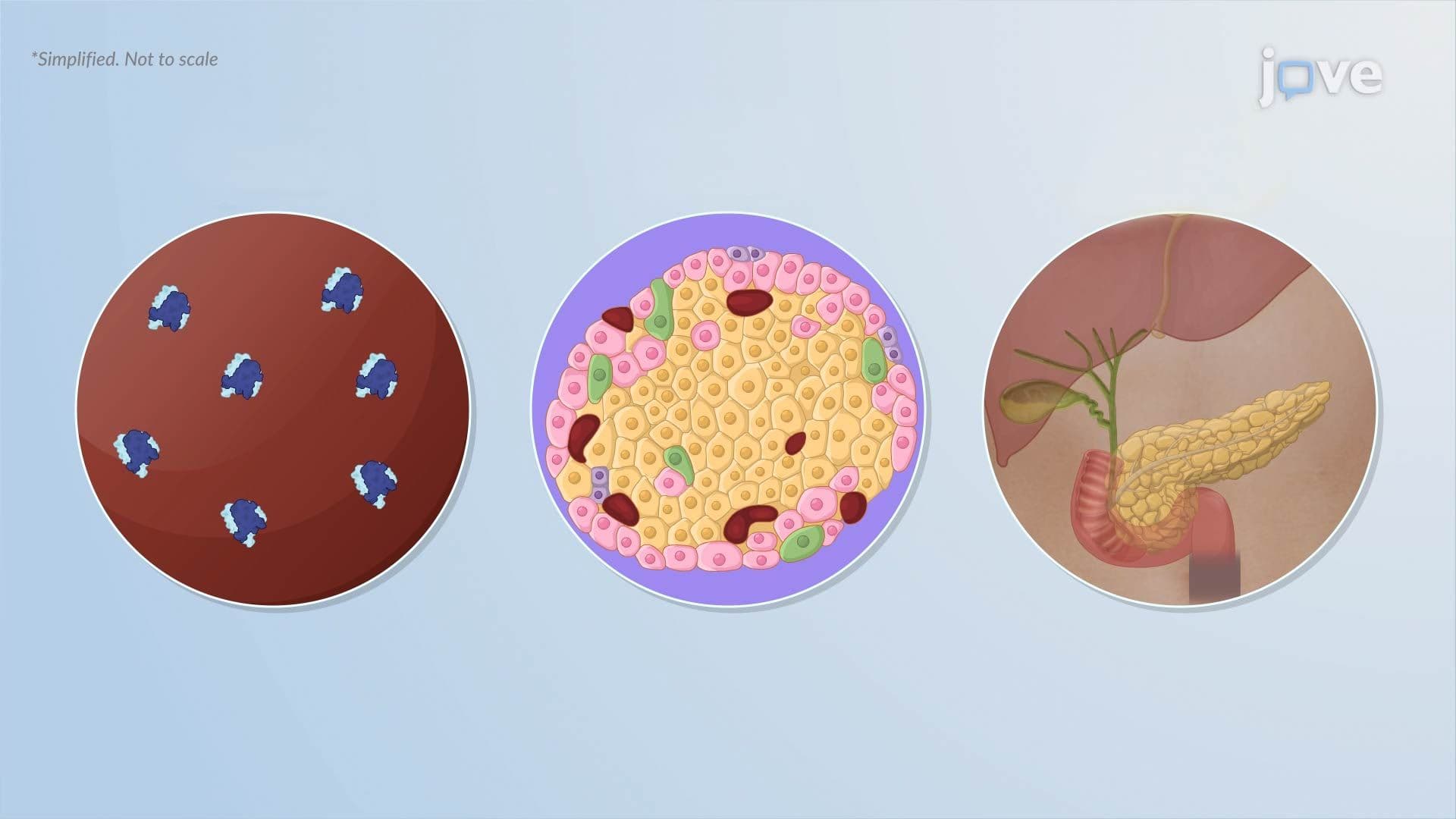
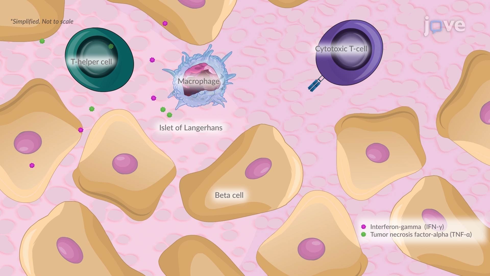
Once triggered, the immune response becomes highly organized and destructive. T helper 1 (Th1) cells are activated and release inflammatory cytokines, most notably interferon-gamma (IFN-γ) and tumor necrosis factor-alpha (TNF-α). IFN-γ plays a central role by activating macrophages and enhancing antigen-presenting cell (APC) function, which in turn supercharges cytotoxic T cell activity. These cytotoxic T cells then infiltrate the islets of Langerhans, the insulin-producing clusters of the pancreas, and systematically destroy beta cells. This localized inflammatory process is called insulitis, and it is the defining histological feature of Type 1 diabetes. On MCAT Biology and USMLE Step 1, students are frequently asked to distinguish insulitis from the mechanisms of Type 2 diabetes, where insulin resistance rather than autoimmune destruction is the primary mechanism.
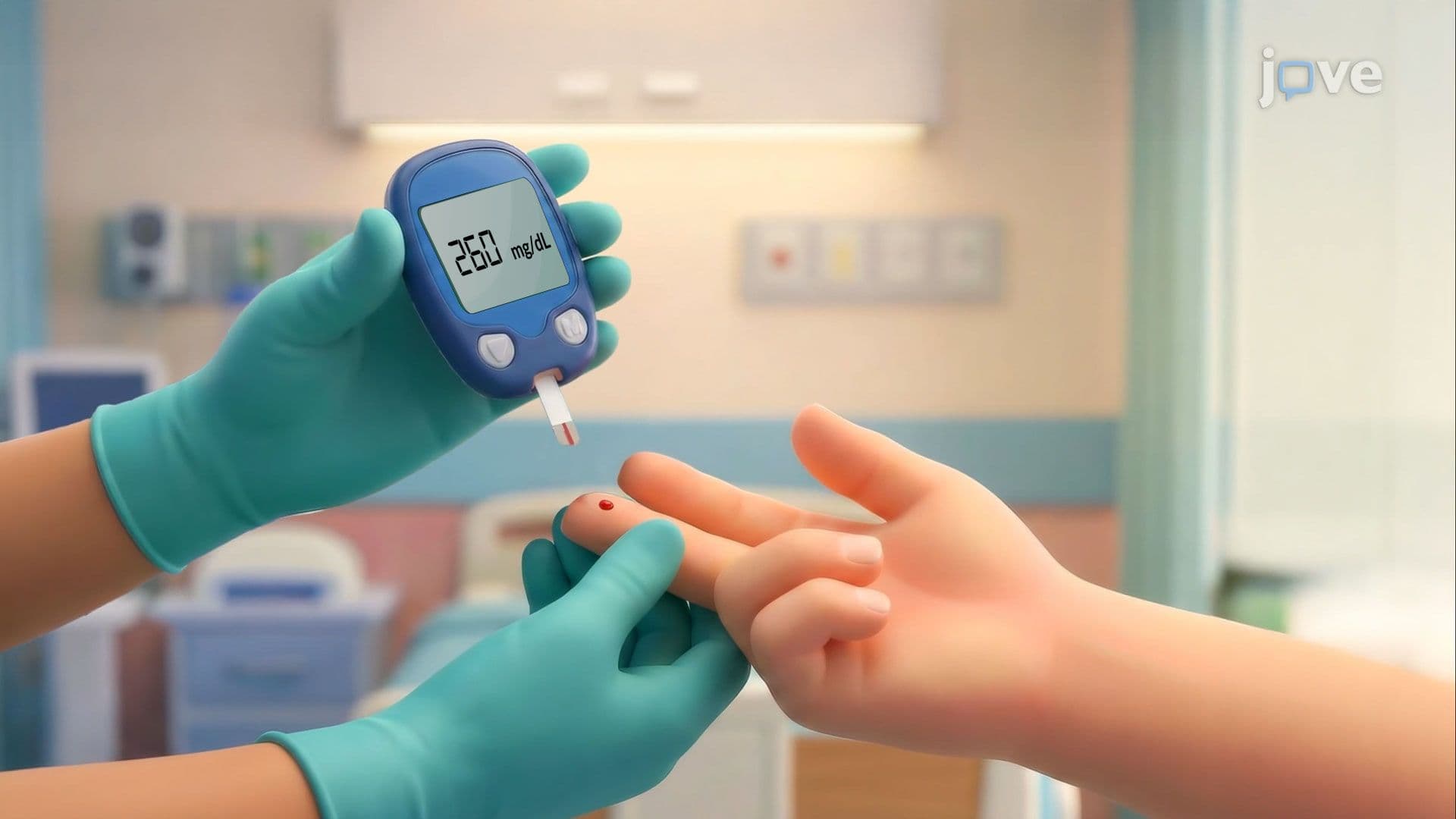
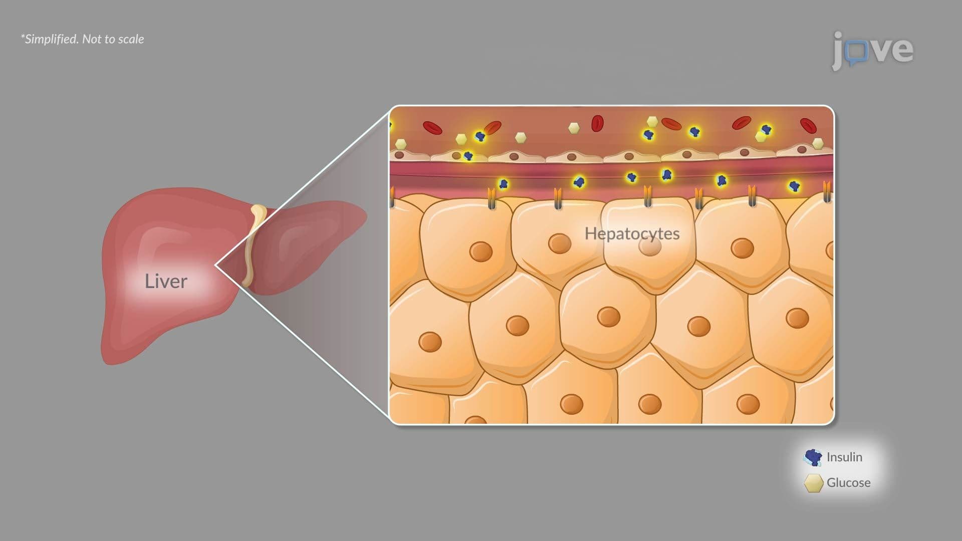
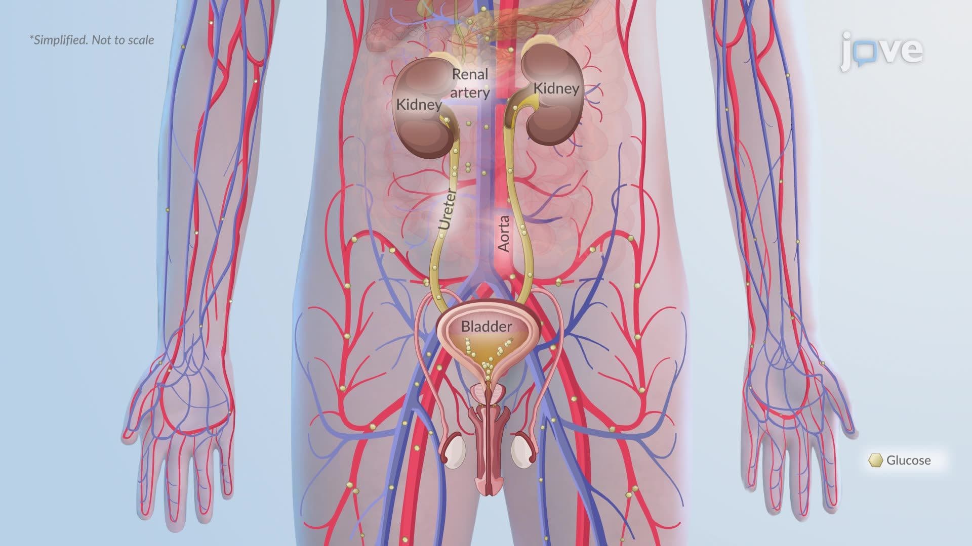
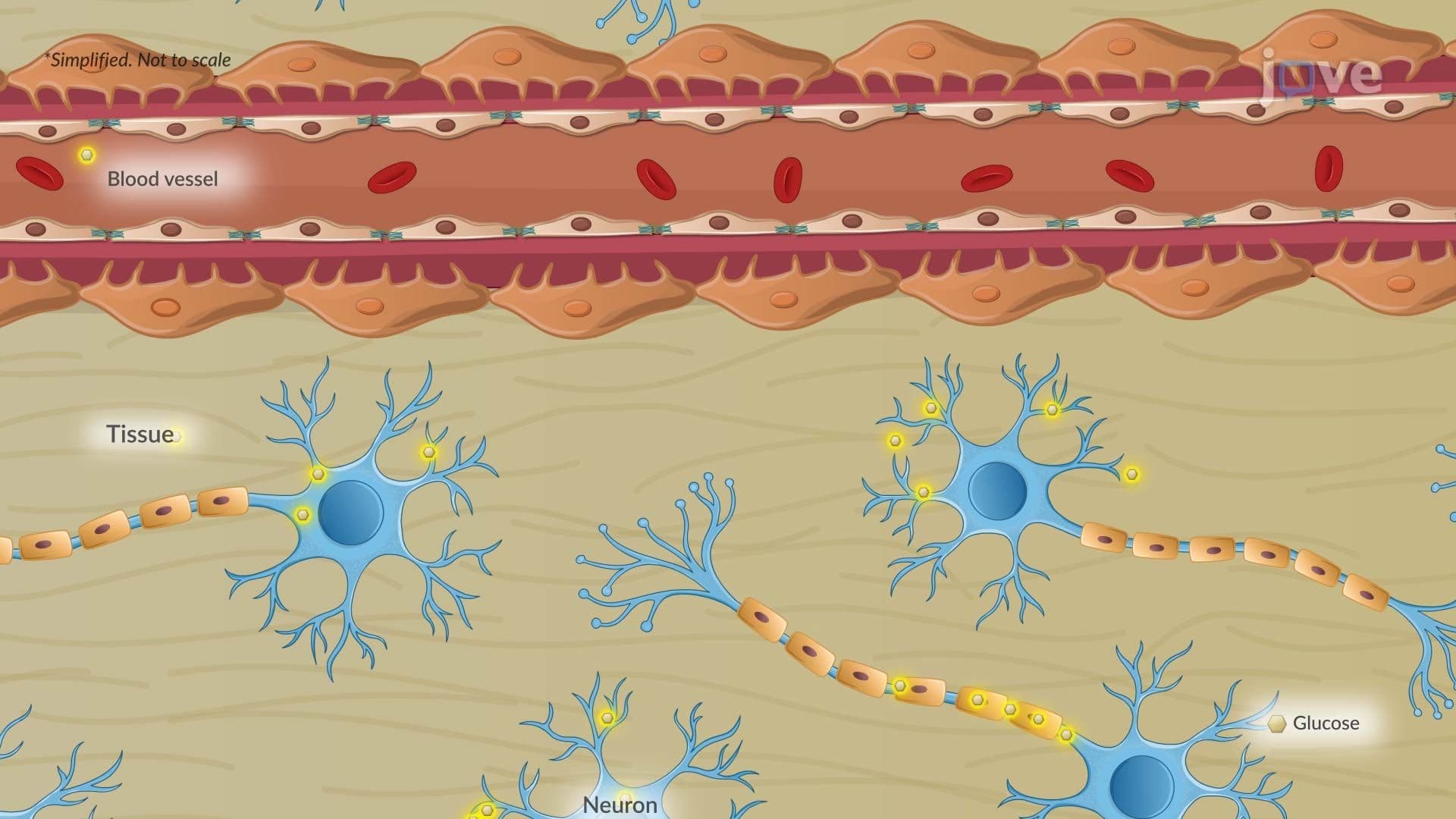
As insulitis progresses and beta cell populations decline, insulin secretion drops dramatically. Insulin is the hormone that signals cells, particularly muscle, liver, and fat cells, to absorb glucose from the bloodstream. Without adequate insulin, glucose cannot enter those cells and accumulates in the blood, causing hyperglycemia. This buildup has cascading effects: cells starve for energy despite abundant blood glucose, triggering fat breakdown and the dangerous production of ketone bodies, potentially leading to diabetic ketoacidosis (DKA), a life-threatening emergency seen frequently in newly diagnosed Type 1 patients across US emergency departments.
Understanding this pathophysiology provides essential context for the wider landscape of endocrine system disorders. Just as Type 1 diabetes results from hormonal imbalances caused by autoimmune destruction, conditions like Hashimoto's thyroiditis (a driver of hypothyroidism), Graves' disease (a driver of hyperthyroidism), Addison's disease (adrenal insufficiency), and even Cushing's syndrome all involve disrupted hormonal regulation, some through immune mechanisms, others through structural or functional gland failure. For students in AP Biology, college anatomy and physiology, or pre-nursing programs preparing for the TEAS or HESI A2, recognizing these connections across endocrine disorders demonstrates the kind of integrative thinking that top exam scores require.
Related Micro-courses