Loading video…

 35 Concepts
 5.1K views

Video Summary: What Is Cerebral Edema L
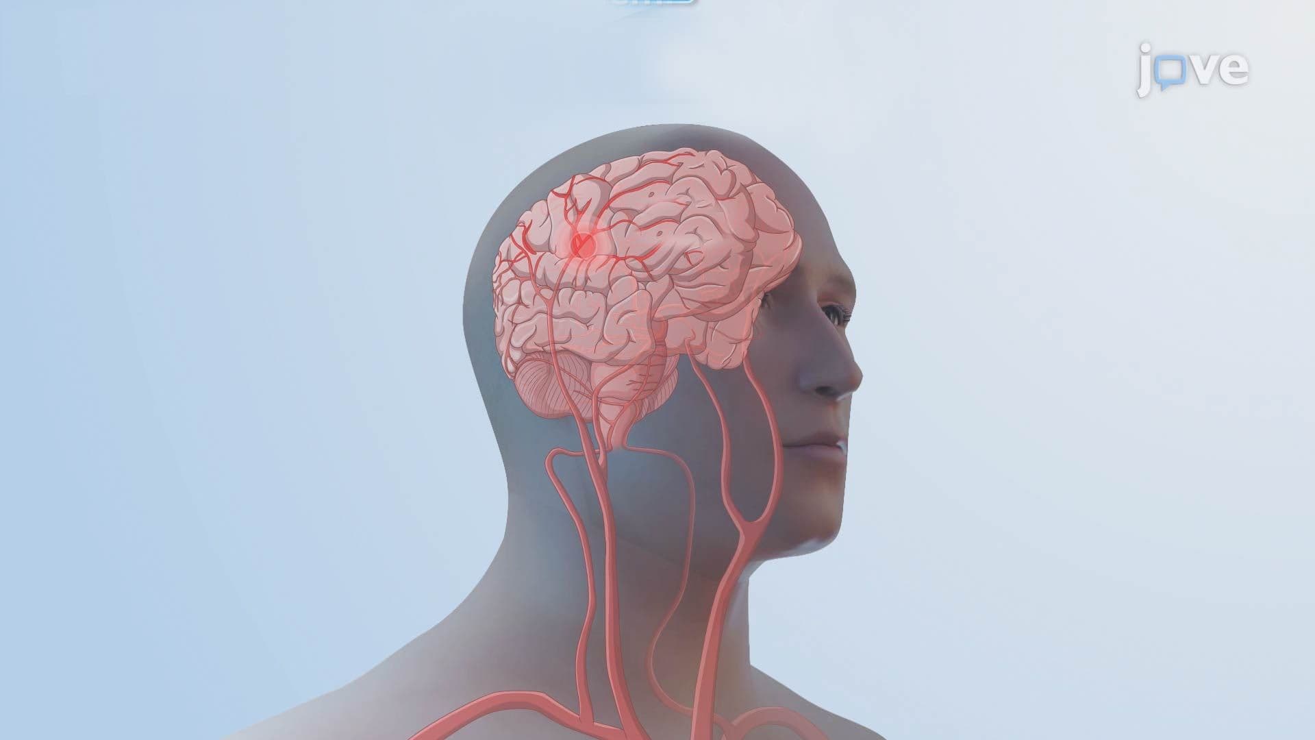
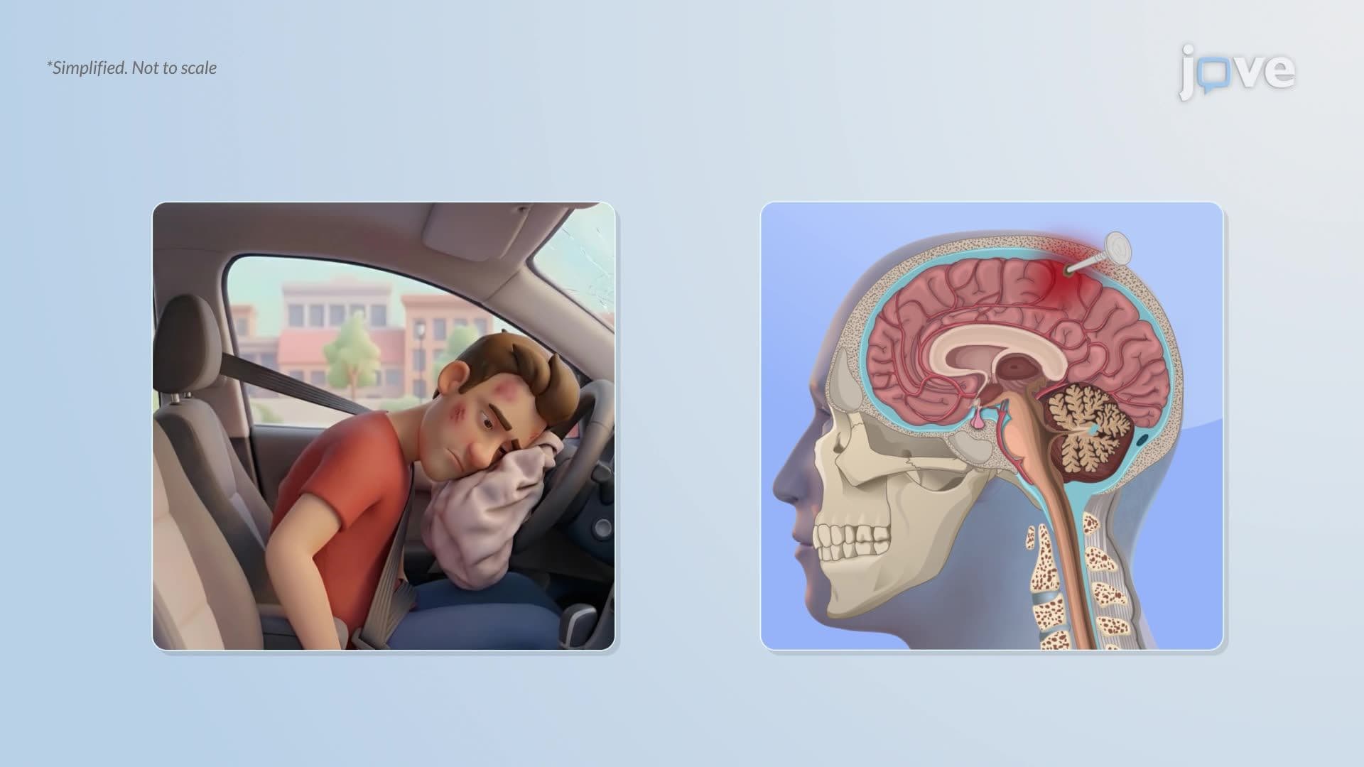
Your brain can swell, and that pressure alone can be life-threatening. Cerebral Edema L describes the abnormal buildup of fluid inside brain tissue, a condition that drives up intracranial pressure and disrupts critical neurological function. In US emergency rooms, it's a leading complication after traumatic brain injury and stroke. Four distinct mechanisms, vasogenic, cytotoxic, interstitial, and ionic, explain exactly how and why this swelling occurs. Watch the full video on JoVE Coach to master this concept with expert-led visuals and step-by-step explanations.
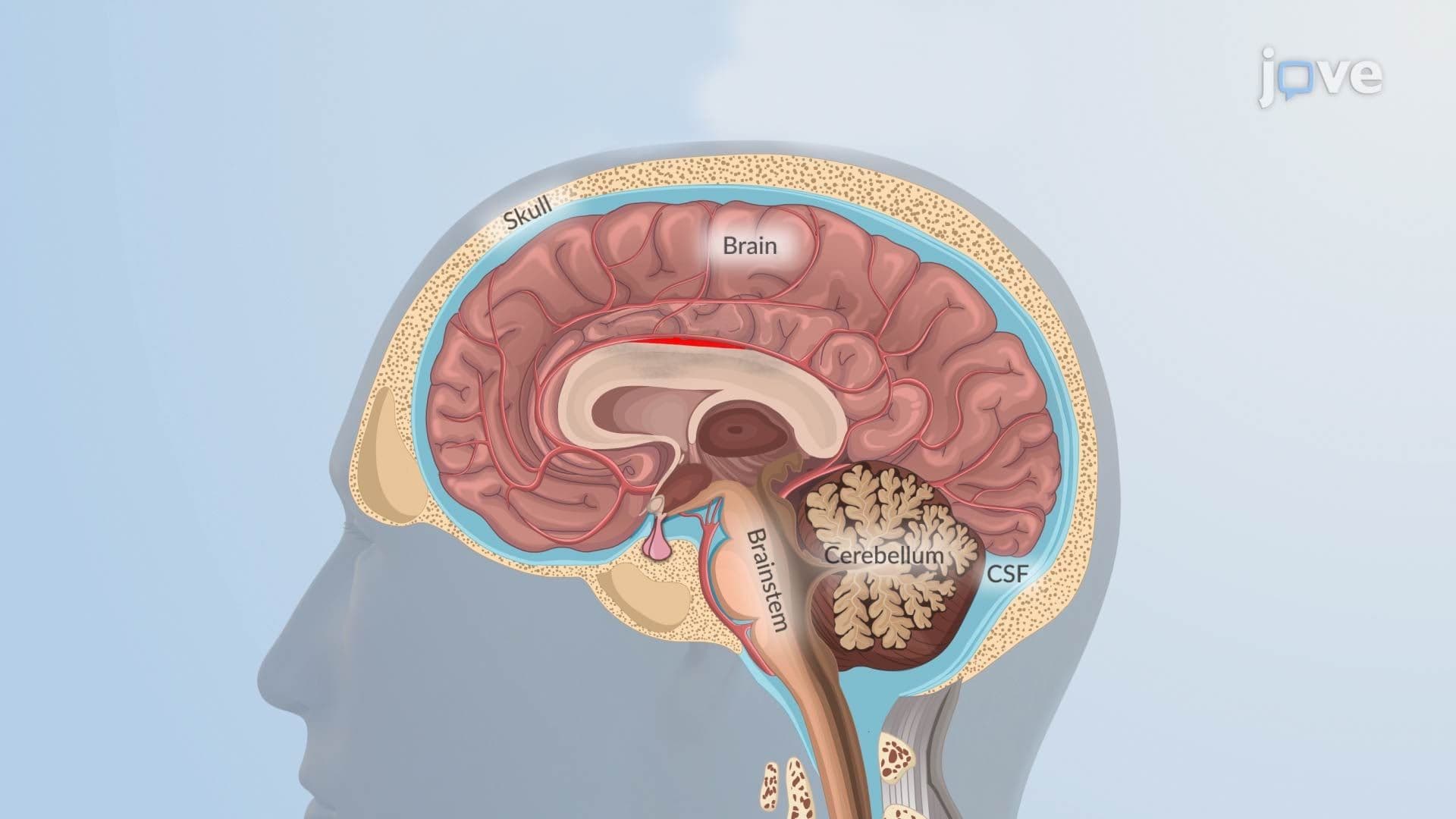
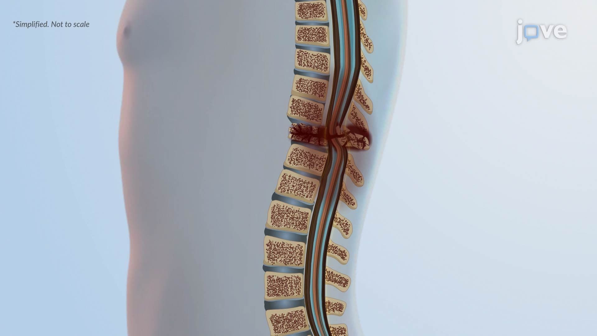
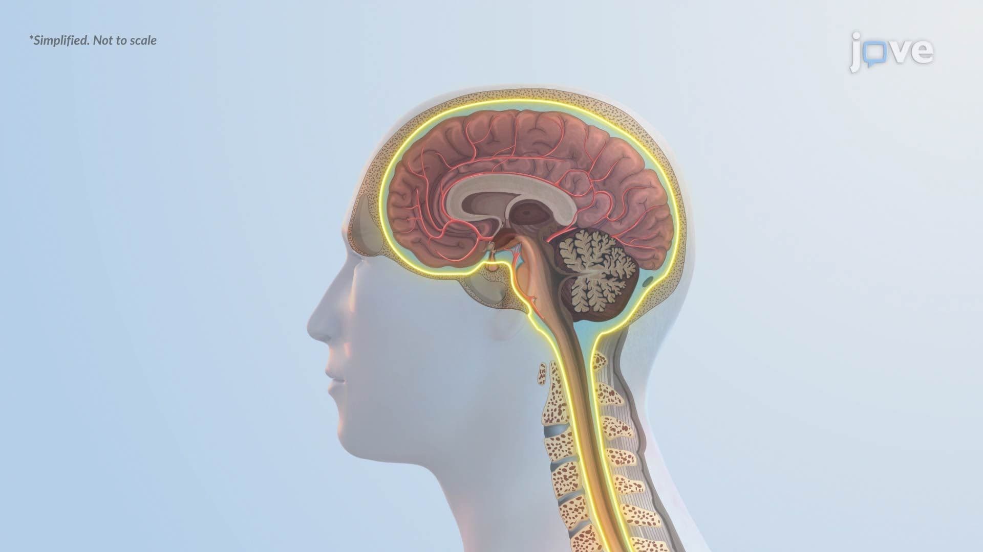
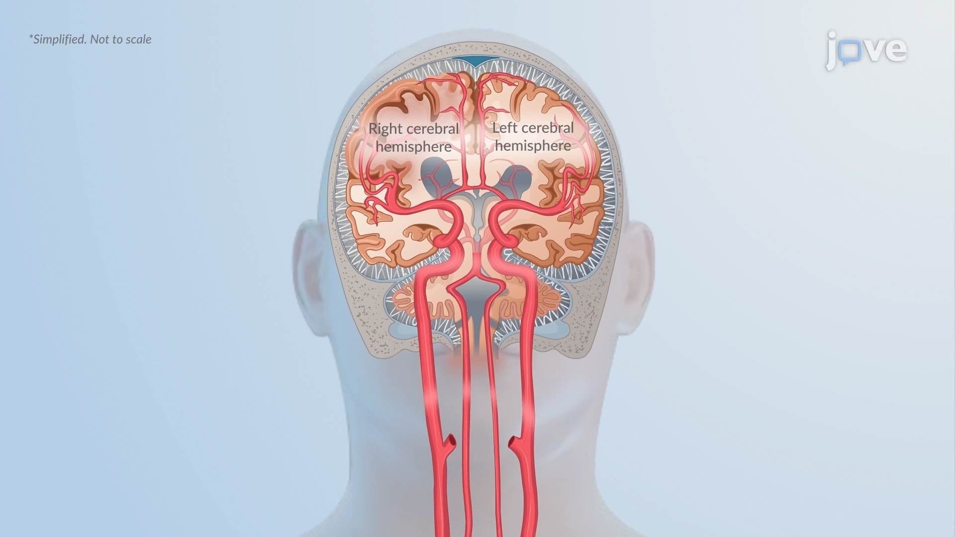
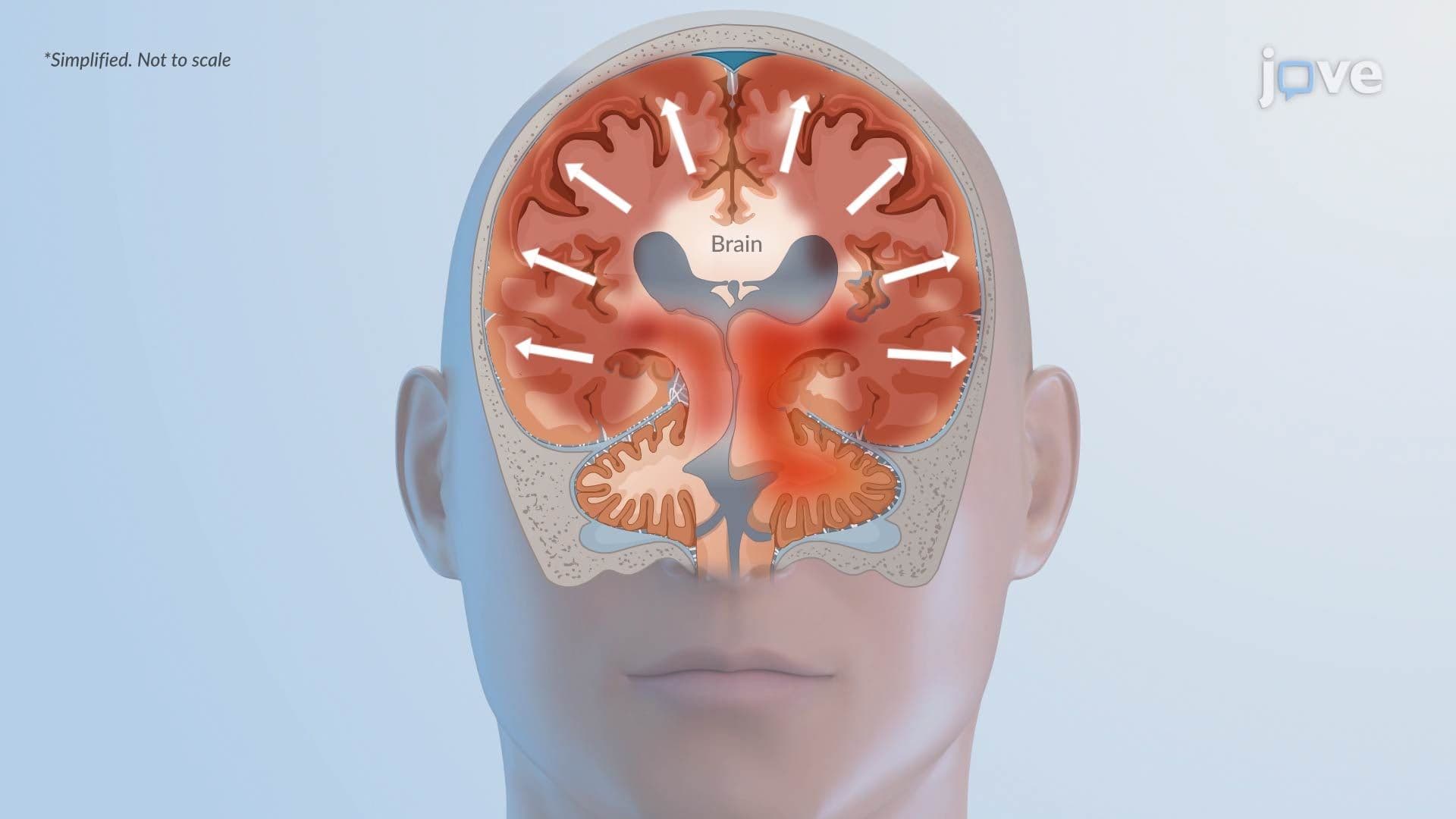
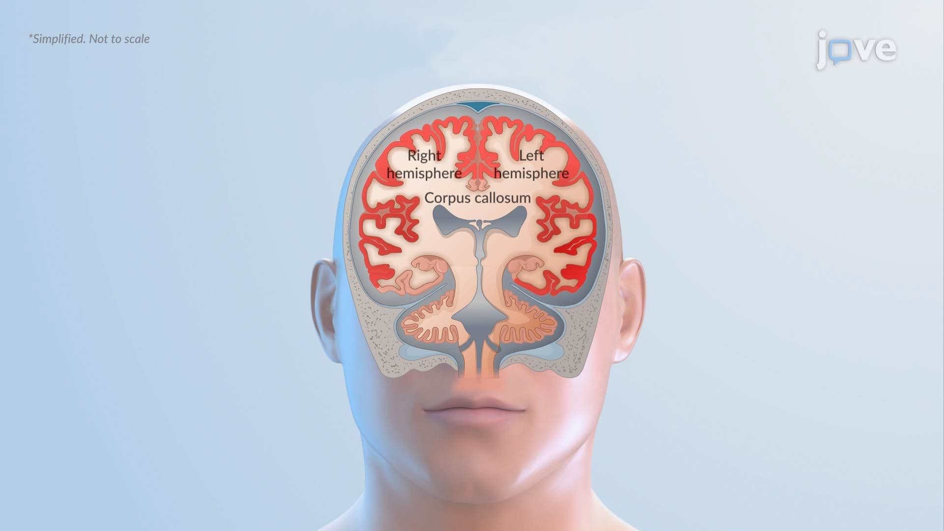
Cerebral Edema L refers to the pathological accumulation of excess fluid within the brain's tissue compartments, a process that increases intracranial pressure (ICP) and can rapidly impair consciousness, motor control, and vital function. Unlike swelling in peripheral tissues, the brain is enclosed within a rigid skull, meaning even small increases in volume can have catastrophic consequences. Understanding the mechanisms behind cerebral edema is essential for students in biology, anatomy, and pre-health pathways, and is directly tested on exams including the MCAT and USMLE Step 1.
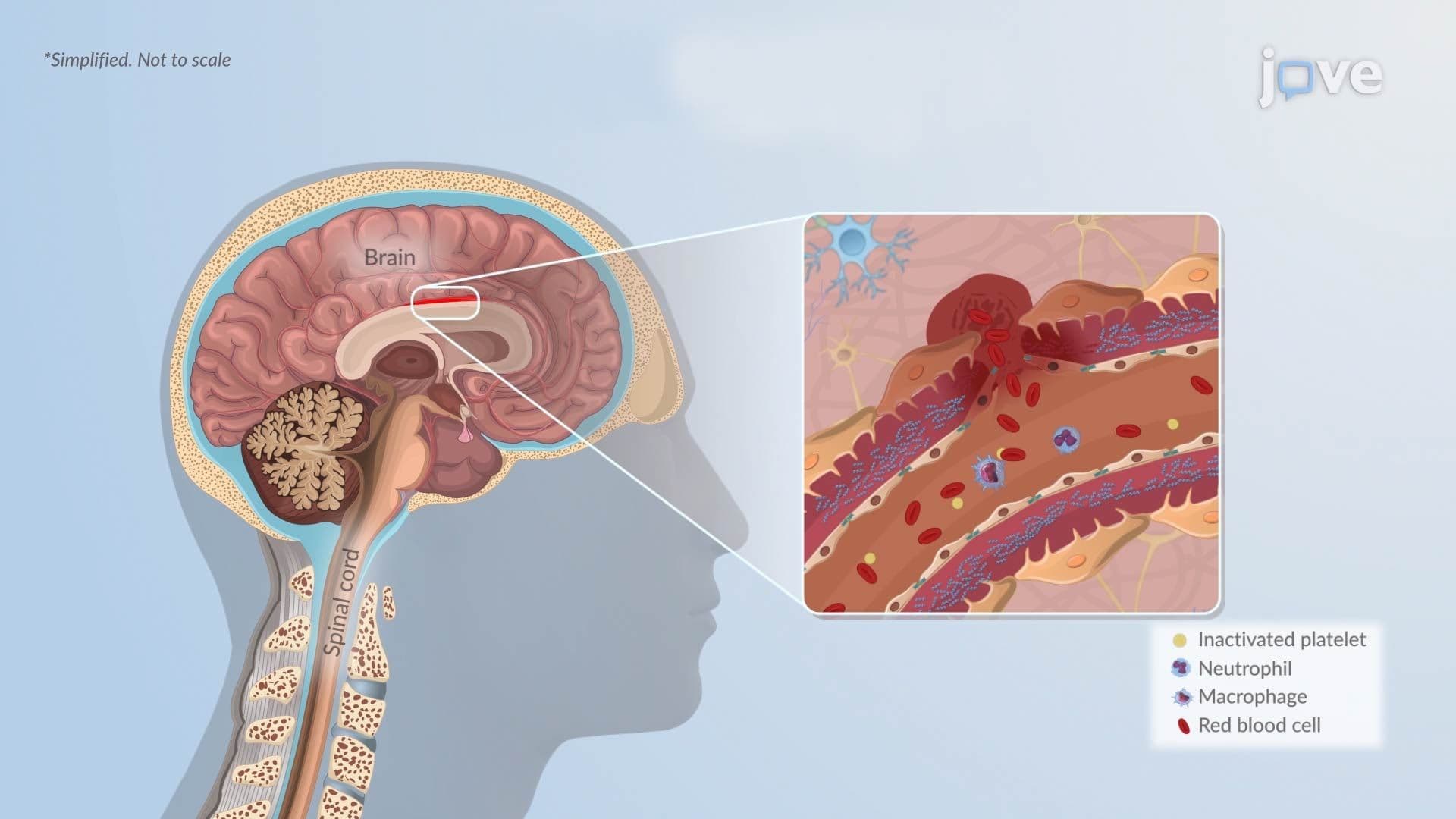
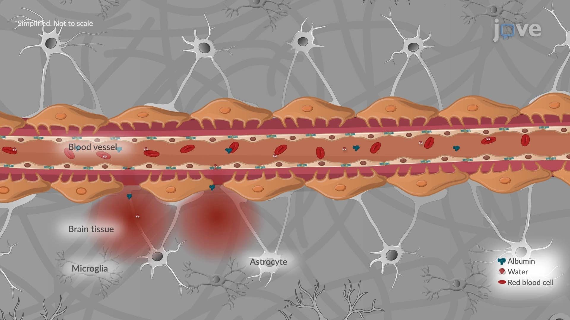
Vasogenic edema is the most clinically common type and occurs when the blood-brain barrier (BBB) breaks down. The BBB is formed by specialized endothelial cells with tight junctions that normally prevent large molecules from entering brain tissue. When trauma, tumors, or severe infections compromise these junctions, protein-rich plasma fluid leaks into the brain's extracellular space. Water follows by osmosis, expanding the interstitial compartment, particularly in white matter. This is frequently seen in patients with brain tumors or bacterial meningitis treated at US trauma centers.
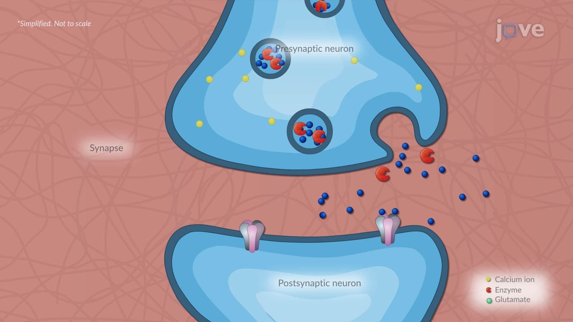
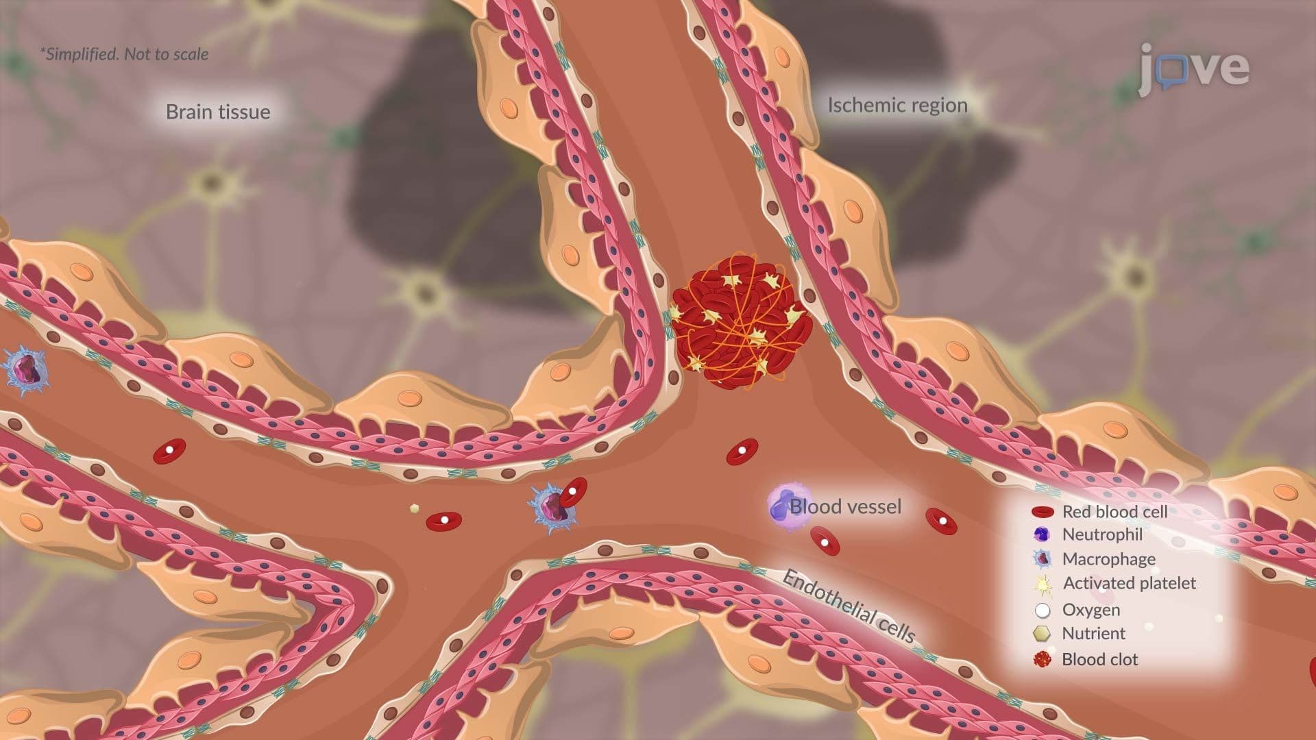
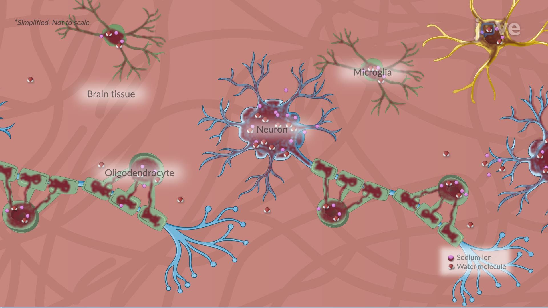
Cytotoxic edema originates at the cellular level. When oxygen or glucose delivery is cut off, as occurs during ischemic stroke, neurons and glial cells can no longer produce sufficient adenosine triphosphate (ATP). Without ATP, the sodium-potassium ATPase pump fails. Sodium accumulates inside cells, and water follows to restore osmotic balance, causing the cells themselves to swell. This process affects both gray and white matter and is the dominant mechanism in ischemic stroke, one of the leading causes of death and disability in the United States.
Interstitial edema is specifically associated with obstructive hydrocephalus. When cerebrospinal fluid (CSF) cannot drain normally, pressure builds within the brain's ventricular system. This elevated pressure forces CSF across the ependymal lining into the surrounding periventricular white matter. On brain MRI scans, commonly ordered in US hospital settings, this appears as characteristic "halo" changes around the ventricles.
Ionic edema is a subtler process. The blood-brain barrier remains structurally intact, but shifts in ionic concentrations and osmotic gradients between blood and brain parenchyma cause water to move passively into neural tissue. This mechanism is often seen in early-stage ischemia before full cytotoxic changes develop.
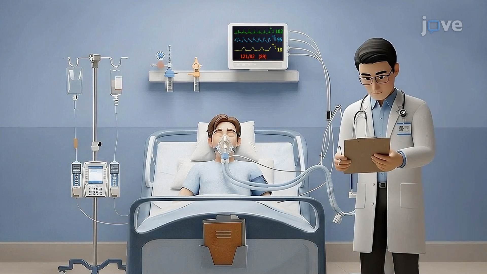
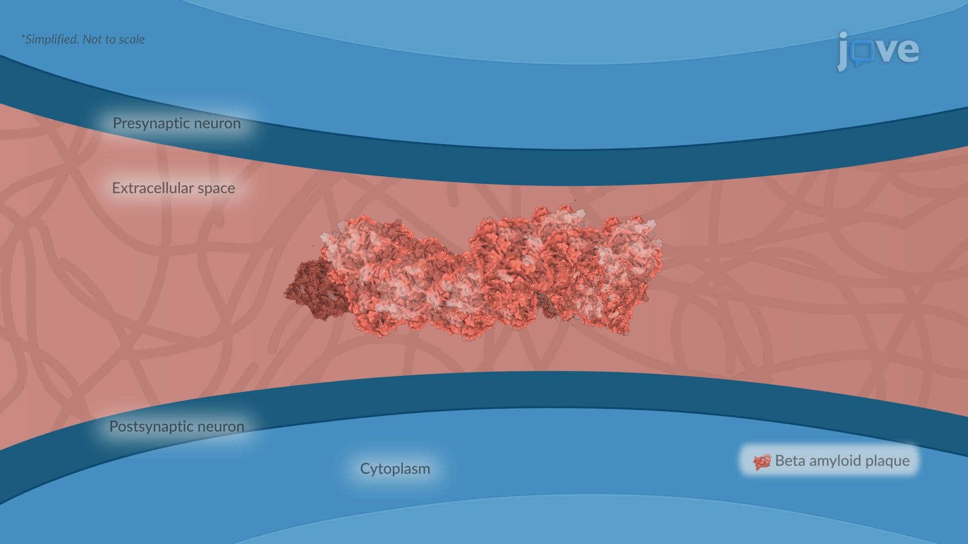
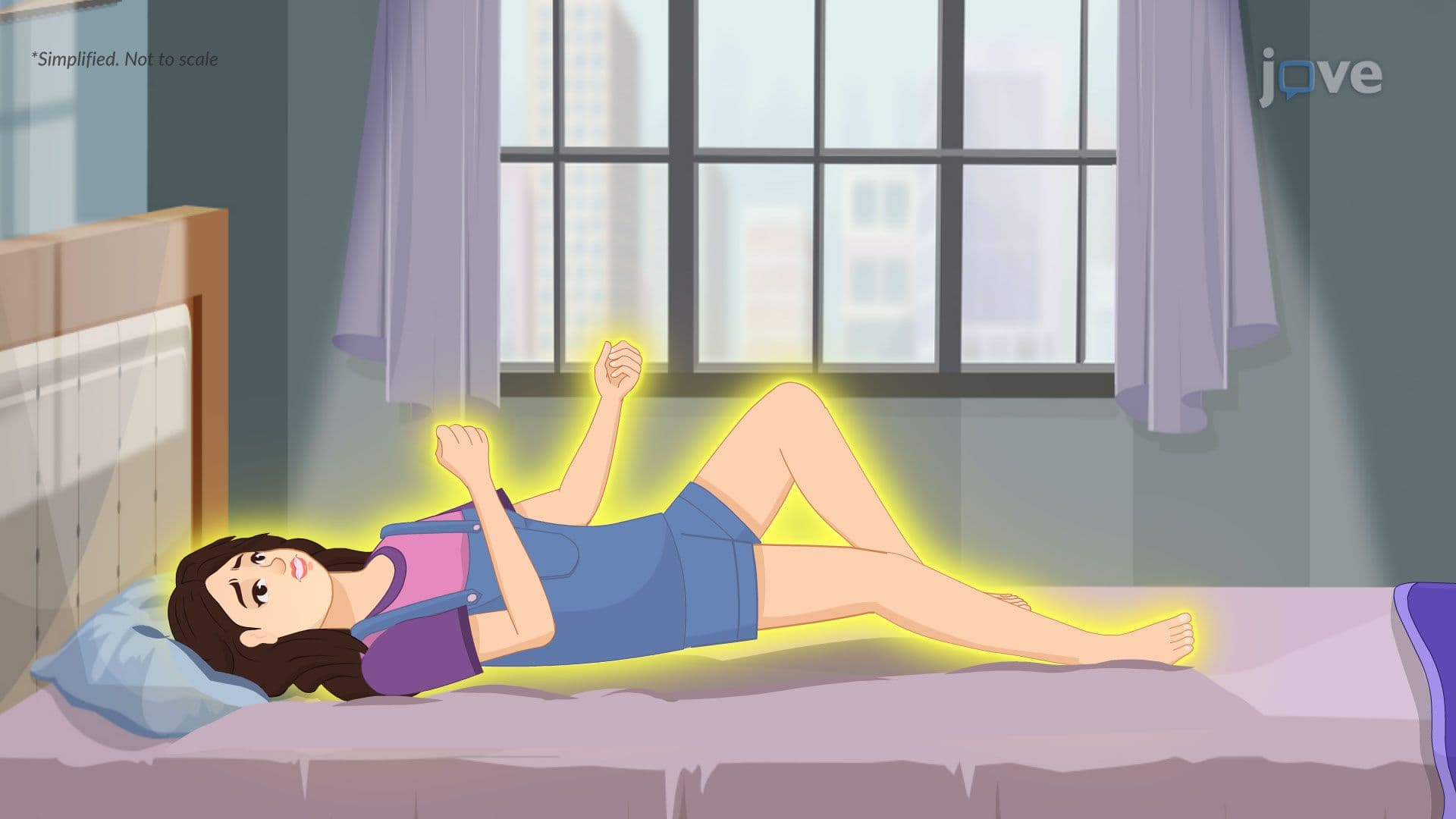
Cerebral edema is not an isolated event, it overlaps with many well-known neurological conditions. In stroke patients, cytotoxic edema develops within hours and can progress to life-threatening herniation. In epilepsy, repeated seizures can transiently disrupt ionic homeostasis. Neurodegenerative diseases like Alzheimer's and Parkinson's disease may involve chronic, low-grade neuroinflammation that gradually compromises barrier integrity. Multiple sclerosis, characterized by immune-mediated demyelination, can also trigger localized vasogenic edema at active lesion sites.
For AP Biology and college-level physiology students, cerebral edema illustrates core concepts including membrane transport, osmosis, and cellular energy metabolism. On the MCAT, passages frequently present clinical scenarios involving ICP elevation or stroke, knowing which edema type is involved helps answer mechanism-based questions efficiently. USMLE and NCLEX learners must distinguish these four types to select appropriate interventions, such as osmotic therapy with mannitol for vasogenic edema or addressing the underlying cause in hydrocephalus-related interstitial edema.
Related Micro-courses