Loading video…

 35 Concepts
 5.1K views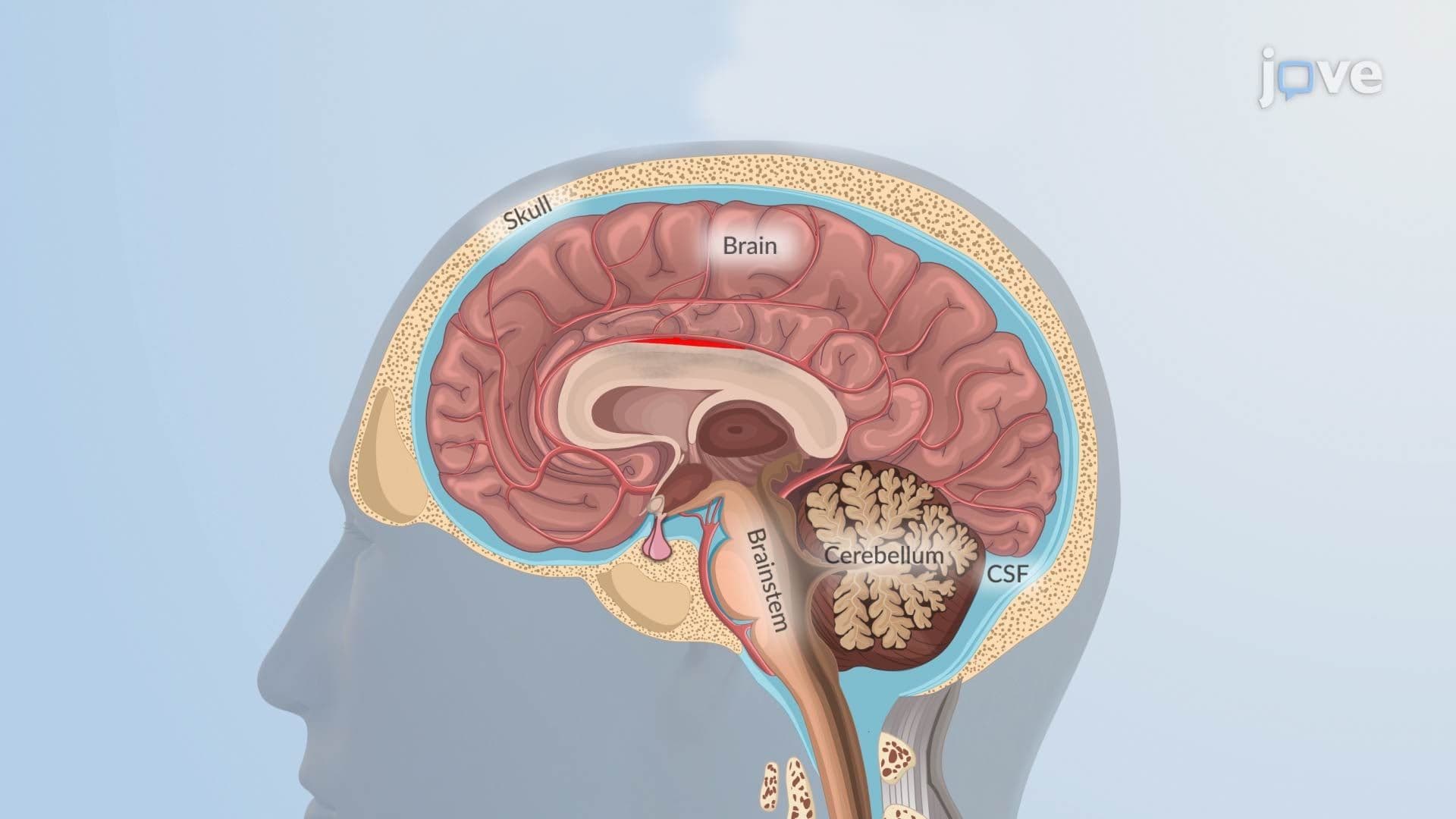

Video Summary: Pathophysiology in Hemorrhagic Stroke Ll
Did you know a single ruptured blood vessel can trigger a chain reaction that permanently changes how the brain functions? The pathophysiology in hemorrhagic stroke II reveals how bleeding, toxic blood components, and dangerous pressure changes combine to devastate brain tissue. In US emergency rooms, hemorrhagic stroke accounts for nearly 20% of all strokes yet causes disproportionately high mortality. Watch the full video on JoVE Coach to master this concept with expert-led visuals and step-by-step explanations.
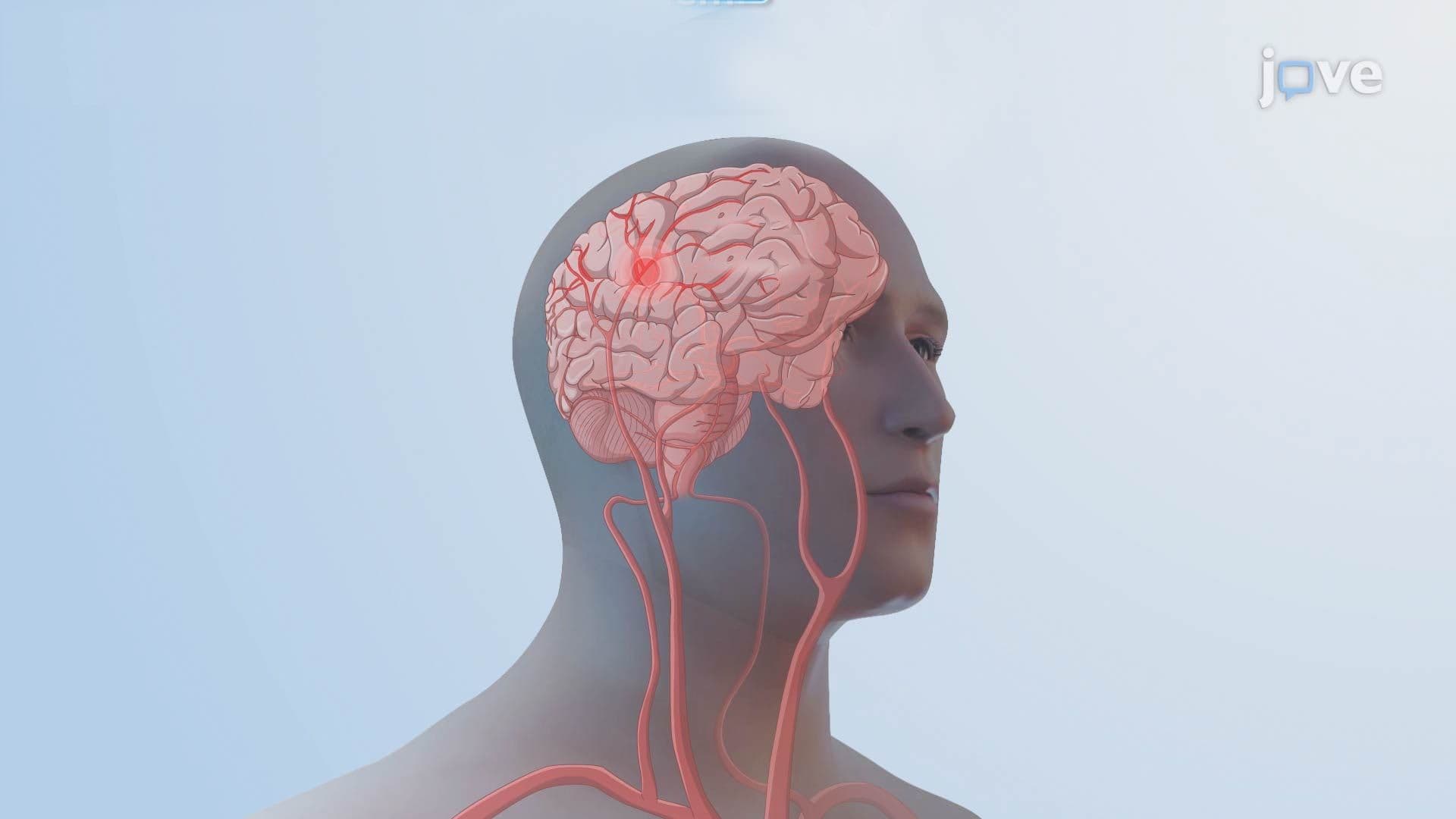
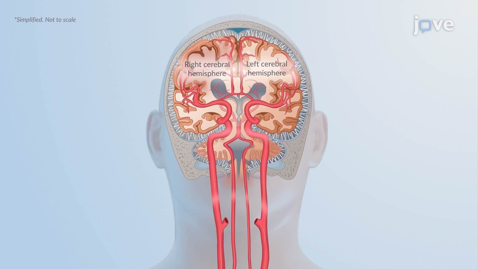
A hemorrhagic stroke is not a single event, it is a rapidly escalating biological crisis. When a blood vessel in the brain ruptures, blood floods areas where it does not belong. Understanding the pathophysiology in hemorrhagic stroke II means tracing two parallel stories: the immediate mechanical damage caused by blood accumulation and the slower, equally destructive biochemical cascade that follows.
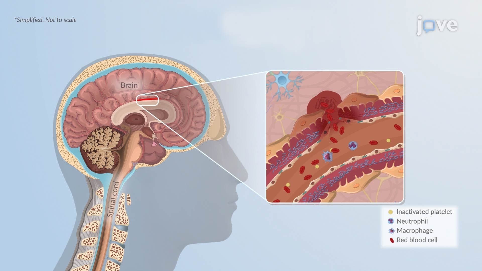
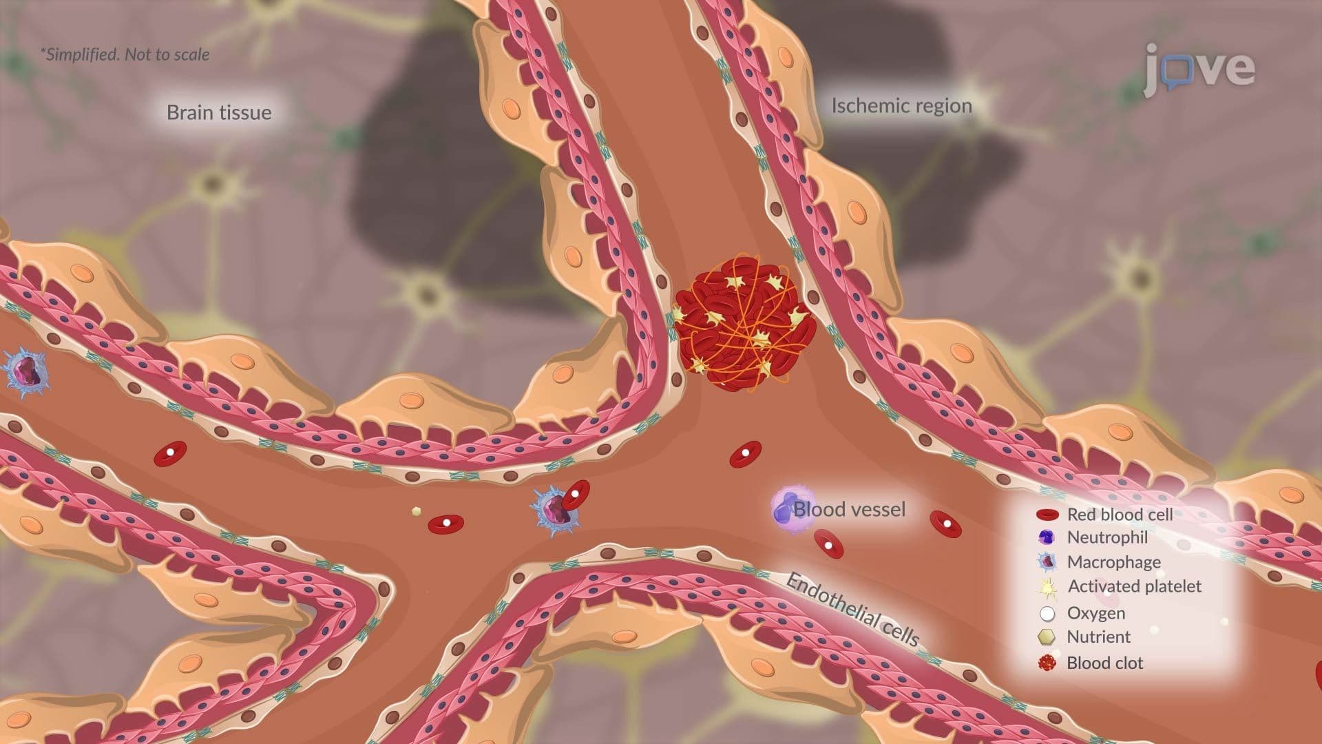
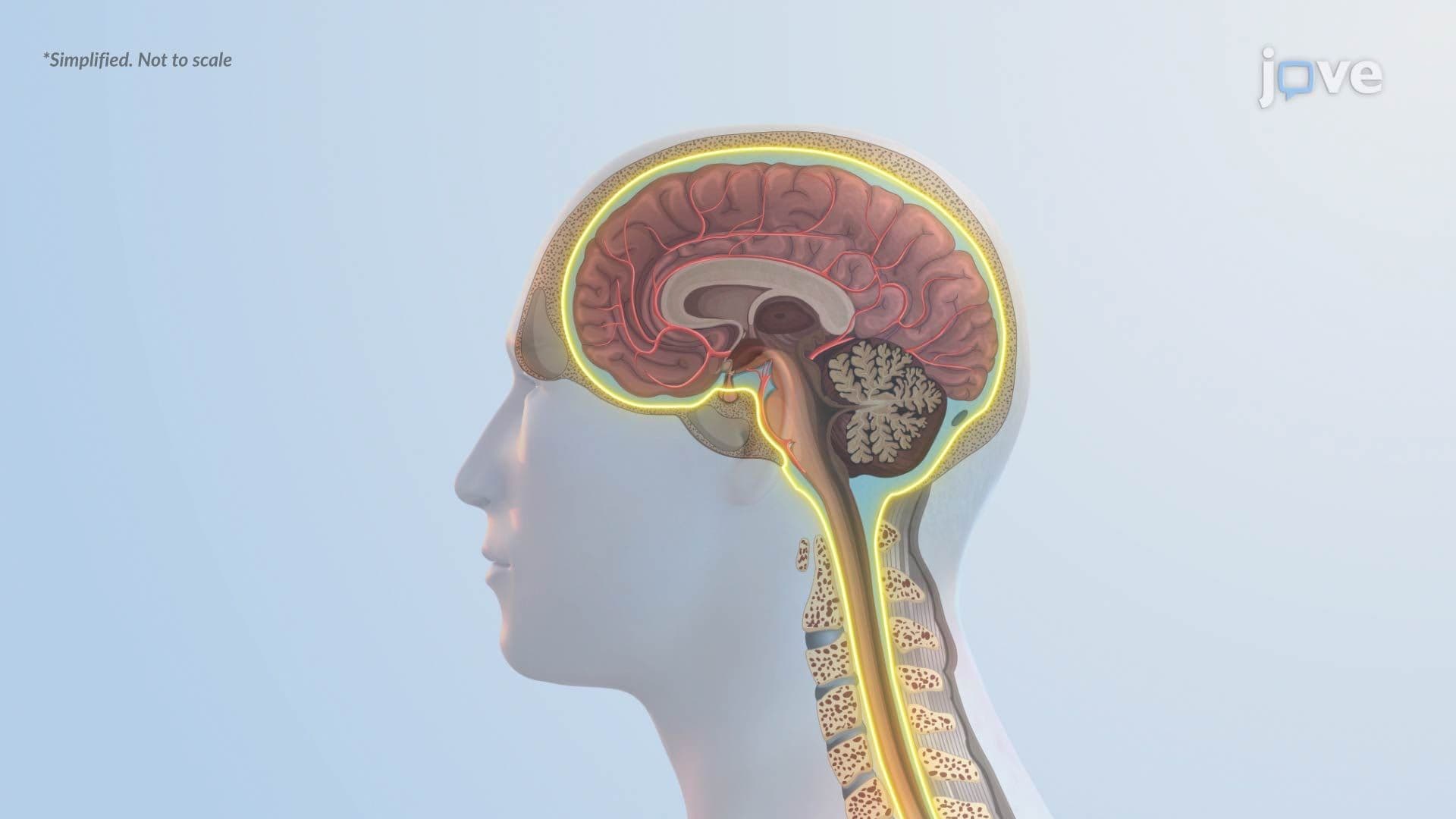
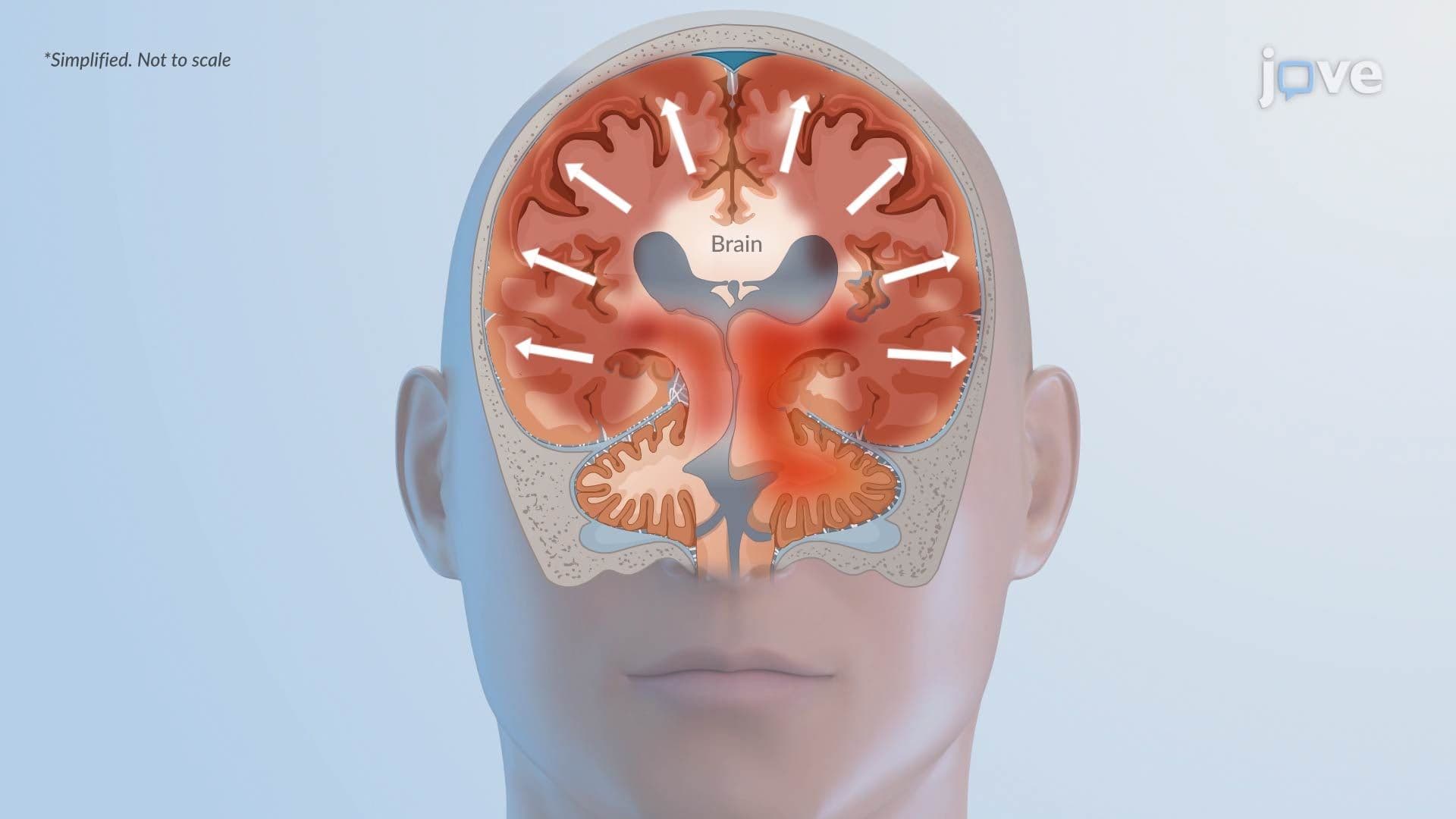
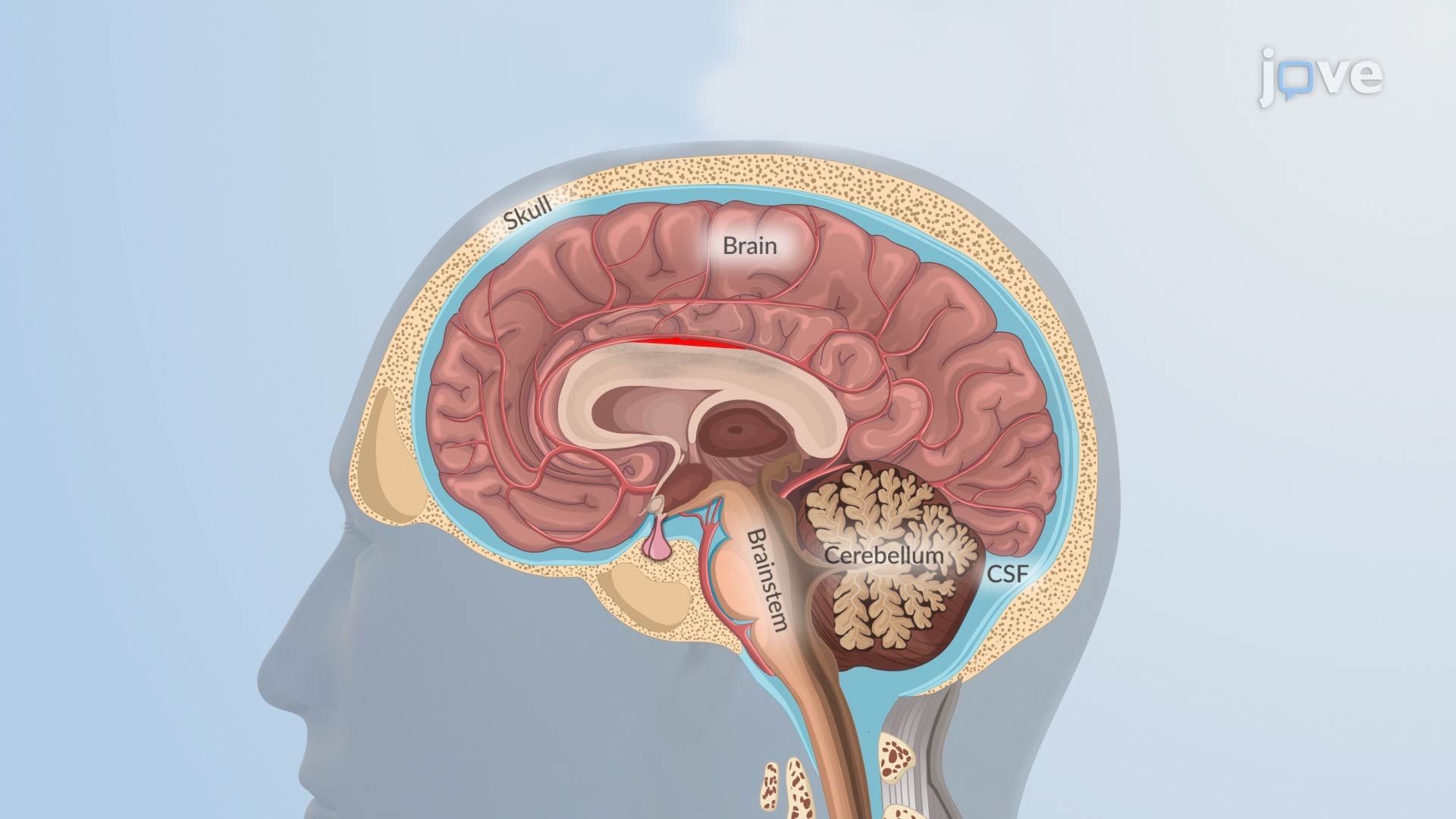
When bleeding occurs into brain tissue, called intracerebral hemorrhage (ICH), or into the fluid-filled subarachnoid space (SAH), blood collects and forms a hematoma. As the hematoma expands, intracranial pressure (ICP) rises sharply. The brain, enclosed in the rigid skull, has almost no room to accommodate this extra volume. Surrounding neurons and supporting cells are compressed, oxygen delivery drops, and localized ischemia develops even in areas far from the original bleed. This pressure-driven injury happens within minutes to hours and is why rapid imaging, such as CT scans performed in US trauma centers, is essential to guide treatment decisions.
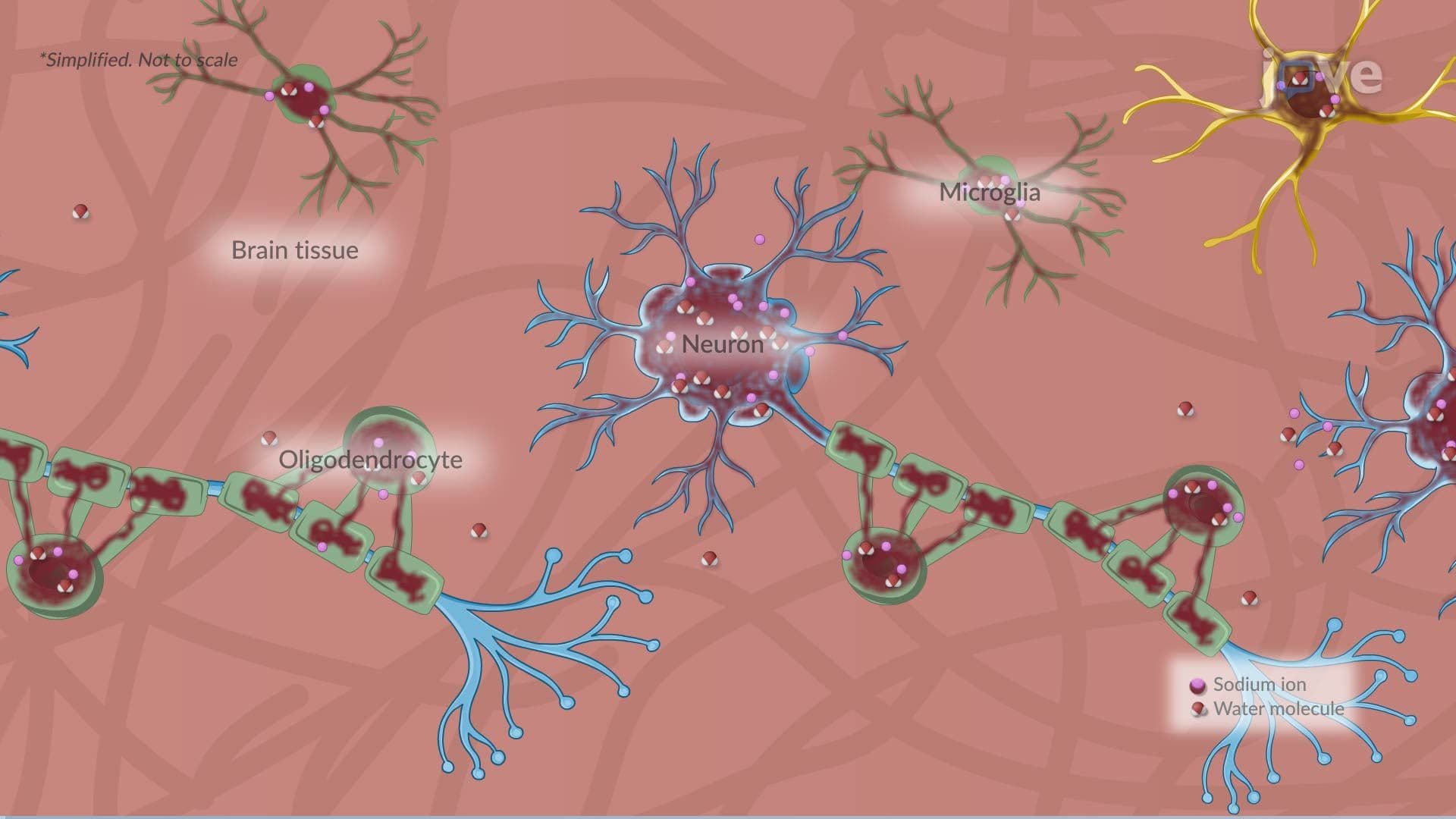
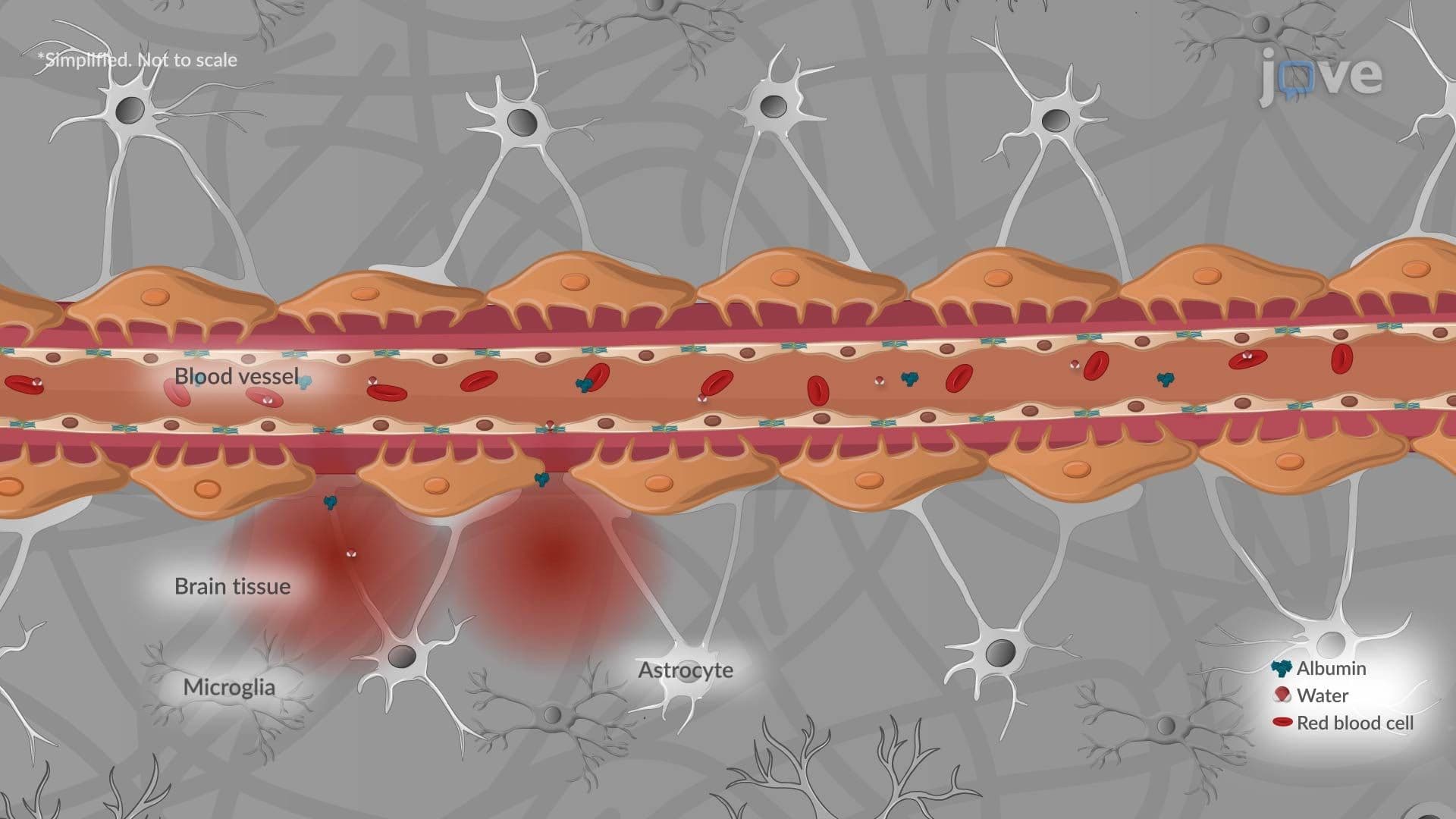
The damage does not stop when the bleeding slows. As red blood cells break apart, hemoglobin degrades into components including free iron. Iron is highly reactive, it catalyzes the Fenton reaction, generating reactive oxygen species (ROS) that attack cell membranes, proteins, and DNA in nearby neurons. Simultaneously, microglia (the brain's resident immune cells) detect the injury signals and release pro-inflammatory cytokines such as interleukin-1β and tumor necrosis factor-alpha (TNF-α). This neuroinflammation amplifies cell death and compromises the blood-brain barrier (BBB), allowing additional harmful molecules to flood brain tissue. Students learning about neurological disorders, from Alzheimer's disease to multiple sclerosis, will recognize oxidative stress and neuroinflammation as recurring mechanisms across many conditions.
Subarachnoid hemorrhage introduces two uniquely dangerous complications. First, vasospasm, a prolonged, abnormal narrowing of cerebral arteries, can develop two to seven days after the initial bleed. Even though the hemorrhage itself may have stopped, vasospasm significantly reduces blood flow, causing delayed cerebral ischemia that injures brain regions that initially survived. In the US, vasospasm is monitored using transcranial Doppler ultrasound and managed with calcium channel blockers like nimodipine. Second, blood entering the cerebrospinal fluid (CSF) can physically clog the arachnoid granulations responsible for CSF drainage. When drainage is blocked, fluid accumulates, a condition called hydrocephalus, which further raises ICP and compounds injury.
On the MCAT, questions about hemorrhagic stroke often target the distinction between ICH and SAH, the role of the blood-brain barrier, and downstream effects on ICP. NCLEX and USMLE Step 1 candidates must understand vasospasm timing and hydrocephalus management. In AP Biology and college neuroscience courses, hemorrhagic stroke serves as a model case for understanding how the nervous system responds to injury. These cascading mechanisms, pressure, oxidative stress, inflammation, and vascular dysfunction, also connect directly to questions about what causes nervous system disorders and how neurological disorders are diagnosed, making this topic foundational across multiple health science pathways.
Related Micro-courses