Loading video…

 35 Concepts
 5.1K views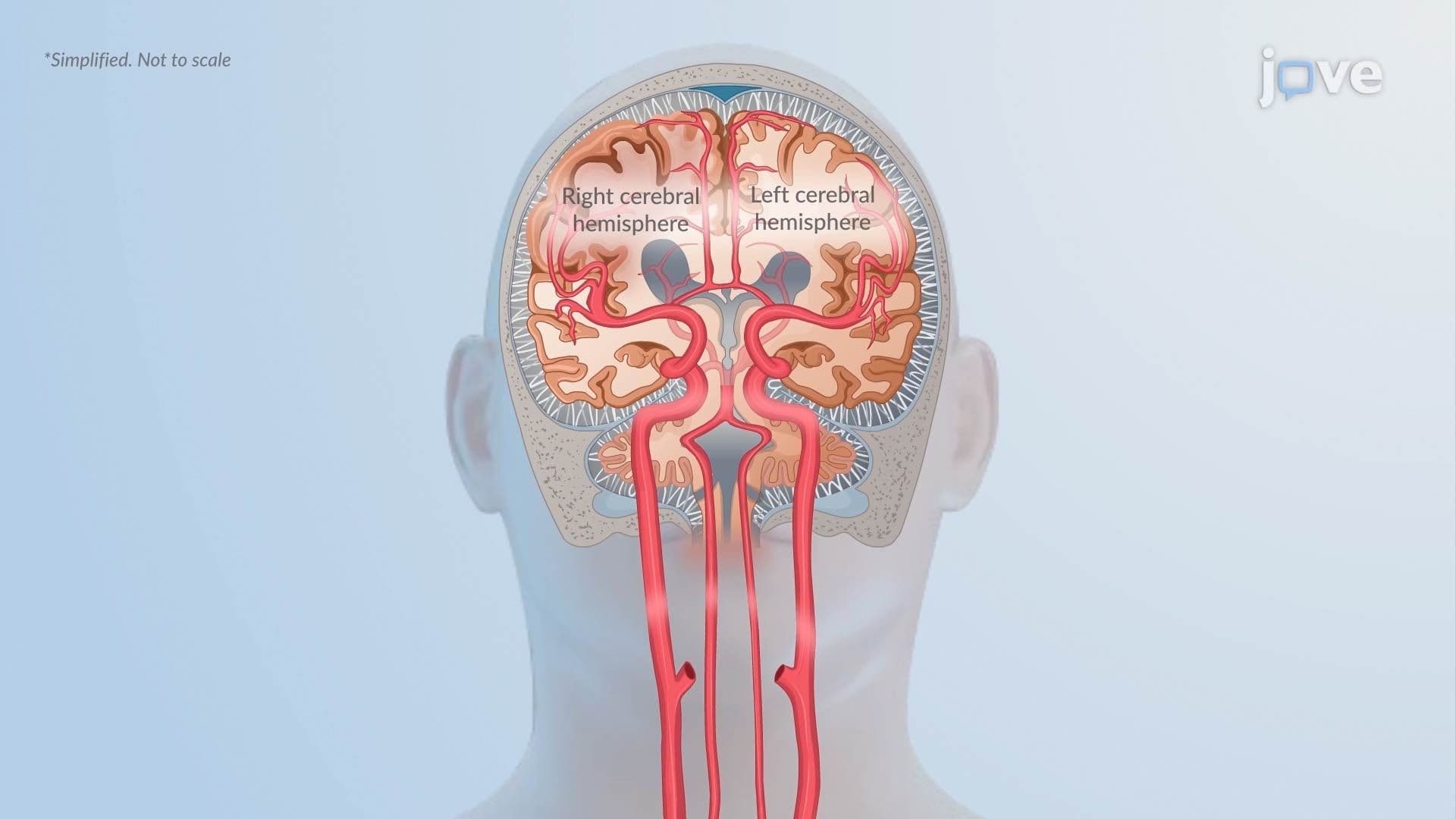

Video Summary: Pathophysiology in Increased Intracranial Pressure Ll
What happens inside the skull when pressure builds faster than the brain can handle? The pathophysiology in increased intracranial pressure ll reveals how a cascade of dangerous events, from cerebral edema to herniation, can quickly become life-threatening. In US neurocritical care units, managing ICP spikes is a round-the-clock emergency priority. When autoregulation fails, brain tissue shifts, brainstems compress, and neurons die. Watch the full video on JoVE Coach to master this concept with expert-led visuals and step-by-step explanations.
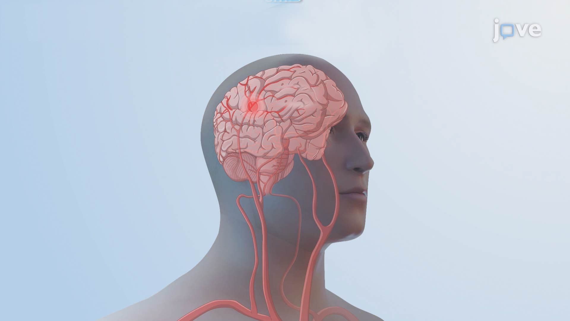
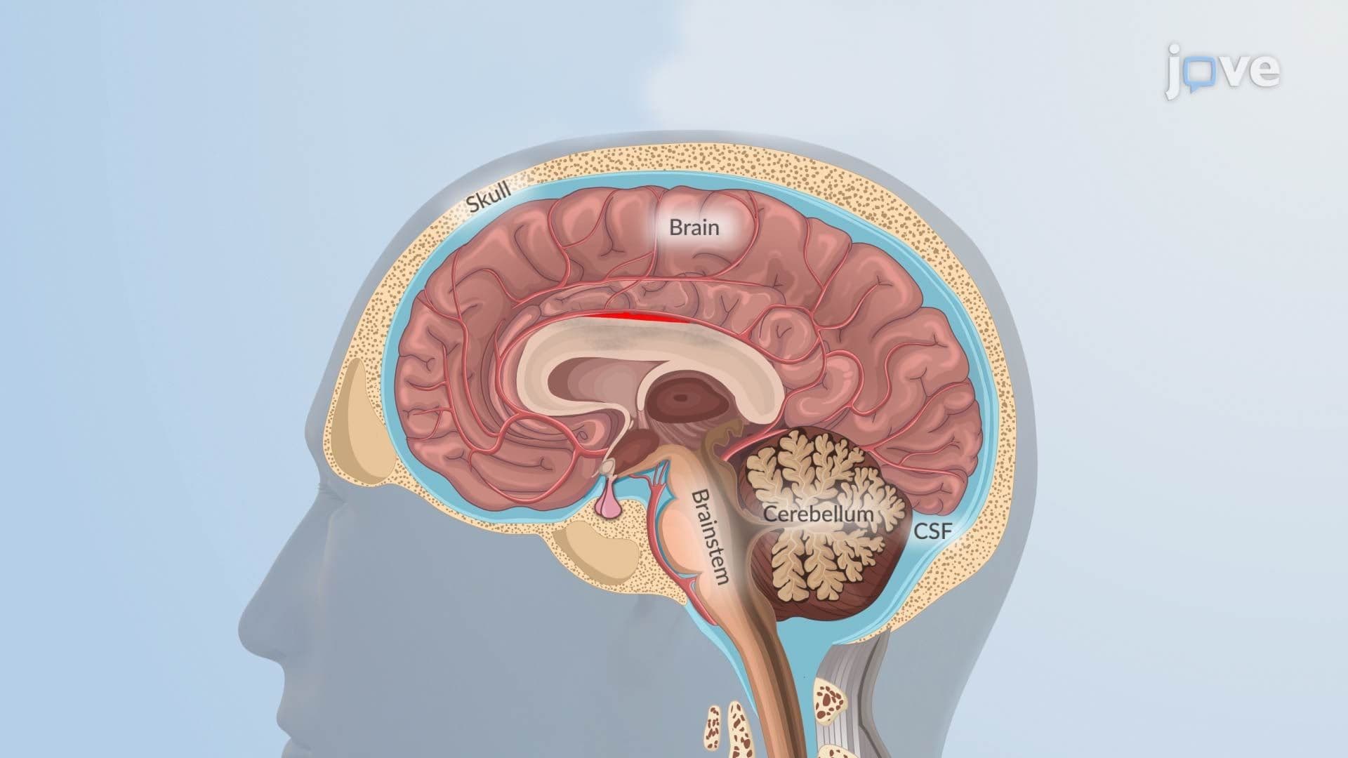
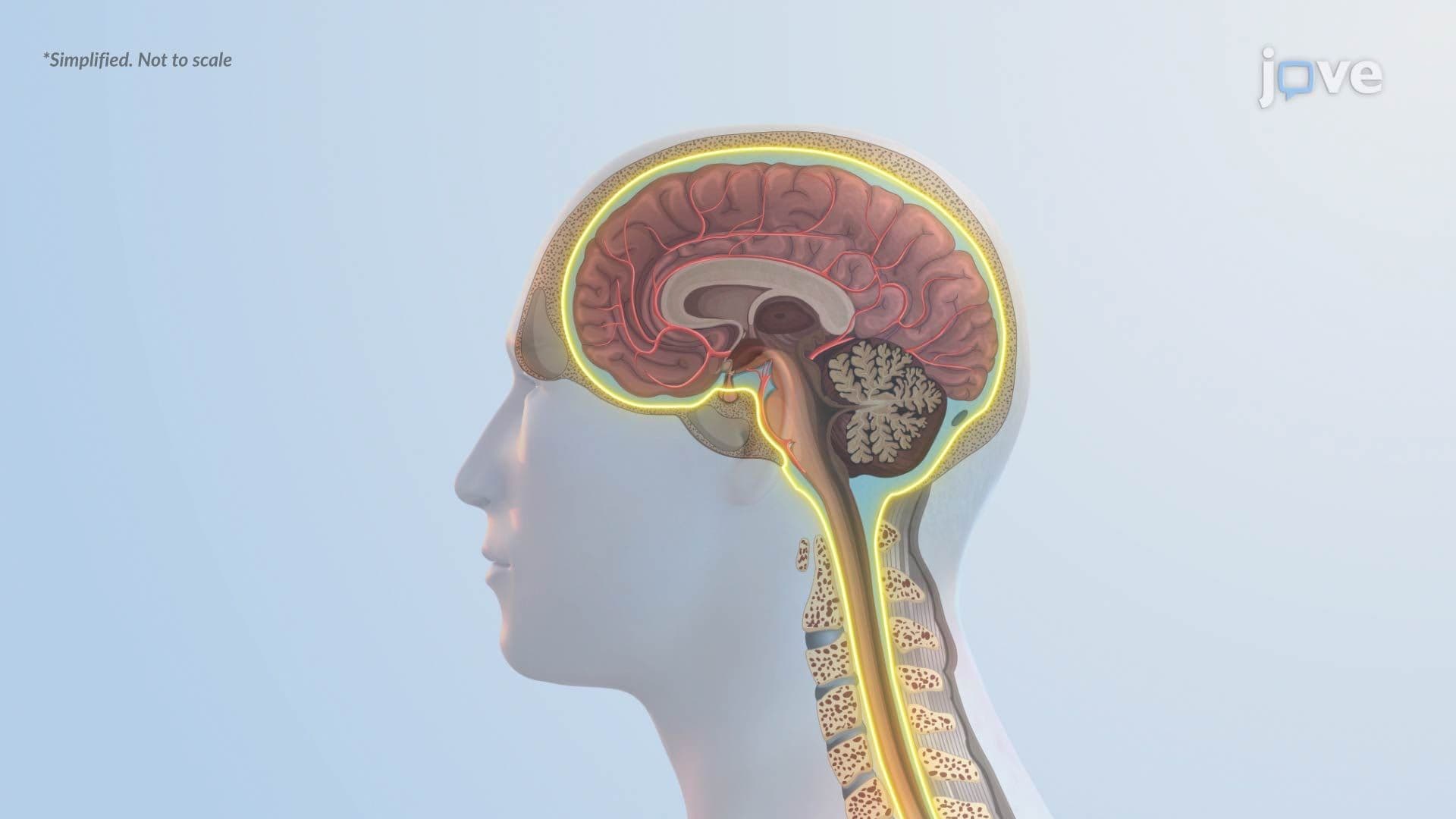
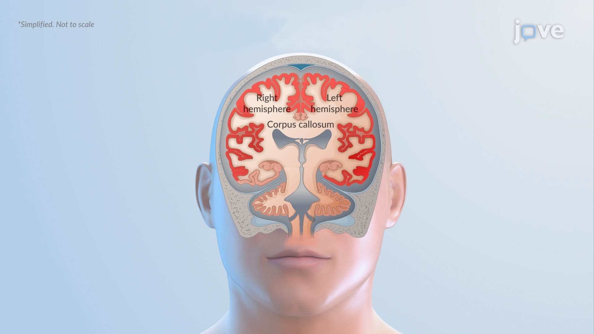
The brain exists inside a closed, rigid container, the skull. Under normal conditions, brain tissue, cerebrospinal fluid (CSF), and blood occupy this space in a carefully balanced ratio. The pathophysiology in increased intracranial pressure ll explains what happens when that balance is disrupted. Whether the trigger is a swelling brain, a bleeding vessel, or a growing tumor, the downstream consequences follow a predictable and life-threatening sequence that every neuroscience and anatomy student must understand.
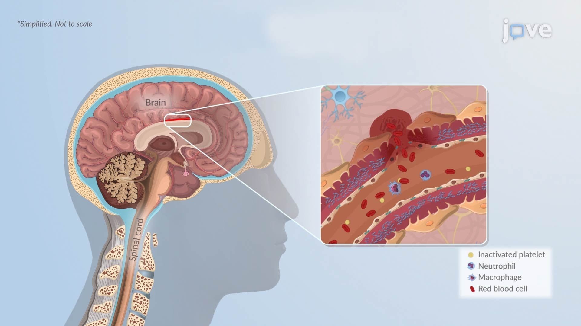
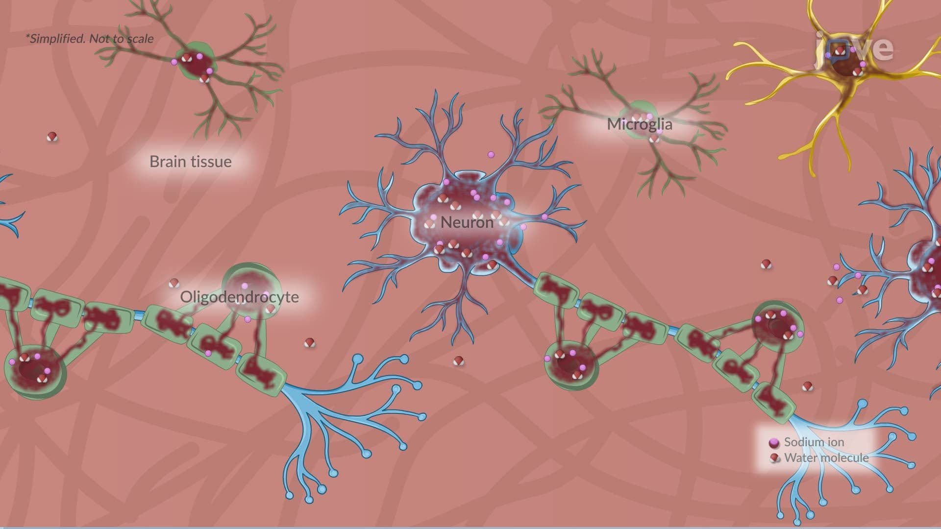
Elevated ICP typically originates from one of three pathological sources: cerebral edema (swelling of brain tissue), a mass effect from hemorrhage or tumor, or obstruction of normal CSF flow, a condition called hydrocephalus. Cerebral edema, for example, is a common complication after traumatic brain injury (TBI), which affects approximately 1.5 million Americans annually according to the CDC. When any of these processes adds volume to the intracranial space, pressure rises because the skull cannot expand to compensate.
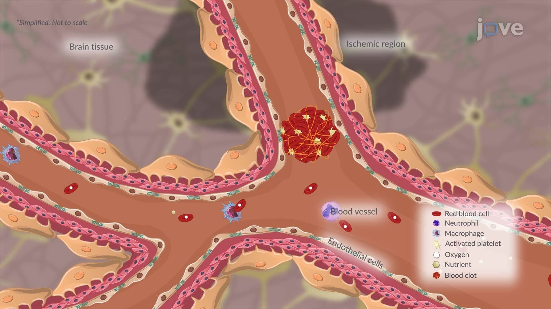
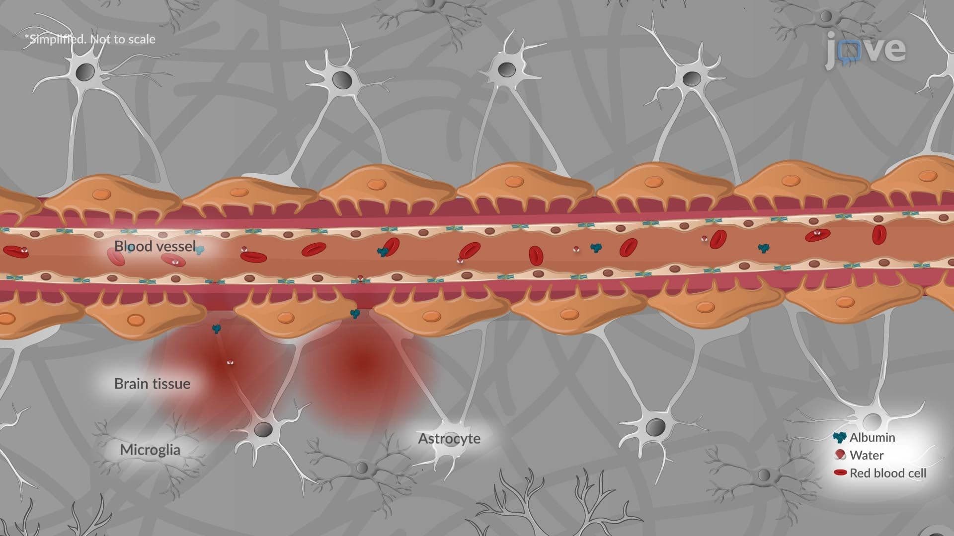
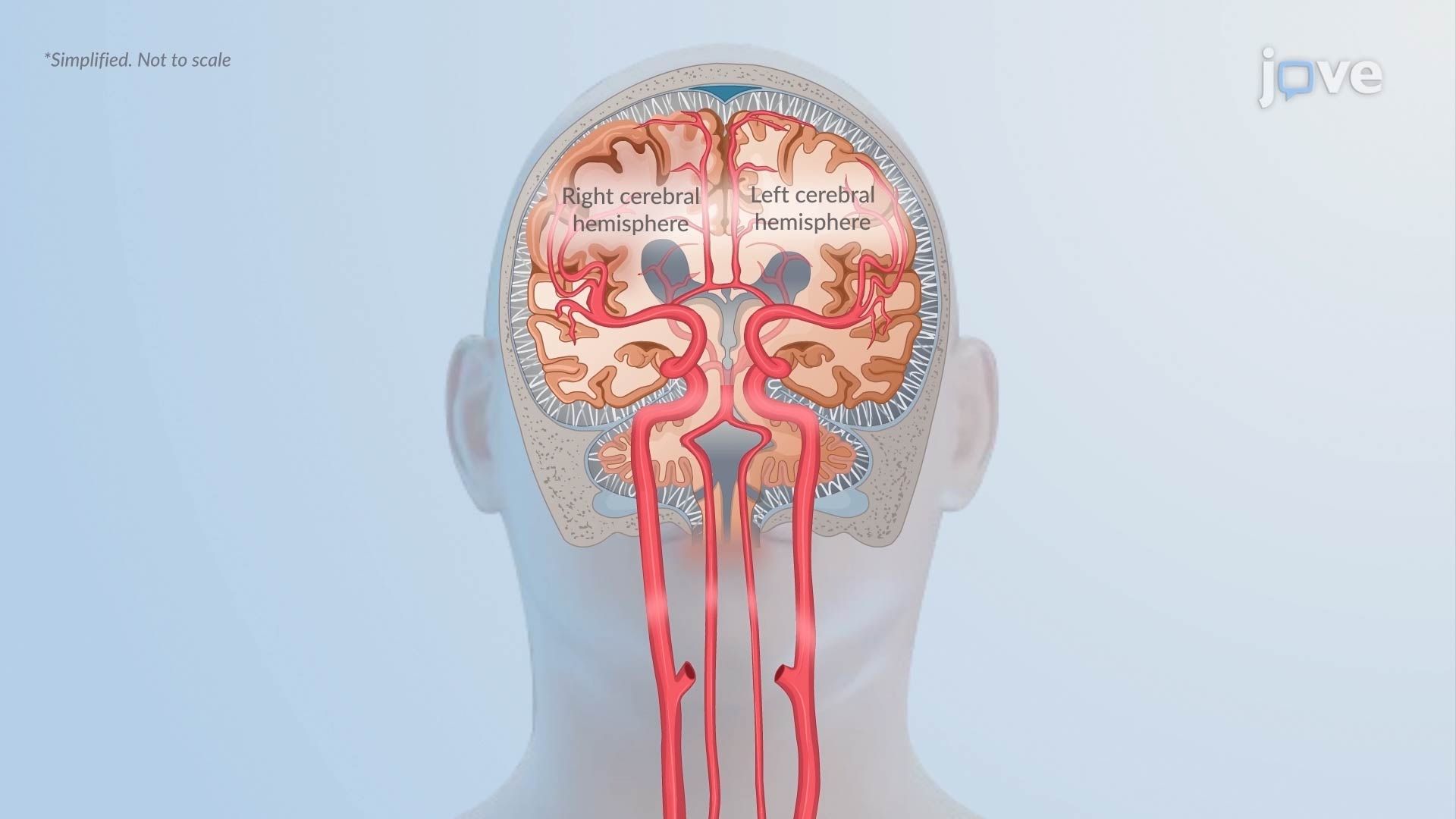
Under healthy conditions, the brain's cerebral vessels actively constrict or dilate to maintain a steady level of blood flow, called cerebral perfusion, regardless of fluctuations in systemic blood pressure. This process is known as cerebral autoregulation. As ICP climbs and edema worsens, this protective mechanism breaks down. Once autoregulation fails, cerebral blood flow becomes entirely dependent on systemic blood pressure. This makes the brain dangerously vulnerable to hyperperfusion, where excess blood flow drives further swelling, creating a self-reinforcing cycle that accelerates injury.
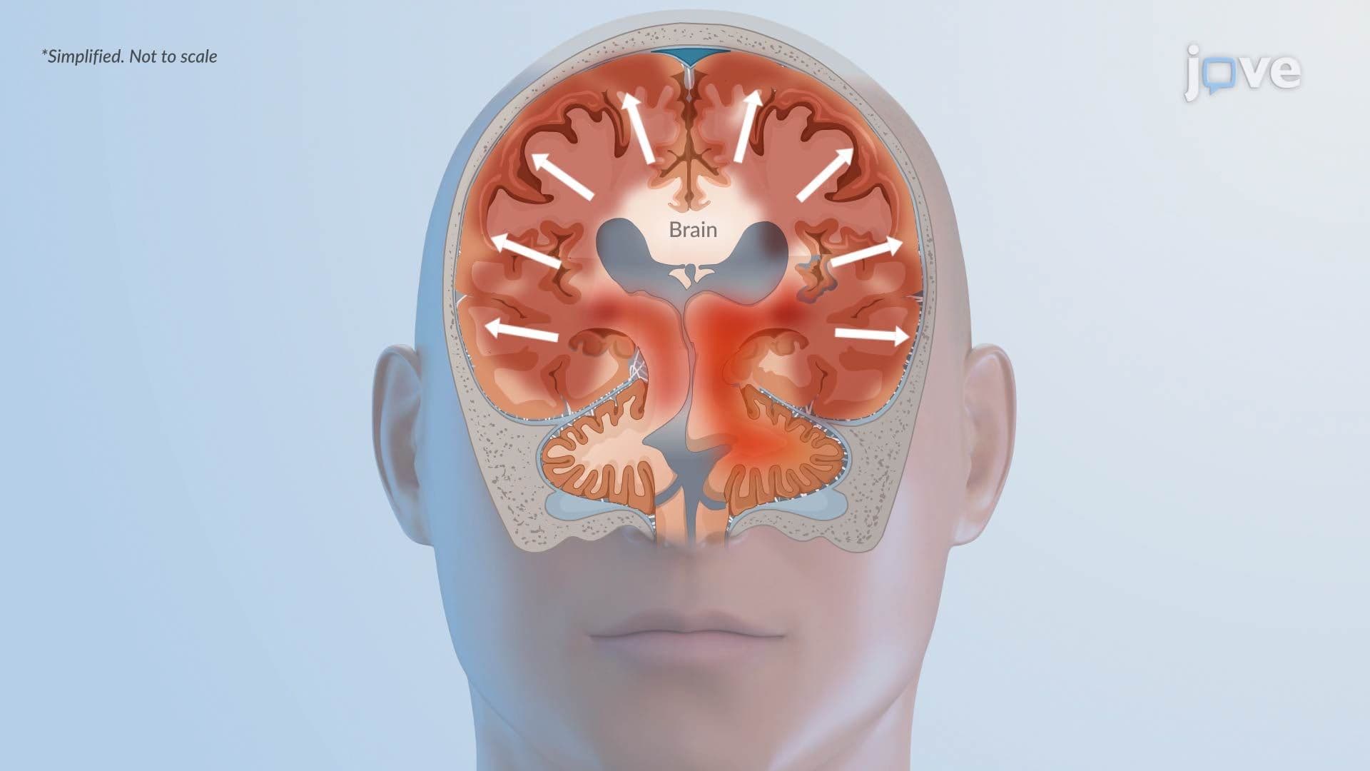
Because the skull and dura mater are non-expandable rigid structures, rising pressure must go somewhere. Brain tissue is physically displaced from high-pressure regions toward lower-pressure regions, a process called herniation. Two major types are tested frequently on exams like the MCAT and USMLE:
Both syndromes can rapidly impair vital functions including respiration, cardiovascular control, and consciousness. These clinical presentations are high-yield topics in AP Biology, college-level neuroscience courses, and health profession licensing exams.
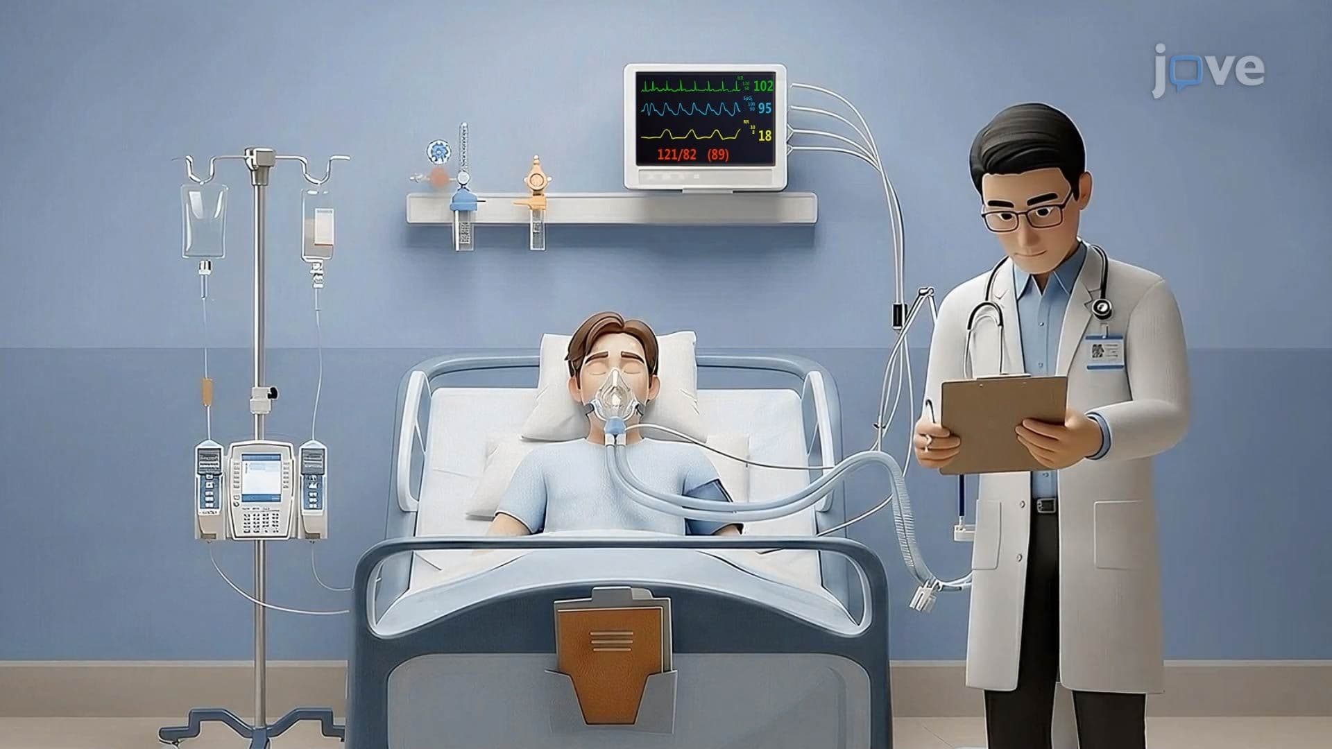
As ICP rises, cerebral perfusion pressure (CPP), calculated as mean arterial pressure minus ICP, drops critically. When CPP falls below approximately 50-60 mmHg, neurons begin experiencing ischemia, meaning they are deprived of the oxygen and glucose they need to survive. Without timely clinical intervention, such as osmotherapy with mannitol, surgical decompression, or CSF drainage, widespread neuronal degeneration occurs. This irreversible damage is the endpoint of the pathophysiology in increased intracranial pressure ll basics, and it underscores why rapid recognition and treatment of elevated ICP is one of the most urgent priorities in neurocritical care medicine across US hospitals.
Related Micro-courses