Loading video…

 35 Concepts
 5.1K views

Video Summary: Pathophysiology in Degenerative Disc Disease Ll
Did you know a spinal disc has no blood supply of its own, yet it must stay alive to protect your entire nervous system? Understanding pathophysiology in degenerative disc disease II reveals exactly how that fragile balance breaks down. At US spine clinics, millions of patients annually experience this cascade, from disc height loss to nerve inflammation. Watch the full video on JoVE Coach to master this concept with expert-led visuals and step-by-step explanations.
Degenerative disc disease (DDD) is not a single event, it is a compounding biological failure involving mechanics, nutrition, and immunity working against the spine simultaneously. At the heart of pathophysiology in degenerative disc disease II is the recognition that symptoms arise from three interconnected mechanisms, each capable of amplifying the others. This layered understanding is essential for students in anatomy, pathophysiology, and pre-health tracks across US colleges.
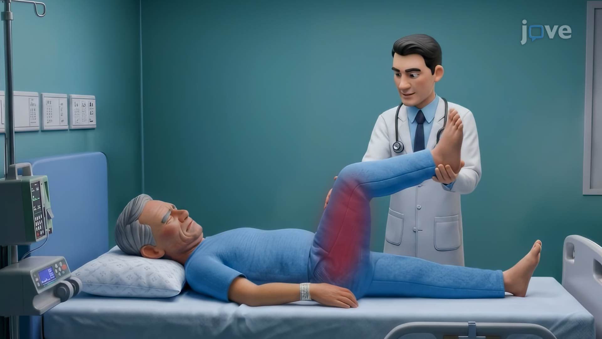
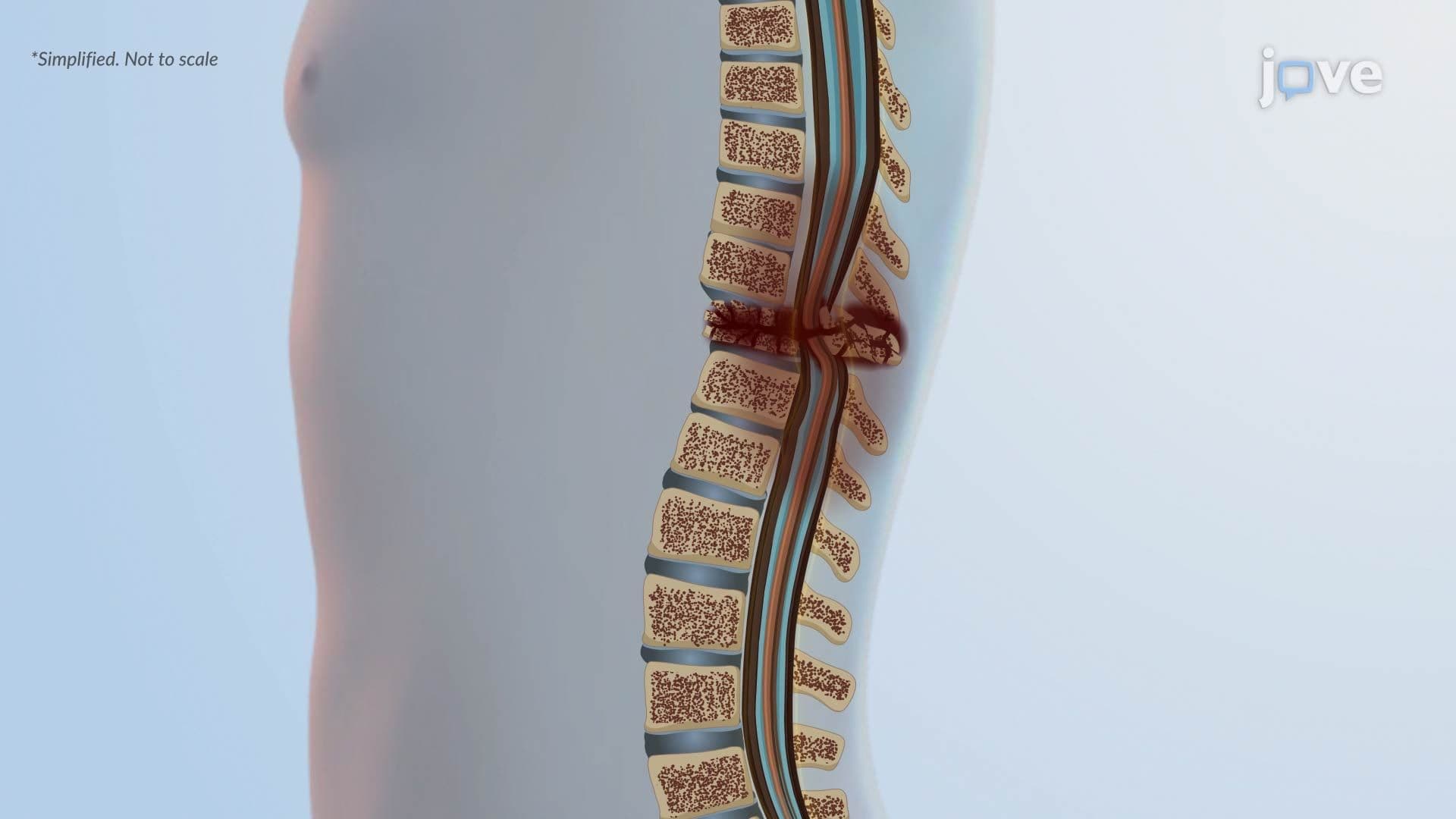
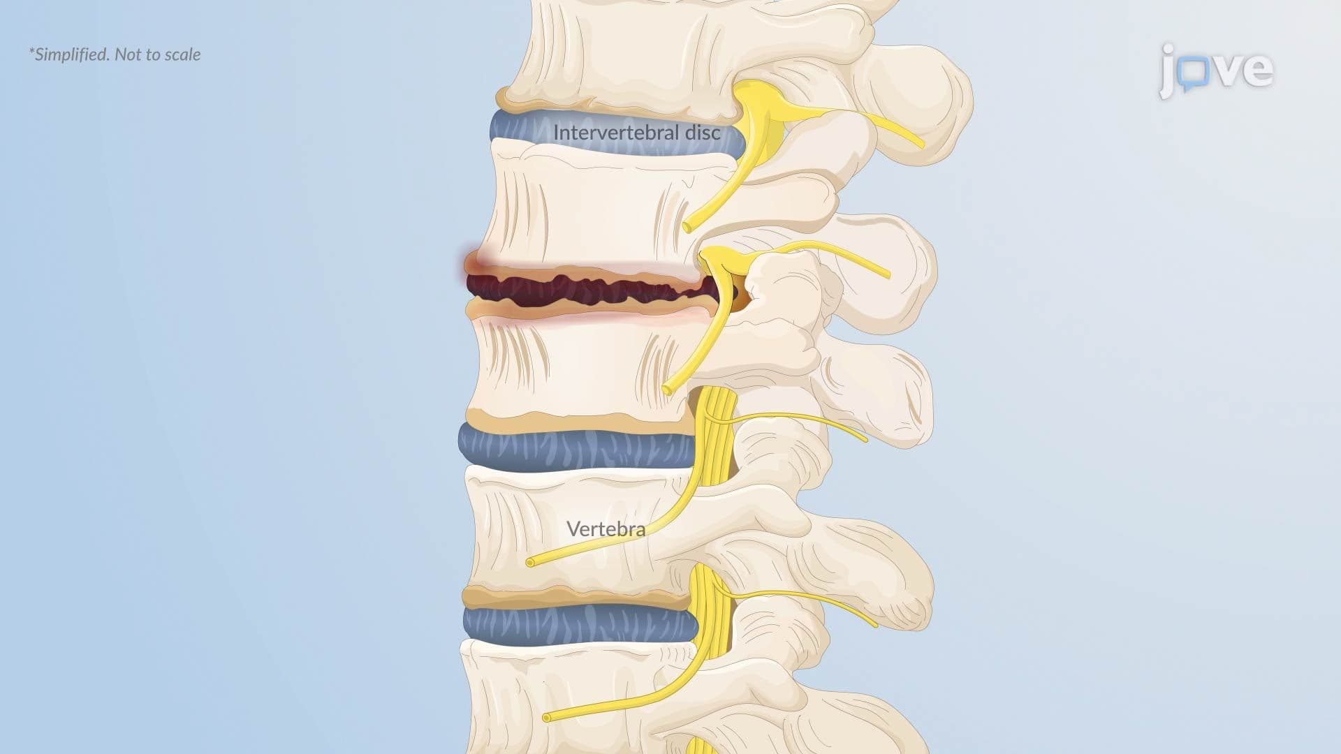
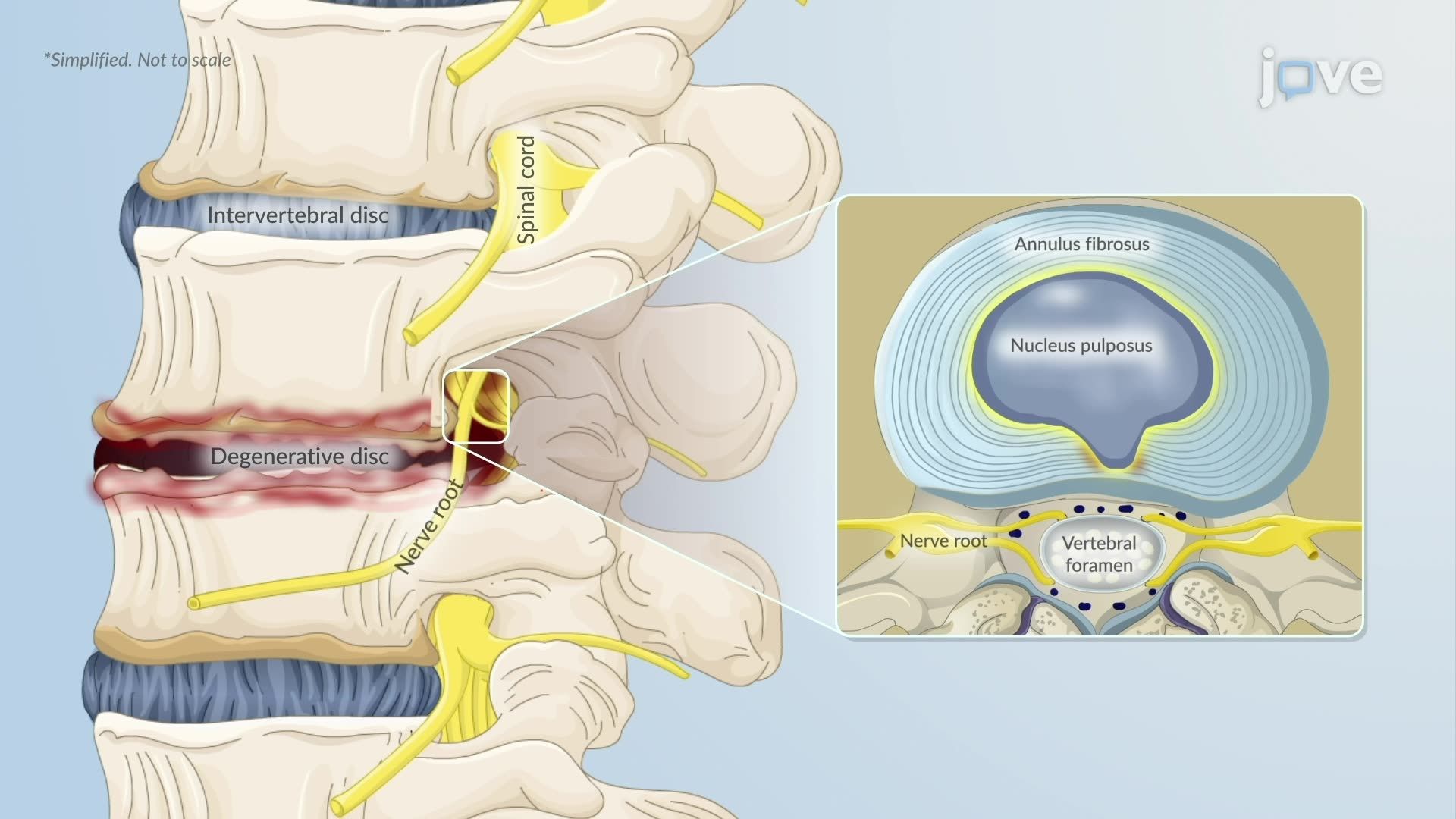
As intervertebral discs lose height and elasticity over time, the cushioning they normally provide between vertebrae diminishes. This structural breakdown increases the probability that the nucleus pulposus, the gel-like inner core of the disc, will herniate outward through weakened annular fibers. When herniated material encroaches on adjacent nerve roots, the result is radicular pain: sharp, radiating discomfort that often travels down the arm or leg depending on spinal level. In clinical settings across the United States, lumbar disc herniations compressing the sciatic nerve are one of the most common causes of lower back and leg pain evaluated by orthopedic and neurology specialists.
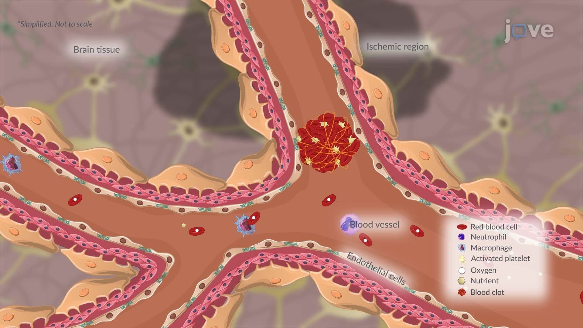
Unlike most tissues in the body, intervertebral discs contain no blood vessels. They depend entirely on passive diffusion of nutrients, glucose, oxygen, through cartilaginous endplates from capillaries in adjacent vertebral bodies. When degenerative changes cause endplate sclerosis (hardening and thickening of the endplate), this diffusion pathway narrows. Disc cells, called chondrocytes and nucleus pulposus cells, become starved of nutrients, triggering apoptosis (programmed cell death) and accelerating the loss of disc matrix integrity. This mechanism is a high-yield concept in US college-level physiology and is frequently tested in pre-medical coursework and on the MCAT under musculoskeletal and connective tissue systems.
Perhaps the most clinically important, and often surprising, insight in DDD pathophysiology is that significant pain can occur without measurable nerve compression. When disc material herniates into the epidural space, the immune system treats it as a foreign body and mounts a local inflammatory response. Cytokines such as TNF-alpha and interleukin-1 are released alongside nitric oxide and matrix metalloproteinases (enzymes that degrade structural proteins). Macrophages are recruited to the site, and nearby nerve roots become chemically sensitized, a process called chemical radiculitis. This explains why some patients with minimal structural compression on MRI still report severe, debilitating pain.
In healthy adults, the outer third of the annulus fibrosus contains some nerve endings, but the inner disc is largely aneural and avascular. In chronic degeneration, however, inflammatory mediators stimulate abnormal ingrowth of both new nerve fibers and blood vessels deep into disc tissue. This neoinnervation and neovascularization transforms previously insensate disc tissue into a persistent pain generator. For students preparing for USMLE Step 1, NCLEX, or upper-division pathophysiology courses at US universities, understanding this distinction between acute mechanical pain and chronic chemical/neurogenic pain is critical for clinical reasoning.
Related Micro-courses