Loading video…


 18 Concepts
 4.3K views

Video Summary: Inflammatory Response in Acute Inflammation I
Ever wonder why a sprained ankle swells up within minutes? The inflammatory response in acute inflammation I explains exactly that, your body's rapid-fire defense system kicking into gear. When US emergency rooms treat trauma patients, this same cascade is happening at the cellular level. Mast cells, macrophages, and cytokines like TNF and IL-1 orchestrate redness, swelling, heat, and pain. Watch the full video on JoVE Coach to master this concept with expert-led visuals and step-by-step explanations.
The inflammatory response in acute inflammation I is one of the most fundamental concepts in biology and medicine, and it is far more organized than the redness and swelling you see on the surface. Acute inflammation is the body's immediate, short-term defense response, typically lasting from minutes to several days. It is triggered by harmful stimuli including bacterial or viral infections, physical trauma like a broken bone, or chemical injury from toxins. Understanding this process is central to courses in AP Biology, college-level Cell Biology, Anatomy and Physiology, and Pathophysiology, and it appears regularly on exams like the MCAT, USMLE Step 1, NCLEX, and even AP Biology free-response questions.
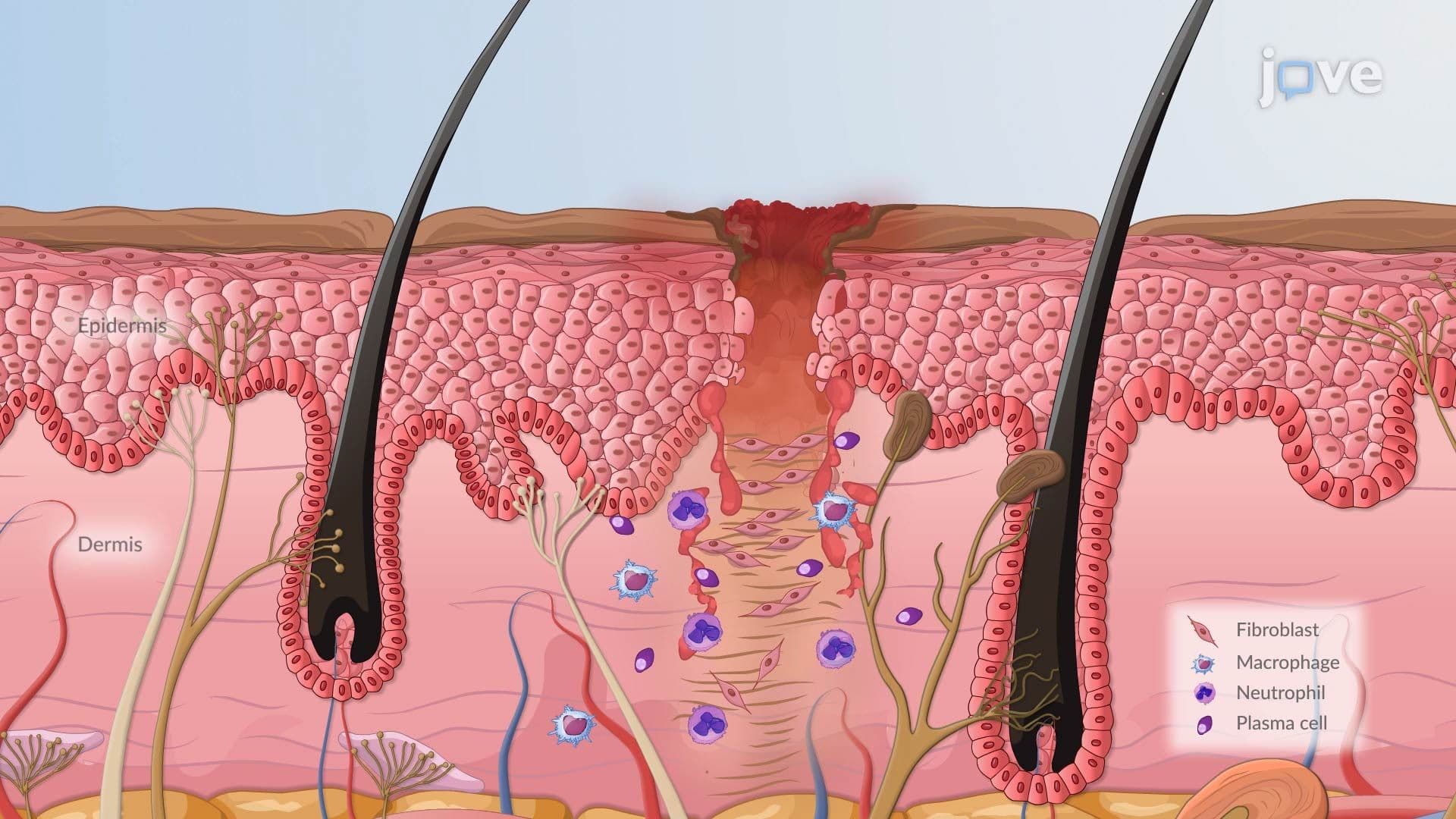
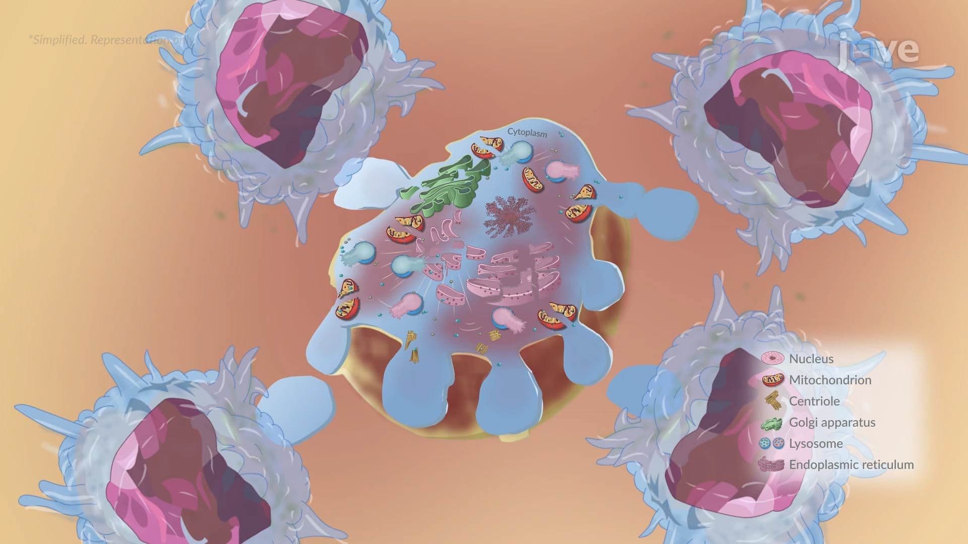
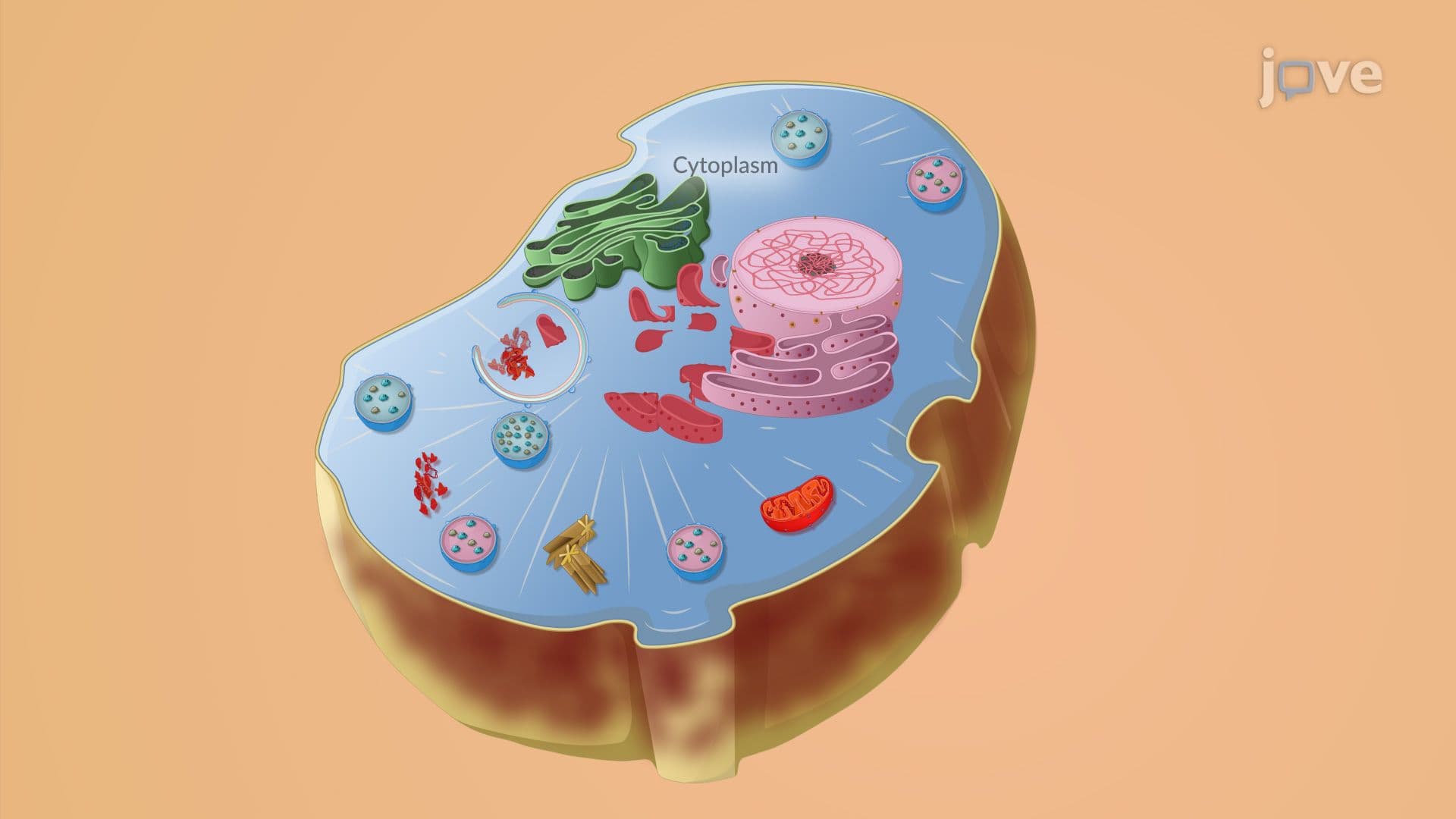
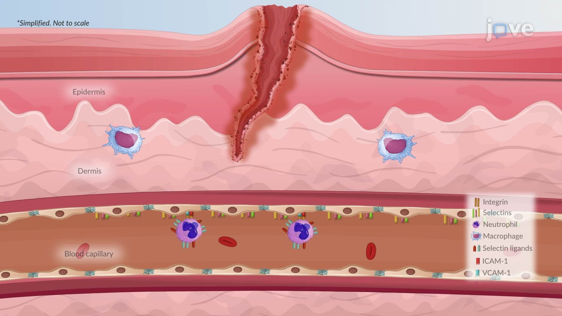
The cascade begins at the molecular level. Resident immune cells, mast cells, macrophages, and dendritic cells, are stationed throughout body tissues like sentinels. They carry surface proteins called pattern-recognition receptors (PRRs), which are specifically designed to detect two categories of danger signals. Pathogen-associated molecular patterns (PAMPs) are molecular signatures found on bacteria, viruses, and fungi, things the human body does not naturally produce. Damage-associated molecular patterns (DAMPs) are distress signals released by injured or dying human cells, a concept directly tied to studying cellular injury and necrosis. When PRRs bind to either type of signal, the immune cell activates and begins releasing powerful chemical messengers known as inflammatory mediators.
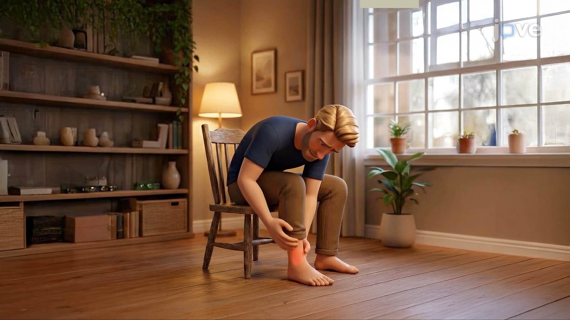
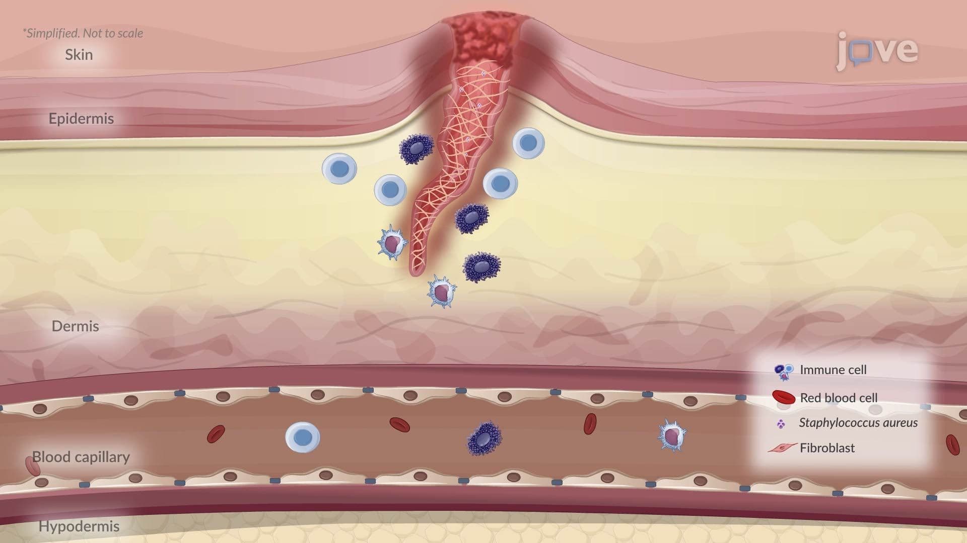
Once activated, mast cells release histamine, and other cells produce prostaglandins, mediators that cause local blood vessels to dilate (vasodilation) and become more permeable. This explains the four cardinal signs of inflammation first described by ancient Roman physician Celsus and still taught in every US medical and nursing school today: *rubor* (redness/erythema from increased blood flow), *calor* (heat from the same cause), *tumor* (swelling/edema from fluid leaking out of permeable vessels), and *dolor* (pain, generated when prostaglandins and bradykinin sensitize local nerve endings). A relatable US example: when a student at a high school football game rolls their ankle, these exact vascular events are responsible for the rapid swelling and throbbing pain that follow within minutes.
Perhaps the most exam-critical part of this topic is leukocyte recruitment, the process by which white blood cells are delivered from the bloodstream into the affected tissue. Cytokines, specifically TNF (tumor necrosis factor) and IL-1 (interleukin-1), signal the endothelial cells lining blood vessels to display surface proteins called selectins and adhesion molecules. These proteins slow passing leukocytes down, causing them to "roll" along the vessel wall, a step literally called rolling. Tighter adhesion molecules then anchor the leukocyte firmly to the endothelium, and finally the leukocyte squeezes between endothelial cells in a process called diapedesis (or transmigration), entering the tissue to destroy pathogens and clear debris. This sequence, rolling, adhesion, diapedesis, is a classic multi-step answer expected on MCAT and USMLE exams. Once in the tissue, leukocytes (especially neutrophils and macrophages) phagocytose bacteria, release enzymes, and work to resolve the inflammation and initiate healing.
Related Micro-courses