Loading video…

 18 Concepts
 4.3K views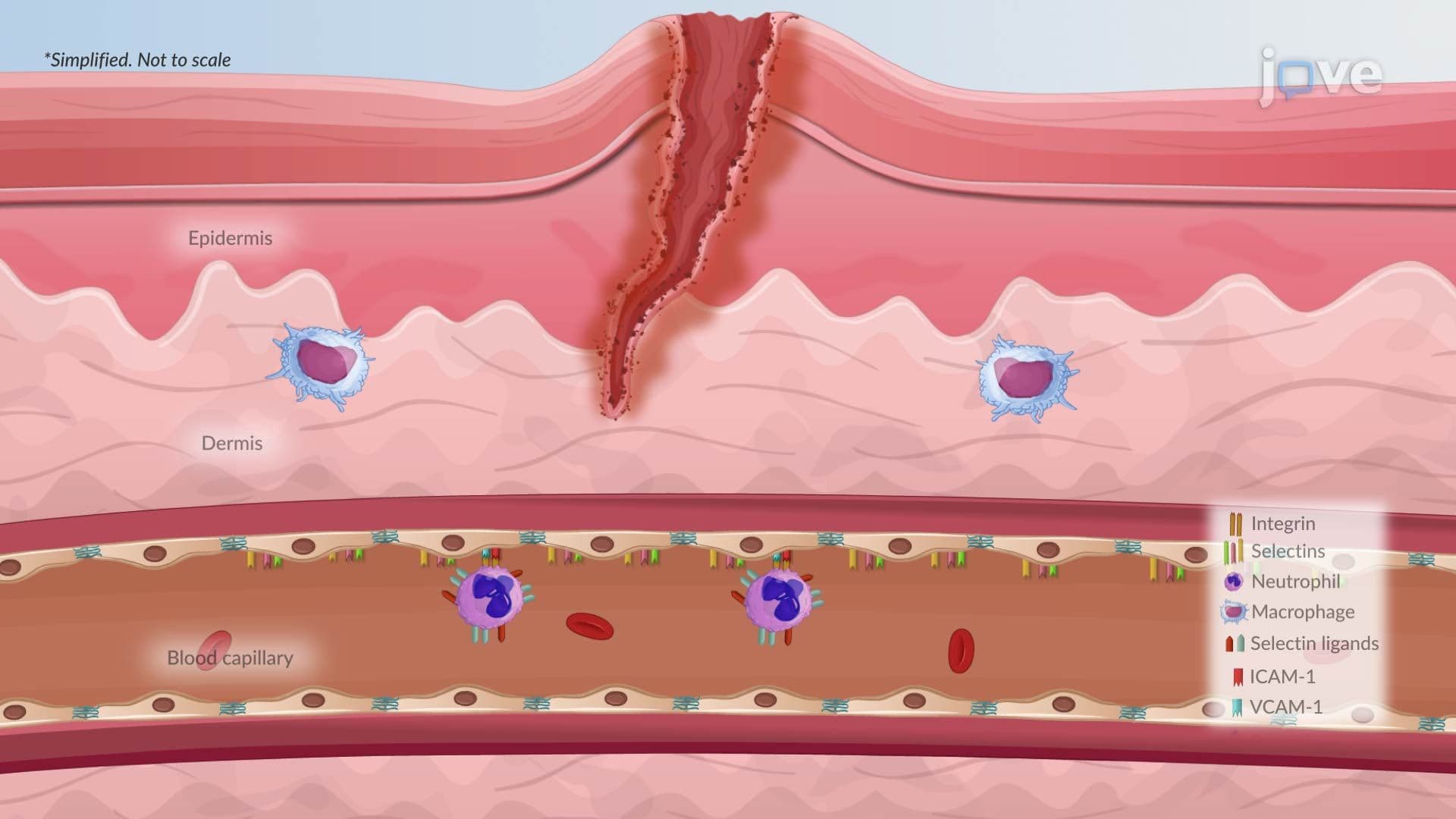

Video Summary: Cellular Phase in Acute Inflammation Ii
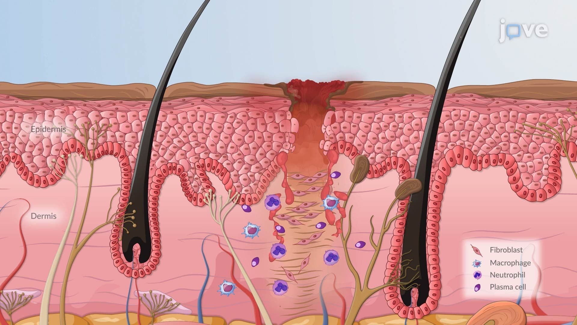
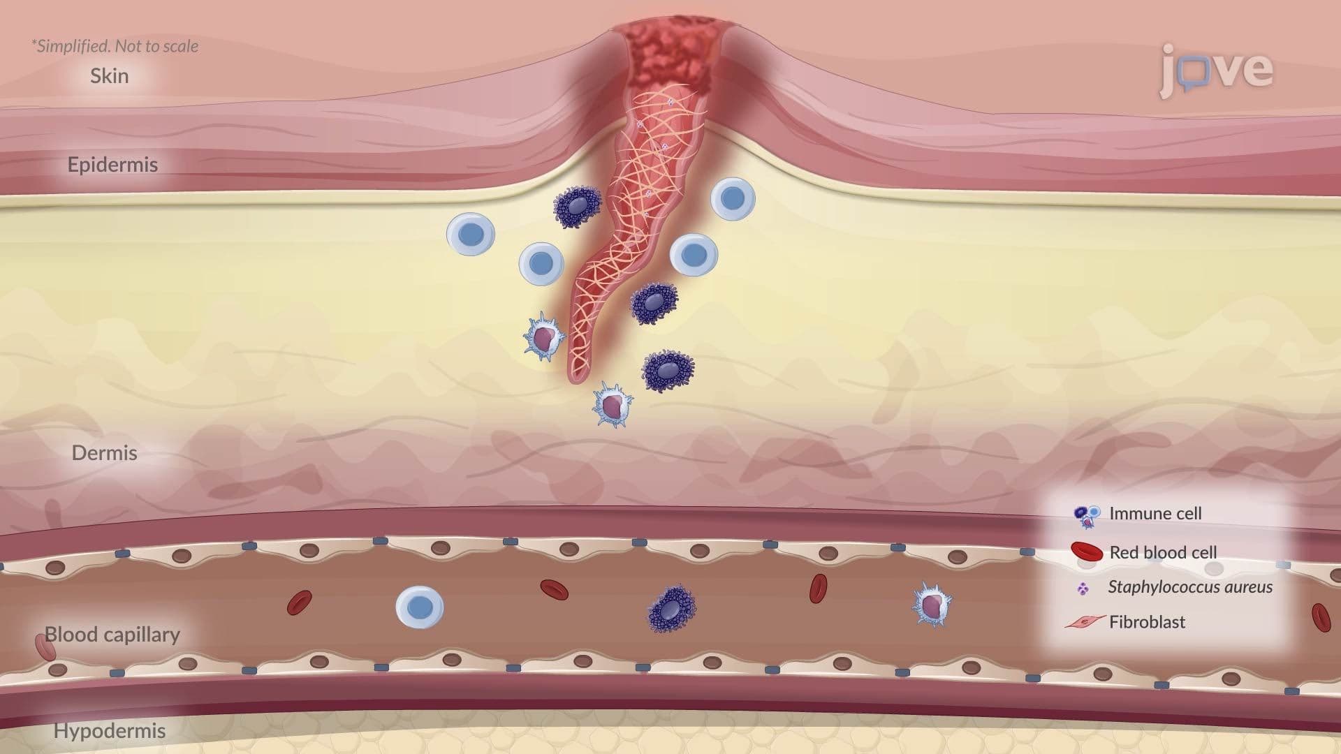
Ever wonder why a simple cut on your finger gets red, swollen, and warm, and then heals? The cellular phase in acute inflammation II explains exactly how your immune cells hunt down pathogens and clean up the damage. In US emergency rooms, this process happens in every infected wound treated daily. Cellular Phase in Acute Inflammation II covers neutrophil recruitment, pathogen destruction, and resolution, or what goes wrong when it doesn't resolve. Watch the full video on JoVE Coach to master this concept with expert-led visuals and step-by-step explanations.
When harmful bacteria invade a tissue, your body doesn't simply send immune cells racing through the bloodstream like firetrucks to a blaze. Instead, a tightly choreographed, multi-step cellular process unfolds, one that determines whether the threat is neutralized quickly and quietly or drags into a prolonged, damaging inflammatory cycle. Cellular Phase in Acute Inflammation II captures this critical sequence, from the moment leukocytes first approach the vessel wall to their ultimate fate after the battle is won, or lost.
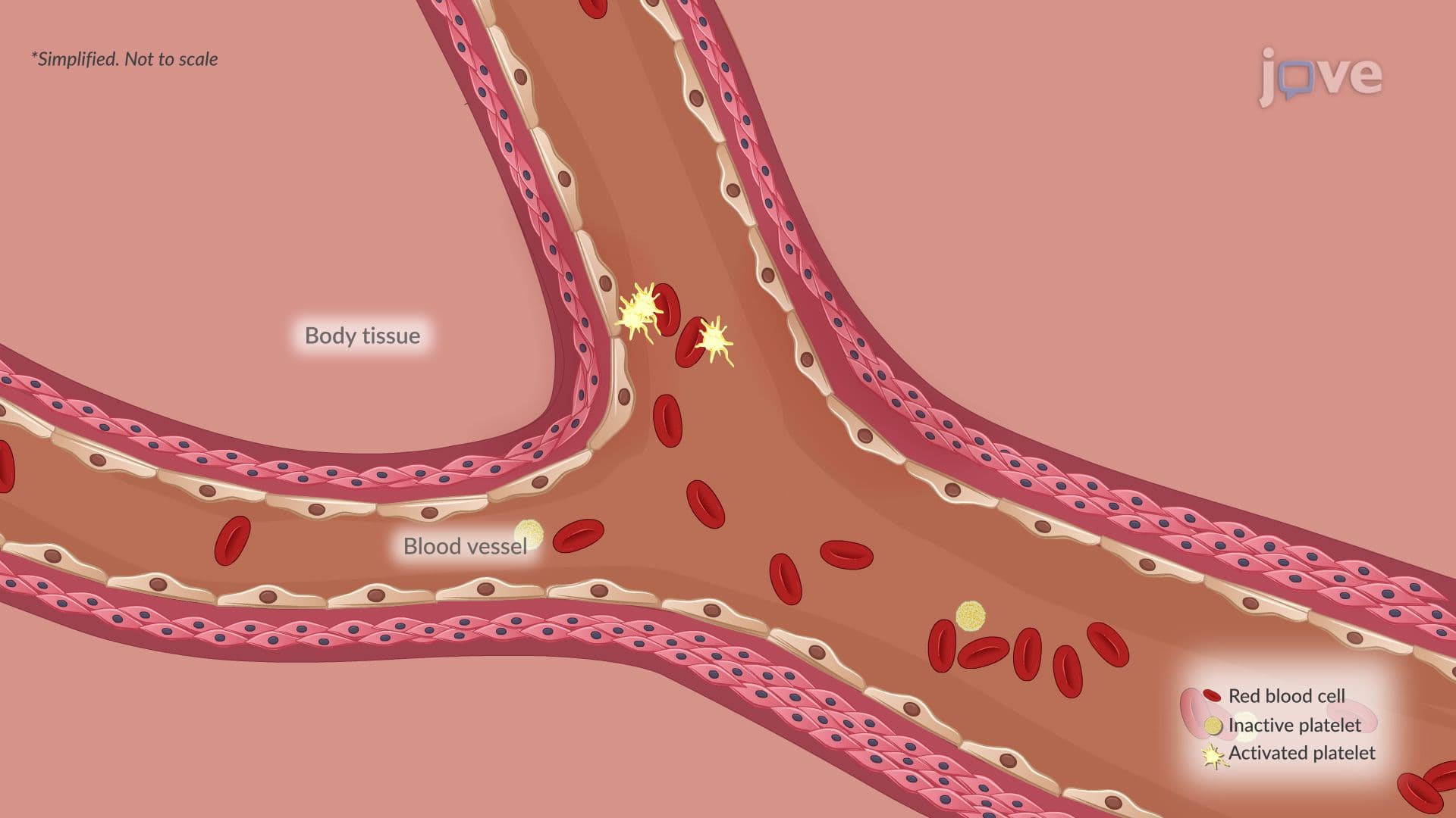
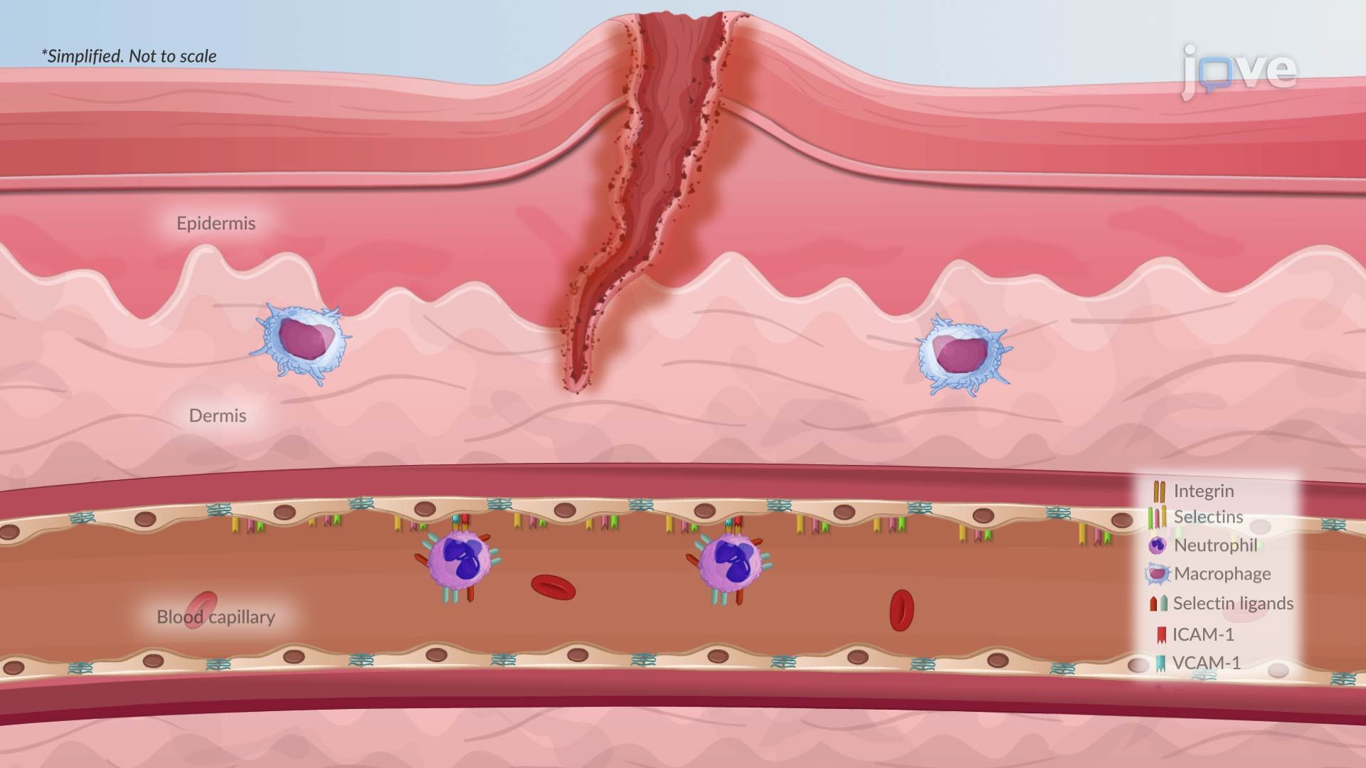
The process begins with margination, a physical shift in blood flow dynamics. As vessels dilate near an injury site, blood flow slows and viscosity rises, pushing larger white blood cells like neutrophils toward the periphery of the vessel. This isn't random; it's a prerequisite for everything that follows.
Next comes rolling, where leukocytes loosely and repeatedly bind to selectin proteins displayed on the activated endothelium. Think of this as slow, tentative contact, the immune cell "sampling" the environment before committing. If the right inflammatory signals are present, the cell transitions to firm adhesion, where integrins on the leukocyte surface lock tightly onto endothelial adhesion molecules, specifically ICAM-1 (Intercellular Adhesion Molecule-1) and VCAM-1 (Vascular Cell Adhesion Molecule-1). This firm grip brings the leukocyte to a complete stop.
Finally, diapedesis occurs, the leukocyte squeezes between neighboring endothelial cells, crosses the basement membrane, and enters the surrounding tissue, guided toward the pathogen by chemical signals in a process called chemotaxis. In US pathology courses at institutions like Johns Hopkins or the University of Michigan, mastering this four-step sequence is fundamental to understanding how diseases develop at the cellular level.
Once a neutrophil reaches its target, it engulfs the pathogen through phagocytosis, forming a sealed compartment called a phagolysosome. Inside this microscopic "kill chamber," two powerful weapons are deployed. Reactive oxygen species (ROS), including superoxide and hydrogen peroxide, chemically destroy the pathogen's cellular components. Proteolytic enzymes such as elastase and myeloperoxidase break down proteins and cell walls. Together, these mechanisms represent the body's frontline cellular defense, a concept directly connected to understanding cellular injury and death in any introductory pathophysiology course.
After eliminating the threat, neutrophils are programmed to self-destruct through apoptosis, a clean, orderly cell death. Nearby macrophages then quietly engulf the dead neutrophils in a process called efferocytosis, preventing their toxic contents from leaking. When this clearance is delayed, which can happen in severe or persistent infections, neutrophil enzymes spill into surrounding tissue, amplifying damage and prolonging inflammation. This failure of resolution is a central concept in understanding how diseases develop, and it explains why chronic inflammatory conditions like COPD or rheumatoid arthritis arise in US patients.
When the injurious stimulus is successfully eliminated, the body releases anti-inflammatory signals and pro-resolving mediators, such as lipoxins and resolvins, that actively restore tissue homeostasis. This is inflammation and repair working as intended. However, if the stimulus persists or resolution mechanisms fail, acute inflammation transitions into chronic inflammation, shifting the cellular players from neutrophils to macrophages and lymphocytes. Understanding this transition is tested on the MCAT, USMLE Step 1, and AP Biology exams, making it one of the highest-yield concepts in introductory biological science and pre-medical curricula.
Related Micro-courses