Loading video…

 18 Concepts
 4.3K views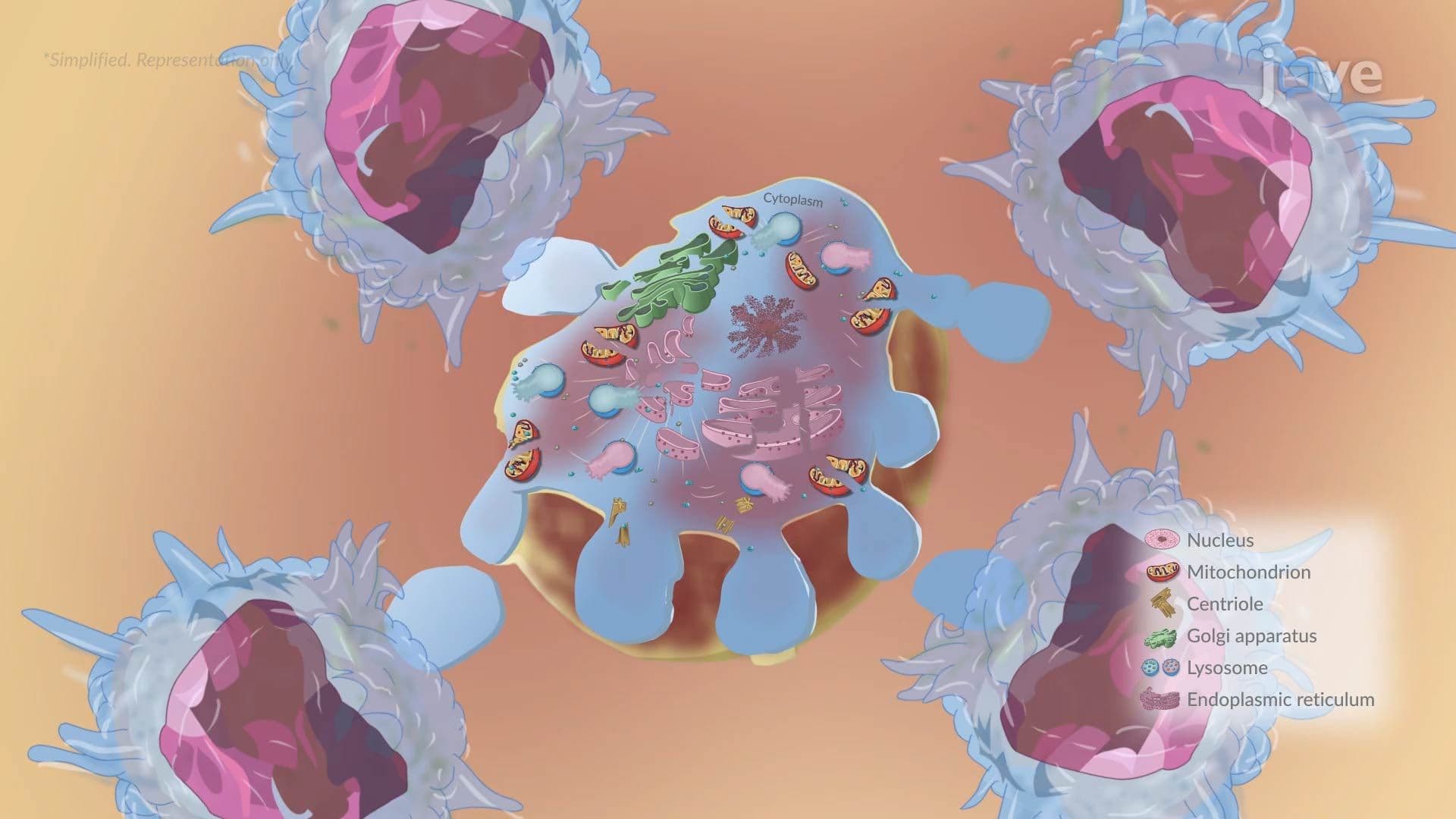

Video Summary: Cellular Death in Cellular Injury Ili
What actually happens inside your cells when they start dying, and can the process be controlled? Cellular Death in Cellular Injury III breaks down the two major pathways of cell death: necrosis and apoptosis. Think of a stroke patient in a US emergency room, their oxygen-deprived brain cells undergo rapid, destructive necrosis. Watch the full video on JoVE Coach to master this concept with expert-led visuals and step-by-step explanations.
Cell death is not a single event, it is a biological outcome that comes in fundamentally different forms depending on the cause and context. Cellular Death in Cellular Injury III focuses on the two core pathways that biology and medicine students must understand: necrosis and apoptosis. Mastering the distinction between these two mechanisms is essential not just for exams, but for understanding how disease progresses in real patients.
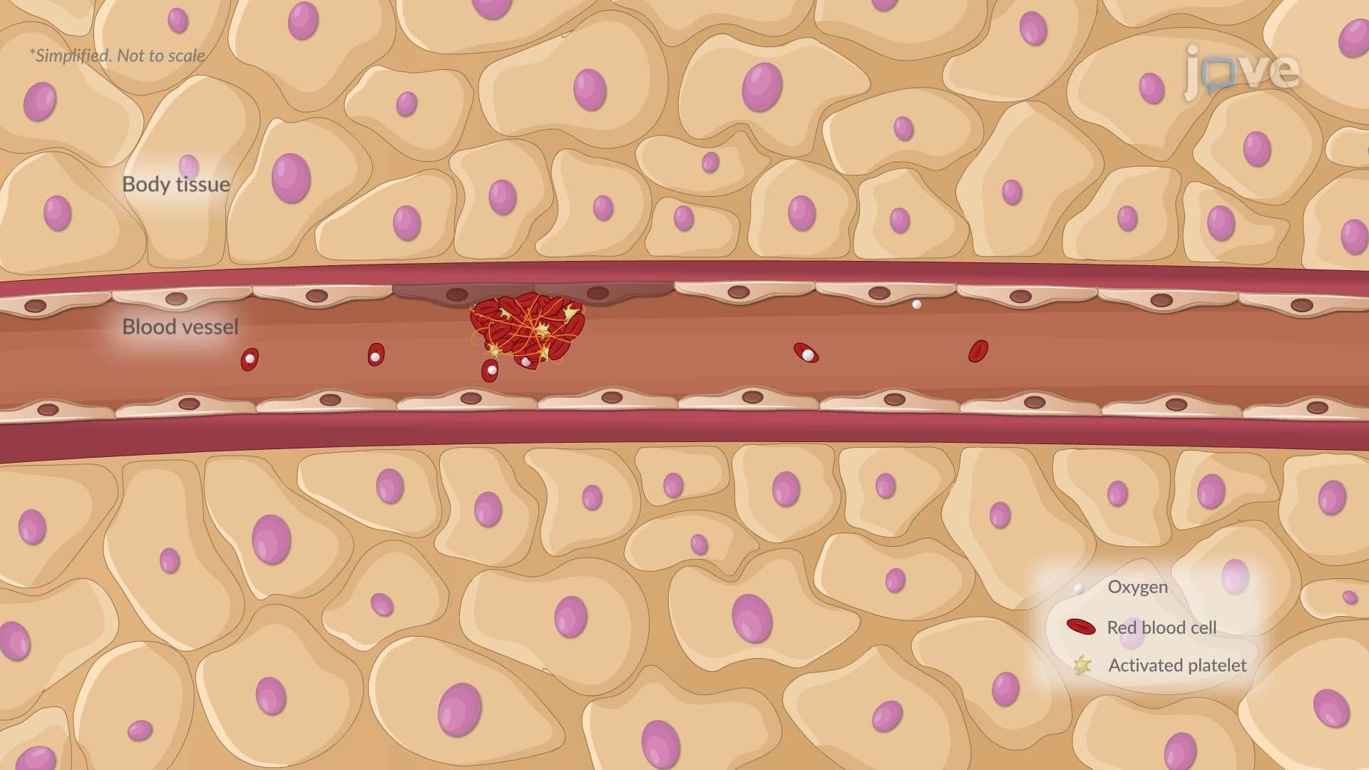
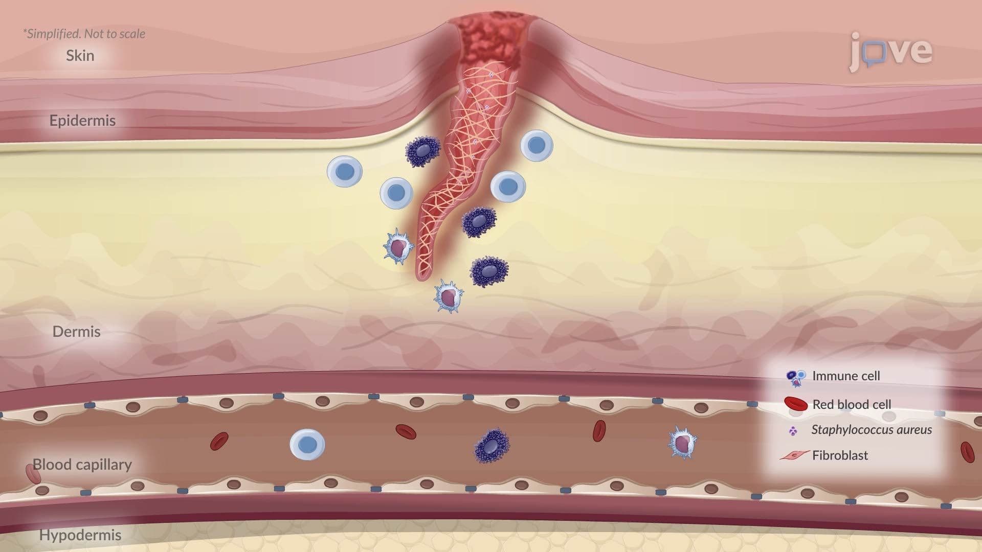
Necrosis occurs when cells are overwhelmed by external injury, such as a lack of blood supply (ischemia), physical trauma, toxin exposure, or infection. When blood flow is cut off to heart tissue during a myocardial infarction (heart attack), for example, the affected cardiac cells rapidly lose their ability to maintain normal membrane function. Sodium and water flood in, causing the cell to swell dramatically. Eventually, the membrane ruptures.
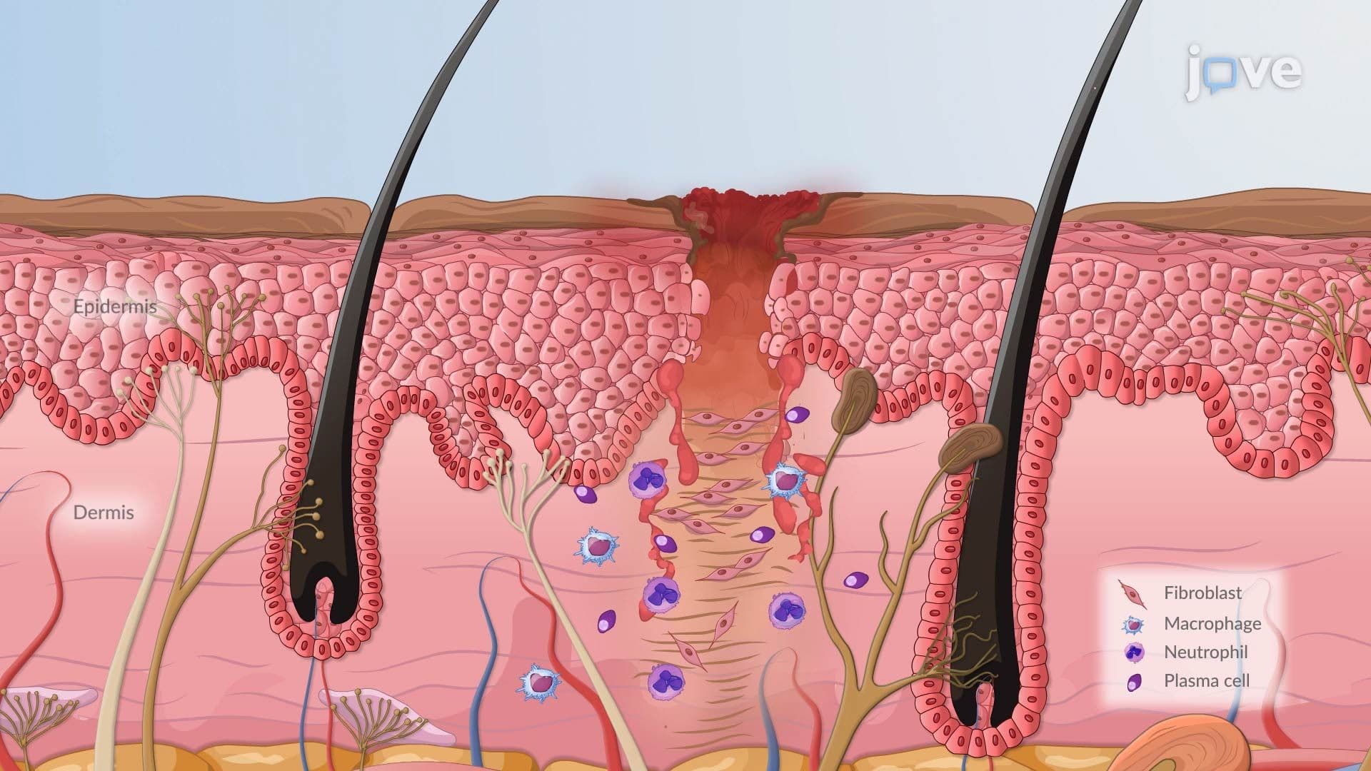
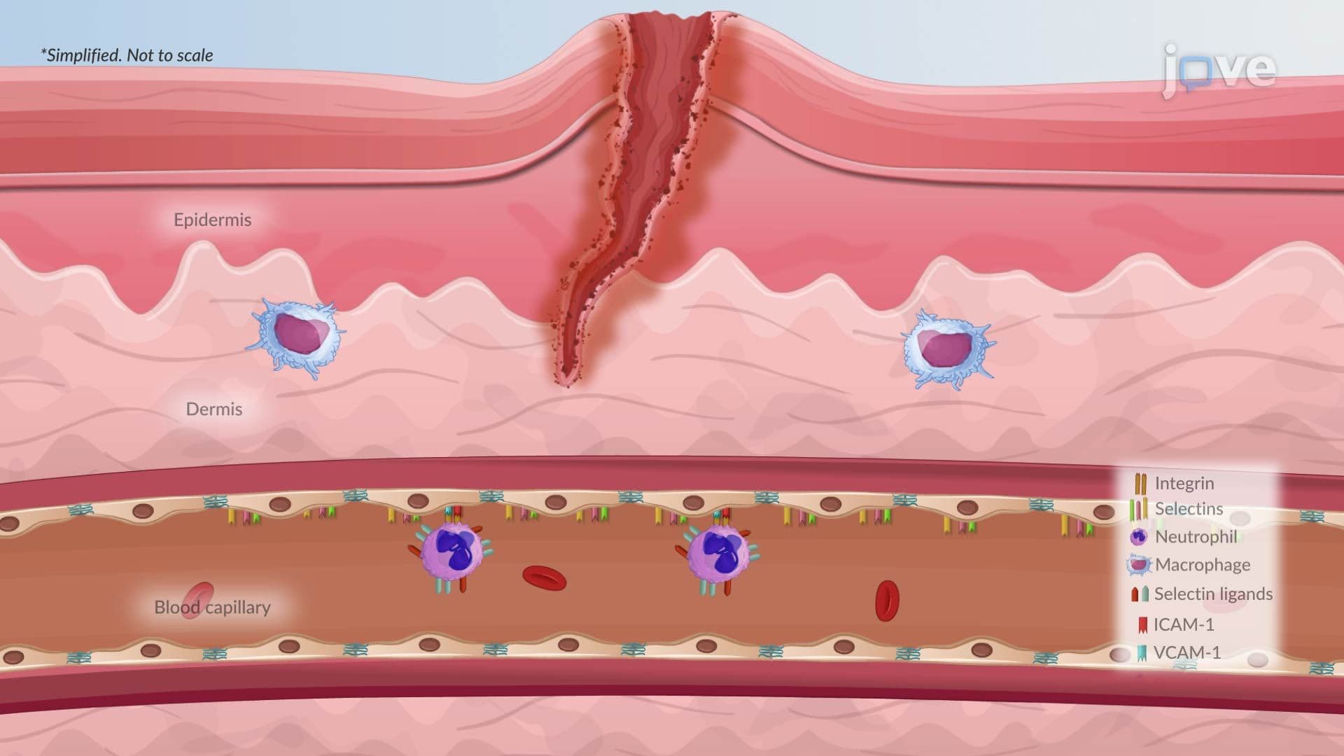
Once membrane integrity is lost, lysosomal enzymes, the cell's own digestive machinery, spill into the surrounding tissue. This triggers a robust inflammatory response. White blood cells, particularly neutrophils and macrophages, rush to the site to clear the cellular debris. This inflammation is what causes the redness, swelling, and pain commonly associated with tissue injury. In the clinical setting, markers like troponin in blood tests help US physicians detect necrotic cardiac cells after a heart attack.
Necrosis is considered passive and unregulated, the cell has no control over its own destruction.
Unlike necrosis, apoptosis is a precisely regulated, energy-dependent process often called "programmed cell death." It is not a sign of catastrophic failure, it is a necessary biological tool. During human embryonic development, for instance, apoptosis is responsible for separating fingers and toes by eliminating the webbing between them. It also eliminates immune cells that would otherwise attack the body's own tissues.
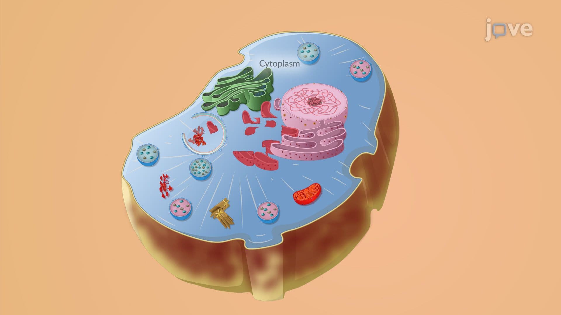
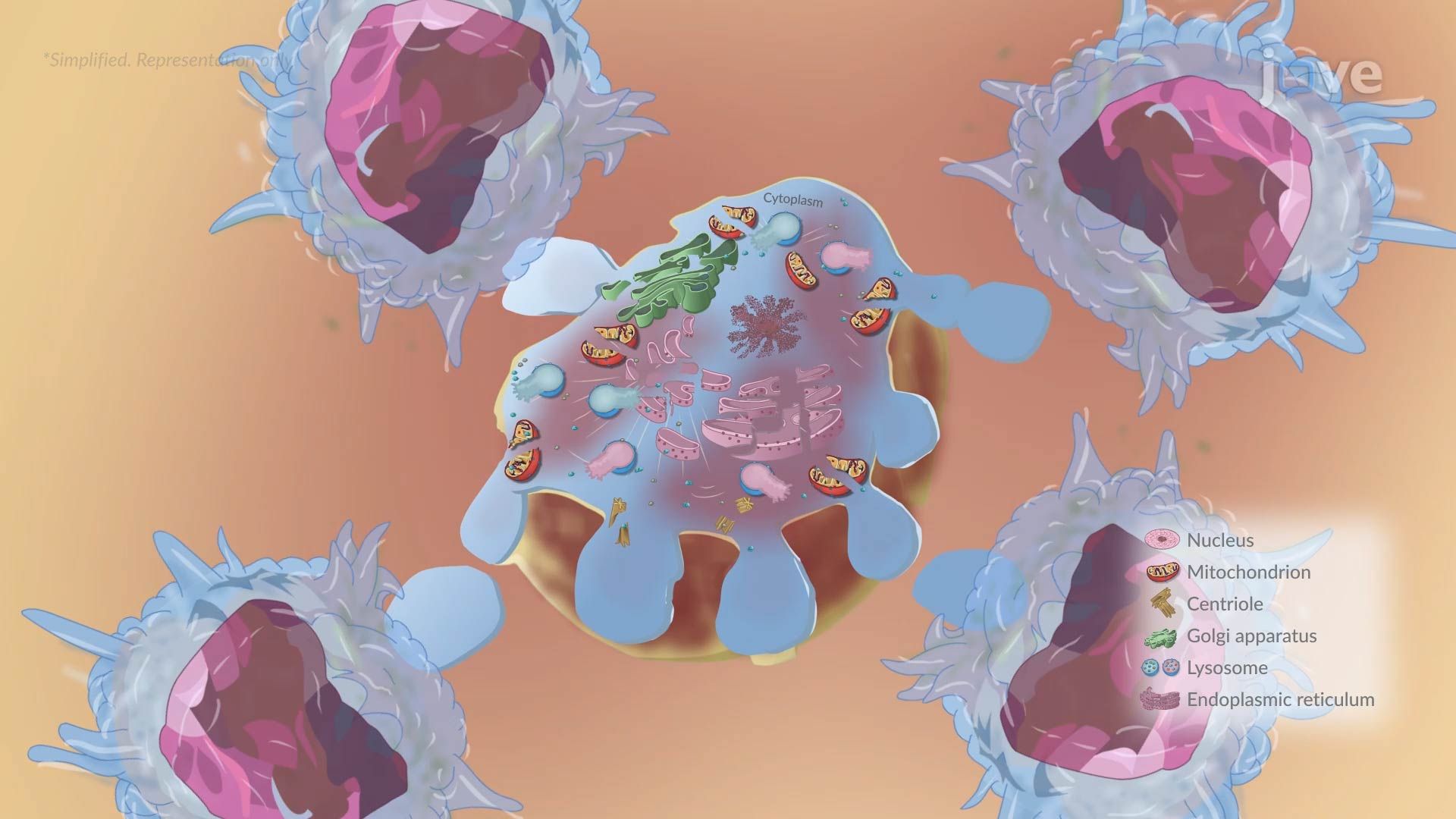
When a cell is targeted for apoptosis, a predictable sequence unfolds: the cell shrinks, its chromatin condenses and fragments within the nucleus, and the cell membrane begins to bubble outward in a process called blebbing. The cell then breaks apart into small, membrane-enclosed packets called apoptotic bodies. Critically, these fragments display surface signals that attract macrophages and neighboring cells, which quietly engulf and recycle the material, all without triggering inflammation.
This clean removal process is what separates apoptosis from necrosis at the biological level.
Understanding the difference between necrosis and apoptosis has direct implications for how diseases develop and how they are treated. In cancer biology, tumor cells often develop mutations that disable apoptosis, allowing damaged or abnormal cells to survive and multiply unchecked. Many cancer therapies, including chemotherapy drugs used in US oncology, work by reactivating apoptotic pathways in tumor cells.
Conversely, excessive or unwanted apoptosis plays a role in neurodegenerative diseases like Alzheimer's disease, where neurons that should survive are instead eliminated. Researchers at institutions like the NIH and major US universities are actively investigating how to modulate these pathways for therapeutic benefit.
In AP Biology, students are expected to understand how cell communication and gene regulation connect to programmed cell death. On the MCAT, necrosis and apoptosis appear in the context of organ system pathology, inflammation, and cell biology. In college-level introductory biology and pathophysiology courses, instructors often test students on the specific morphological differences between the two, such as whether inflammation is triggered (necrosis: yes; apoptosis: no). Creating a simple side-by-side comparison chart of the two pathways is one of the most efficient study strategies for locking in these distinctions before an exam.
Related Micro-courses